Vol.2, No.3, 200-203 (2010) Health
doi:10.4236/health.2010.23029
Copyright © 2010 SciRes Openly accessible at http://www.scirp.org/journal/HEALTH/
Left ventricular noncompaction associated with
hypertrophic cardiomyopathy and
Wolff-Parkinson-White syndrome
Luis Alday1, Eduardo Moreyra1, Eva Bruno2, Norma Rossi3, Hector Maisuls2
1Divisions of Pediatric Cardiology and Cardiology, Sanatorio Allende, Córdoba, Argentina; lealday@arnet.com.ar
2Division of Cardiology, Children’s Hospital, Cordoba, Argentina
3Division of Genetics, Private Hospital, Cordoba, Argentina
Received 14 October 2009; revised 10 December 2009; accepted 14 December 2009.
ABSTRACT
We report a 35-year-old female patient with hy-
pertrophic cardiomyopathy, left ventricular non-
compaction, and Wolff-Parkinson-White EKG patt-
ern. Several other family members present the
same clinical condition. We speculate that this
phenotype is related to the genotypes PRKAG2
and LAMP2 represented by mutations of the
genes encoding AMP-activated protein kinase
(PRKAG2) and lysosome associated membrane
protein 2 (LAMP2 ).
Keywords: Left Ventricular Noncompaction;
Hypertrophic Cardiomyopathy;
Wolff-Parkinson-White Syndrome
1. INTRODUCTION
Left ventricular noncompaction (LVNC) is the result
from the a rrest in m yocardi al dev elopm ent w ith pe rsist ent
sinusoid tracts interspersed with prominent muscular
trabeculae. Affected patients usually develop congestive
heart failure associated with arrhythmias and systemic
thromboembolism.W e report a proband with mental retar-
dation and peculiar somatic findings, associated LVNC
and hypertrophic cardiomyopathy (HCM). Other family
members were also involved, all of them, the proband
included, had Wolff-Parkinson-White (WPW) syndrome,
suggesting a relationship regarding the etiology of this
LVNC - HCM phenotype overlapping.
2. CASE REPORT
The proband is a 35-year-old female who had been fol-
lowed since infancy. She was referred to us for a heart
murmur and congestive heart failure. Clinical examina-
tion at presentation was compatible with moderate to
severe mitral regurgitation. There was cardiomegaly and
signs of pulmonary venous hypertension on the chest
X-ray. The EKG showed a WPW pattern with extremely
high voltage. Cineangi ography reveal ed a hypercontractile
left ventricle without outflow tract obstruction, moderate
to severe mitral regurgitation, and a never-seen-before
cont our wh ich made i t rese mble a “porcu pine ” since there
were heavy trabeculations leaving thin spaces between
them as if they were wheel spokes radiating from the
center to the periphery of the ventricular cavity. On the
basis of the a n gi o gra phic findings this pat i ent’s heart was
called a “spongy” heart because was thought to be the
result of persistence of embryonic myocardium [1].
The patient was treated for heart failure until early
childhood. There was clinical i mprovement with decreasi ng
mitral regurgitation as judged by auscultation. She re-
mained symptom free for several years and she had
non-obstructive HCM with mild mitral insufficiency
upon echocardiographic examination. She continued
regular follo w- up visits and the echocardiograms showed
progression to a dilated stage. Eight years ago she was
admitted with pulmonary edema and frequent ventricular
extrasystoles. Following treatment she was in NYHA
functional class II and was discharged. Occasional re-
missions occurred caused by treatment non-com pliance.
Presently, she is in functional class III and has slight
mental retardation. She has lower than normal stature, a
short neck, low posterior hairline, and a high palate.
Clinical genetics evaluation suggests the diagnosis of
Noonan syndrome. Cardiovascular examination shows, a
mitral regurgitation murmur and gallop rhythm. Chest
x-ray a nd E KG pr ese nt e d s e ve re ca r di omegaly wi th si g ns
of pulmonary venous hypertension and WPW pattern,
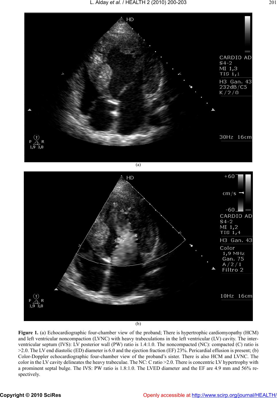
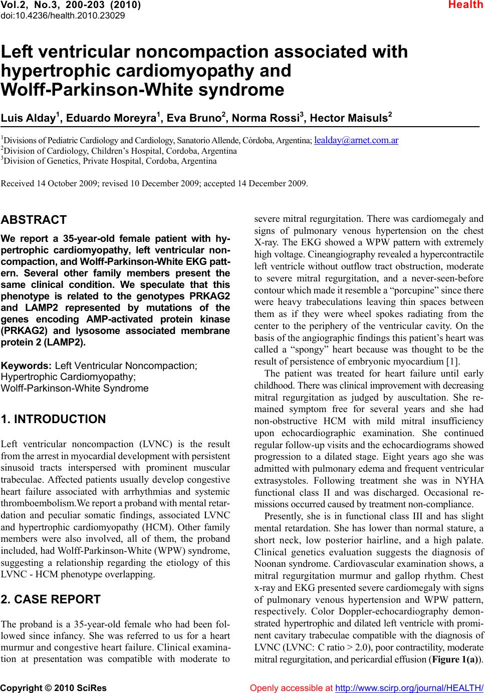
respectively. Color Doppler-echocardiography demon-
strated hypertrophic and dilated left ventr icle with promi-
nent cavitary trabeculae compatible with the diagnosis of
LVNC (LVNC: C ratio > 2.0), poor contractility, moderate
mitral regur gitation, and pericardial ef fusion (Figur e 1(a)).
L. Alday et al. / HEALTH 2 (2010) 200-203
Copyright © 2010 SciRes Openly accessible at http://www. scirp.org/journal/HEALTH/
201
(a)
(b)
Figure 1. (a) Echocardiographic four-chamber view of the proband; There is hypertrophic cardiom yopathy (H CM)
and left ventricular noncompaction (LVNC) with heavy trabeculations in the left ventricular (LV) cavity. The inter-
ventricular septum (IVS): LV posterior wall (PW) ratio is 1.4:1.0. The noncompacted (NC): compacted (C) ratio is
>2.0. The LV end d iasto lic (ED ) dia meter is 6. 0 and the e jection frac tion (E F) 23 %. Peri cardia l ef fusio n is pr esent ; (b)
Color-Doppler echocardiographic four-chamber view of the proband’s sister. There is also HCM and LVNC. The
color i n the LV c avity d eline ates t he heav y trab eculae . The NC: C r atio > 2.0. T here i s conce ntric LV hypert rophy w ith
a prominent septal bulge. The IVS: PW ratio is 1.8:1.0. The LVED diameter and the EF are 4.9 mm and 56% re-
spectively.
L. Alday et al. / HEALTH 2 (2010) 200-203
Copyright © 2010 SciRes Openly accessible at http://www.scirp.org/journal/HEALTH/
202
I
II
III
Unaffected status
Hypertrophic Cardiomyopathy
Hypertrophic Cardiomyopat hy and Left Ventricular Noncompaction
Unknown clinical status
1 2 345
1 2 3 4 5 6 78 910 11 12 13 14 15 16
1 2 34 56 7
Figure 2. Pedigree of the kindred with hypertrophic cardiomyopathy (HCM), Wolff-Parkinson-White syndrome (WPW),
and left ventricular noncompaction (LVNC) inherited as a Mendelian autosomal dominant trait.The proband is pointed by
the ar row. I4: Pr oband’s grandmother with unknown clinical statu s. II7 and 8: Proband’s mother and aunt both of them with
HCM and WPW. III4: Proband’s sister with HCM, WPW, and LVNC.
The better resolution of the new echocardiographic
technology allowed confirmation of the initial cinean-
giography. An updated family tree (Figure 2) showed an
interesting family pattern where the proposita had a
37-year-old sister also with non-obstructive HCM with
non-compaction and normal left ventricular function
(Figure 1(b)). External features and mental status were
normal. She also had a WPW pattern on the EKG with
normal voltage. She had occasional episodes of su-
praventricular tachycardia. Both the mother and her
identical twin had HCM, WPW, and atrial fibrillation.
They might have had also LVNC but could not be con-
firmed. They died one year apart at the ages of 59 and 60
years due to congestive heart failure and stroke, respec-
tively. The mother’s sister had no descendants. The twins
had 3 siblings and half- siblings each with no history of
HCM. The maternal grandmother died suddenly at a
young age of unspecified causes.
3. DISCUSSION
LVNC was a poorly recognized entity that began to gain
interest from the medical community in the 90’ s. Recently ,
it has become clear that is more pre valent than previ ously
recognized [2]. H owever, its etiology remained unknow n
until recently, when molecular genetic studies showed
that mutations of cytoskeletal genes like those encoding
taffazin (TAZ) and alpha-dystrobrevin (DTNA) causing
Barth syndrome were associated with LVNC [3]. More
recently , it was found that LV NC was also associated with
mutations of sarcomeric genes shared by patients with
HCM and dilated cardiomyopathy, like the alpha-cardiac
actin gene (ACTC), beta-myosin heavy chain (MYH7),
and cardiac troponin T (TNNT2) [4,5]. Cases of patients
sharing both the LVNC and HCM phenotypes have been
published rece ntly but there was no m ention of a mutation
[6,7].
The patient reported here, had other family members
with the same disease, and all had the associated WPW
syndrome. Such overlapping phenotype suggests a strong
genetic association.
WPW syndrome is infrequently associated with HCM.
It has been found in patients with HCM phenotypes with
mutations of the genes encoding AMP-activated protein
kinase (PRKAG2) and lysosome associated membrane
protein 2 (LAMP2). PRKAG2 is an important enzyme
involved in th e production of cellular energy and its mu-
tations might cause HCM, WPW syndrome, conduction
disorders requiring pacing, skeletal myopathy, and gly-
cogen storage disease [8]. About 30% of affected indi-
viduals have atrial fibrillation or supraventricular tachy-
cardia. This clinical pattern was seen in three of the fam-
ily members of our proposita.
LAMP-2, an X-linked gene, encodes proteins that
regulate the integrity and function of lysosomes. Muta-
tions of this gene cause Danon disease characterized by
HCM, WPW syndrome, skeletal myopathy and mental
retardation. Female carriers present later in life with less
severe disease than in males [9].
L. Alday et al. / HEALTH 2 (2010) 200-203
Copyright © 2010 SciRes Openly accessible at http://www. scirp.org/journal/HEALTH/
203
In the family under discussion, the proband and the
sister had a phenotype of HCM, WPW syndrome, and
LVNC. The proband had also somatic features suggestive
of Noonan syndrome and mental retardation. The dis-
eased mother and her twin, with normal external pheno-
type, had ha d HCM, WPW syndr ome, an d late onset at rial
fibrillation, but LVNC could not be confirmed.
The members of this family had features shared with
patients affected with the PRKG2 and LAMP-2 mutations.
It is then possible that any of the two might be the in-
volved gene. There are no systematic molecular genetic
studies available showing the association of these genes
with the H CM, WPW and LVNC phenotypes.
The complex overlapping phenotypes of HCM with
WPW syndrome and LVNC seen in this fam ily is possibly
due to a previously not described mutation which should
be studied further.
REFERENCES
[1] Van Praagh, R., Personal communication.
[2] Pignatelli, R.H., McMahon, C.J., Dreyer, W.J., et al.
(2003) Clinical characterization of left ventricular non-
compaction in children: a relatively common form of
cardiomyopathy. Circulation, 108, 2672-2678.
[3] Ichida, F., Tsubata, S., Bowles, K.R., et al. (2001)
Novel gene mutations in patients with left ventricular
noncompaction or Barth syndrome. Circul ation, 103,
1256-1263.
[4] Monserrat, L., Hermida-Prieto, M., Fernandez, X., et al.
(2007) Mutation in the alpha-cardiac actin gene associ-
ated with apical hypertrophic cardiomy opathy, left ven-
tricular non-compaction, and septal defects. European
Heart Jour na l, 28, 1953-1961.
[5] Klaassen, S., Probst, S., Oechslin, E., et al. (2008) Muta-
tion in sarcomere protein genes in left ventricular non-
compaction. Circulation, 117, 2893-2901.
[6] Pantazis, A.A., Kohll, S.K., and Elliott, P.M. (2006)
Images in cardiology. Hypertrophic cardiomyopathy
and left ventricular hypertrabeculation: evidence for an
overlapping phenotype. Heart, 92, 349.
[7] Kelley-Hedgepeth, A., Towbin, J.A., and Maron, M.S.
(2009) Images in cardiovascular medicine. Overlapping
phenotypes. Left ventricular noncompaction and hy per-
trophic cardiomyopathy. Circulati on, 119, 588-589.
[8] Murphy, R.T., Mogensen, J., McGarry, K., et al. (2005)
Adenosine monophosphate-acti va ted protein kina se dis-
ease mimicks hypertrophic cardiomyopathy and Wolff-
Parkinson-White sy ndrome. Natural history. Journal of
the American College of Cardiology, 45, 922-930.
[9] Yang, Z., McMahon, C.J., and Smith, L.R. (2005) Danon
disea se as an unde rrecogni zed cause of hypertrop hic car-
diomyopathy in children. Circulation, 112, 1612-1617.