International Journal of Clinical Medicine
Vol.3 No.1(2012), Article ID:16818,2 pages DOI:10.4236/ijcm.2012.31013
Double Appendicitis
![]()
1Department of General Surgery, Sheri Kashmir Institute of Medical Sciences, Srinagar, India; 2Department of Radiology, Sheri Kashmir Institute of Medical Sciences, Srinagar, India.
Email: drmajidmushtaque@gmail.com
Received October 20th, 2011; revised November 28th, 2011; accepted December 24th, 2011
Keywords: Appendix; Duplication; Appendicitis
ABSTRACT
Duplication of the vermiform appendix is extremely rare with reported incidence of 0.004% patients operated on for acute appendicitis. It is important to recognize this condition which may have serious clinical and medico legal consequences. A 16-year-old male presented with clinical features typical of acute appendicitis. Surgical exploration revealed a perforated gangrenous double appendix which was dealt by double appendicectomy.
1. Introduction
Duplication of the vermiform appendix is rare, with a reported incidence of 0.004%. About 100 appendiceal anomalies have been reported in the literature. Most anomalies of the appendix have been observed in adults and most were noticed incidentally during surgery not primarily involving the appendix [1]. Picoli (1892) reported the first case of appendix duplex in a female patient who had associated anomalies of duplication of the entire large bowel, two uteri with two vaginae, ectopia vesicae and exomphalos [2]. Double appendix are usually asymptomatic, the majority of them are diagnosed at surgery or on post-mortem examination, some of them can be picked up preoperatively on barium enema. Symptoms are usually the result of obstruction and inflammation. The clinical presentation can vary according to the location of the appendices [3].
2. Case Report
A 16 year old male presented with a migratory right iliac fossa pain of two day duration. There was also history of fever and anorexia. On examination, he was febrile (100˚F), with pulse of 100/min. Abdominal examination revealed features of localised peritonitis confined to right lower quadrant. Complete blood count documented elevated total white cell count (12,500/mm3) with neutrophilia (90%). X-ray chest and abdomen were normal as was the urine examination. On abdominal ultrasonography a dilated, noncompressible, thickened-walled vermiform appendix, 13 mm in diameter was found. Exploration for appendicectomy via McBurney’s incision was done which revealed a double appendix with gangrenous changes distally (Figure 1). In addition to a normal appendix arising from the caecum at the usual site, there was a second, appendix arising from caecum along the lines of the taenia about 3 cm away at from the first (B2 duplex appendix). Appendices were matted together and
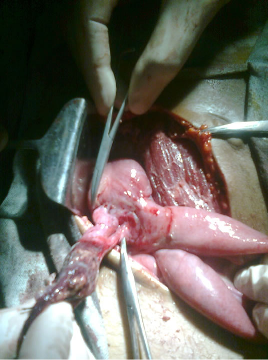
Figure 1. B2 duplex appendix with gangrenous changes distally. Mosquito forceps pointing towards base of two appendices.
surrounded by omentum with copious inflammatory exudates in the right lower abdomen. There were no other noteworthy surgical findings and the remaining abdominal viscera were normal. Double appendectomy was performed. Histopathological examination of both appendices revealed inflammatory changes with lymphoid follicles and smooth muscle, confirming our diagnosis. Patient was discharged on 3rd day after an uneventful postoperative period.
3. Discussion
Duplication of part of the alimentary tract, in particular of the vermiform appendix, is of embryological curiosity and may be associated with other congenital duplications. Histologically the appendix can be distinguished from other intestinal duplications by the presence of a complete and separate inner and outer longitudinal muscle layer and the amount and arrangement of lymphoid tissue [4].
Wallbridge [5] modified Cave’s original classification [6] of duplicated vermiform appendix was again modified by Biermann in 1993 as follows:
. Type A: Single caecum with one appendix exhibiting partial duplication.
. Type B: Single caecum with two obviously separate appendices.
. B1: The two appendices arise on either side of the ileocaecal valve in a “bird-like” manner.
. B2: In addition to a normal appendix arising from the caecum at the usual site, there is also a second, usually rudimentary, appendix arising from caecum along the lines of the taenia at a varying distance from the first.
. B3: The second appendix is located along the taenia of the hepatic flexure of the colon.
. B4: The location of the second appendix is along the taenia of the splenic flexure of colon.
. Type C: Double caecum, each bearing its own appendix and associated with multiple duplication anomalies of the intestinal tract as well as the urinary tract.
The horseshoe anomaly of the appendix might be considered a type D anomaly [7]. Our case had a type B2 appendiceal duplication with gangrenous appendicitis of both.
All these anomalies are of great practical importance, and a surgeon has to bear them in mind during an operation, since in case he/she overlooks them the operated patient may experience grave consequences. They also may be the forensic issue in cases when repeated explorative laparotomy reveals “previously removed” vermiform appendix [8]. It may also remain totally asymptomatic or mimic other intra-abdominal conditions such as carcinoma, caecal diverticulum, diverticulosis of the appendix or stump appendicitis [9]. In patients with appendicular duplication, when only one of them is found to be inflamed on exploration or laparoscopy, both of them should be removed so as to avoid diagnostic confusion that may arise on removal of single appendix. However, non-inflamed duplication detected when exploration or laparoscopy is performed for some other condition need not be subjected to appendicectomy [10].
In conclusion, surgeons, especially junior surgical residents, should be aware of the potential anatomical anomalies and malpositions of the vermiform appendix and careful inspection of the caecum should be performed to avoid missing any other appendiceal anomalies which may result in serious clinical and medico-legal conesquences.
REFERENCES
- E. Eroglu, E. Erdogan, G. Gundogdu, et al., “Duplication of appendix vermiformis: a case in a child,” Techniques in Coloproctology, Vol. 6, No. 1, 2002, pp. 55-57. doi:10.1007/s101510200010
- A. K. Khanna, “Appendix vermiformis duplex,” Postgraduate Medical Journal, Vol. 59, 1983, pp. 69-70.
- A. A. Kothari, K. R. Yagnik and V. P. Hathila, “Duplication of vermiform appendix,” Journal of Postgraduate Medical, Vol. 50, No. 4, 2004, pp. 285-286.
- I. Chamisa, S. Nikolov and T. Q. Bam, “Duplex Appendicitis,” South African Medical Journal, Vol. 97, No. 9, 2007, pp. 842-843
- P. H. Wallbridge, “Double Appendix,” British Journal of Surgery, Vol. 50, No. 221, 1963, pp. 346-347. doi:10.1002/bjs.18005022124
- A. J. E. Cave, “Appendix Vermiformis Duplex,” Journal of Anatomy, Vol. 70, 1936, pp. 283-292.
- T. Mesko, “Horseshoe Anomaly of the Appendix: A Previously Undescribed Entity,” Surgery, Vol. 106, No. 3, 1989, pp. 563-566.
- E. Drino, D. Radnić, B. Kotjelnikov and G. Aksamija, “Rare Anomalies in the Development of the Appendix,” Acta Chir lugo, Vol. 38, No. 1, 1991, pp. 103-111.
- A. Samee, M. Alibhai, et al., “Appendicitis in a duplex appendix mimicking intussusceptions,” BMJ Case Reports, 2010.
- B. C. Lin, R. J. Chen, J. F. Fang, et al., “Duplication of the Vermiform Appendix,” European Journal of Surgery, Vol. 162, 1996, pp. 589-591.