International Journal of Otolaryngology and Head & Neck Surgery
Vol.3 No.1(2014), Article ID:42037,2 pages DOI:10.4236/ijohns.2014.31012
Clinical Photograph: “A Third Arythenoid?”
Itzhak Braverman1,2*, Yuri Pastuhov1,2
1Otolaryngology—Head and Neck Surgery Unit, Hillel Yaffe Medical Center, Hadera, Israel 2The Technion—The Israel Institute of Technology, Haifa, Israel
Email: *braverman@hy.health.gov.il
Copyright © 2014 Itzhak Braverman, Yuri Pastuhov. This is an open access article distributed under the Creative Commons Attribution License, which permits unrestricted use, distribution, and reproduction in any medium, provided the original work is properly cited. In accordance of the Creative Commons Attribution License all Copyrights © 2014 are reserved for SCIRP and the owner of the intellectual property Itzhak Braverman, Yuri Pastuhov. All Copyright © 2014 are guarded by law and by SCIRP as a guardian.
Received December 2, 2013; revised January 2, 2014; accepted January 12, 2014
ABSTRACT
A case of anatomical variation of the hyoid bone in a girl is presented. Over-extended bending of the elongated and curved right side of the hyoid bone, may project to the lumen of the supraglottic area as the patient presented. She had a foreign body sensation in her throat, and on fiber optic laryngeal examination, a bulge’s appearance as a “third arythenoid” was seen. We present the clinical finding and the picture of a “third arythenoid” with literature review.
Keywords: Arythenoid; Hyoid Bone; Anatomic Variation; Larynx
1. Case Presentation
A 15-year-old girl with a foreign body sensation in her throat was referred for examination.
No foreign body was observed and her vocal cords showed good movement.
Following an endoscopic examination, we found a round bulge near the right arythenoid: Is this a “third arythenoid?” (Figure 1).
CT scan of the neck showed an elongated and curved right side of the hyoid bone, extending to the right arythenoid area (Figure 2 and 3).
This finding did not disturb the vocal box. Despite the bulge’s appearance as a “third arythenoid”, it actually was an anatomical variant projection of the hyoid bone into the supraglottic area.
2. Discussion
The hyoid bone is a horseshoe-shaped bone which lies between the thyroid cartilage and the mandible, consisting of the body and two pairs of cornua—the greater and the lesser cornu. The greater cornu projects posterosuperiorly as it extends laterally from the body.
It is the only bone in the human skeleton with no articulation to any other bone [1].
Over-extended bending of the greater cornu may project to the lumen of the supraglottic area as the patient presented.
Anomalies of the digastric muscle and the thyro-hyoid articulation have been described [2,3] in a single report from France [4]. The authors reported the developmental

Figure 1. Endoscopic view of the larynx of the patient showing a rounded bulging near the right arythenoid, that does not move as arythenoids do during speaking but moves wile swallowing.
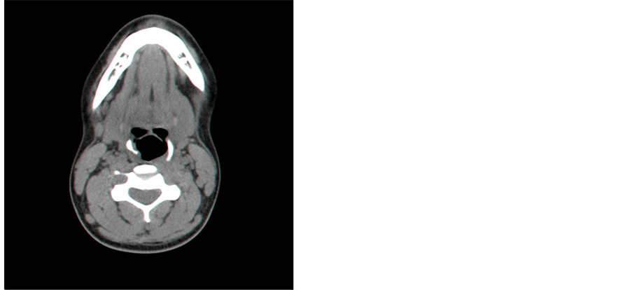
Figure 2. Axial CT scan of the neck shows an elongation and curve on the right side of the hyoid bone, extending medially to the laryngeal area close to the right arythenoid.

Figure 3. Reconsrtuction of axial CT scan of the neck shows an elongation and curve on the right side of the hyoid bone.
anomaly of the hyoid bone with an unusual cause of dysphagia. They described a hyoid syndrome caused by a developmental anomaly of the second branchial cleft, presenting in an adult with dysphagia.
3-D CT of the hyoid bone anomaly showed an uncurvated and elongated lesser cornu, causing persistent impingement on the lateral oropharynx wall.
This case is similar to our patient’s case, which is the second case report on the topic, to the best of our knowledge. It is the first report on greater cornu anomaly, presented as “a third arythenoid.”
Acknowledgment
Reuven Schreiber, Radiology Department, Rambam Medical Center, Haifa.
[1] REFERENCES
[2] H. Gray, “The Hyoid Bone,” Anatomy of the Human Body, II. Osteology, 5b. 9.
[3] A. Holiblova and L. Machalek, “A Report on Anomalies of the Digastric Muscle,” Acta Universitatis Palackianae Olomucensis Facultatis Medicae, Vol. 142, 1999, pp. 57- 59.
[4] V. Ilankovan, “An Anomaly of the Thyro-Hyoid Articulation,” The Journal of Laryngology & Otology, Vol. 101, No. 9, pp. 959-961. http://dx.doi.org/10.1017/S0022215100103068
[5] A. Boyadjian, K. Marsot-Dupuch, E. Scmitt, C. Chouard and J. Tubiana, “Developmental Anomaly of the Hyoid Bone: An Unusual Cause of Dysphagia,” Journal of Radiology, Vol. 82, No. 4, 2001, pp. 491-494.
NOTES
*Corresponding author.