Advances in Bioscience and Biotechnology
Vol.5 No.4(2014), Article ID:43676,10 pages DOI:10.4236/abb.2014.54044
In Vitro Regeneration of Curcuma caesia Plantlets from Basal Part and via Somatic Embryogenesis
Ab. Rahman Zuraida1*, Kamaruddin Fatin Liyana Izzati1, Othman Ayu Nazreena1, Che Mohd Zain Che Radziah2, Sheikh Ghadzi Siti Nur Asyikin2, Subramaniam Sreeramanan3
1Biotechnology Research Centre, MARDI Headquarters, Persiaran MARDI-UPM, Serdang, Malaysia
2Faculty of Science and Technology, Universiti Kebangsaan Malaysia, Bangi, Malaysia
3School of Biological Sciences, Universiti Sains Malaysia (USM), Minden Heights, Malaysia
Email: *azuraida@mardi.gov.my
Copyright © 2014 by authors and Scientific Research Publishing Inc.
This work is licensed under the Creative Commons Attribution International License (CC BY).
http://creativecommons.org/licenses/by/4.0/


Received 17 January 2014; revised 19 February 2014; accepted 6 March 2014
ABSTRACT
Plantlets of Curcuma caesia were produced in vitro from newly sprouting vegetative buds of tubers. Segments of the plantlets from the junction between the root and the basal portion of the stem were subsequently used as explants to investigate factors affecting callus induction and plant regeneration via somatic embryogenesis. The explants were placed on Woody Plant Medium (WPM) together with different concentrations of 2,4-dichlorophenoxyacetic acid(2,4-D) and benzyl aminopurine (BAP) in the presence of light. The growth medium supplemented with 5 mg/L BAP and 2 mg/L 2,4-D promoted callus induction after 70 days of culture. Sub-culturing on the same medium enhanced the production of friable callus. Culture media containing higher concentrations of agar promoted the development of green somatic embryos from the callus. Respond of somatic embryogenesis was most successful with MS medium in 6.0 g/L agar supplemented with 5 mg/L BAP and 0.2 mg/L 2,4-D whereby the callus developed into green somatic embryos with an efficiency of 53%. This culture medium also produced the largest number plantlets.
Keywords:In Vitro; Regeneration; Somatic Embryogenesis; Curcuma caesia

1. Introduction
Curcuma caesia L., known as black turmeric, is a perennial herb belonging to the family Zingiberaceae. The plant is native to North-East and Central India [1] , growing in the Papi Hills of East Godavari, West Godavari, and Andhra Pradesh [2] . The rhizome of various curcuma species has long been used by various tribal communities for medicinal. Extracts from C. caesia have high economic potential owing to their putative medicinal properties. There have been claims for their medicinal actions against leukoderma, epilepsy, cancer and HIV/AIDS [3] . The rhizomes are traditionally used in treating leucoderma, asthma, tumours, piles, bronchitis, bruises, etc. [2] .
In vitro production of many plant species can be achieved either through direct shoot multiplication or via somatic embryogenesis, although neither technique has been published for C. caesia. Of the two approaches, somatic embryogenesis is thought to be more efficient [4] . According to [5] , some of the advantages somatic embryogenesis has over direct in vitro propagation systems include the former’s high multiplication rates, possibility of cryopreservation of embryogenic callus, the potential for scale-up in liquid suspension cultures, amenability to the use of bioreactors and the application of somatic synthetic seed technologies. In addition, somatic embryogenesis can be a very useful tool in genetics and breeding research [6] . Plantlets derived from this technique can be used to produce genetic lines that are free of viruses [7] and to introduce genes by genetic transformation [8] .
In Zingiberaceae, embryogenesis success is highly dependent on the interaction among genotype, explants source and culture medium. Hence, each Zingiberaceae cultivar requires a medium suited to it. Plant regeneration systems that make use of embryogenic callus have been developed for numerous species of curcuma including Curcuma soloensis, Curcuma kwangsiensis [9] , and Curcuma aromatic [10] . However, as stated above, the regeneration of Curcuma caesiavia somatic embryogenesis has not been reported. In this report, we describe the in vitro regeneration of C. caesia plantlets through callus production and somatic embryogenesis.
2. Materials and Methods
2.1. Plant Materials
Rhizomes of Curcuma caesia were cultivated in the glasshouse to allow the sprouting of vegetative buds that served as explants. Sprouted buds of 3 - 4 cm were cleaned under running tap water for an hour and then washed with commercial laboratory detergent (Decon 5%, v/v) before rinsing thoroughly with water. The buds were immersed in 1% (v/v) fungicide solution for one hour before being rinsed thoroughly under running tap water for 5 min. Subsequently, the explants were surface sterilized with 10% - 20% Clorox® and a few drops of Tween-20 under sterile conditions. They were then rinsed several times with sterile distilled water before inoculating onto Woody Plant Medium (WPM) supplemented with 3% sucrose and 5.0 mg/L benzyl aminopurine (BAP) to produce plantlets in vitro.
2.2. Callus Induction
The basal part of the stem of Curcuma caesia plantlets were used as explants. Roots and leaves were first removed from these plant segments (Figure 1(a)). The culture medium used was solidified with 0.3% gelrite agar and contained WPM basal medium, 3% sucrose and 0.5 - 5 mg/L 2,4-dichlorophenoxyacetic acid(2,4-D) alone or in combination with 0.5 - 5 mg/L BAP. Recordings of callus induction were carried out at weekly intervals and the results were expressed as percentage of callus induction after 4 months of culture. In order to test and optimize callus proliferation, the calli from the best callus induction medium were sub-cultured onto fresh medium with different concentrations of BAP and 2,4-D. Fresh and dry weights of calli were recorded after 45 days. The morphology of the calli that developed was observed and examined.
2.3. Plant Regeneration
Friable calli produced on callus induction medium containing 5 mg/L BAP and 2 mg/L 2,4-D (the medium yielding optimal callus induction) were used to establish an appropriate medium for shoot formation. Small pieces of calli (approximately 200 mg) were transferred to light culture on different medium combinations containing BAP (0, 2 and 5 mg/L), 2,4-D (0.2, 1 and 2 mg/L) and agar (3.0, 4.5, 6.0, and 9.0 g/L) as listed in Table 1. The efficiency by which calli developed green somatic embryos was noted. Embryos that were produced were then sub-cultured on to the same medium for further development. The number of plantlets regenerated per callus was recorded after4 weeks of culture. Regenerated shoots were then transferred to WPM basal medium
 (a)
(a)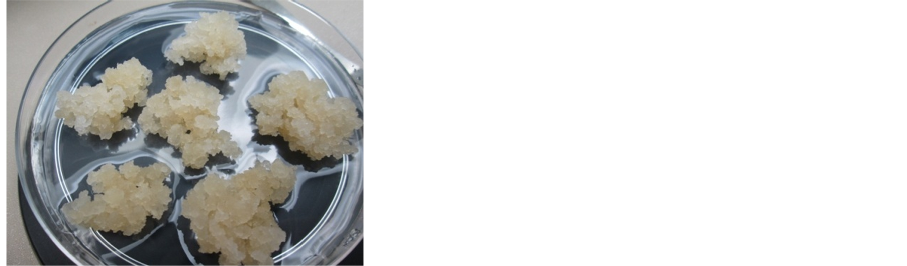 (b)
(b)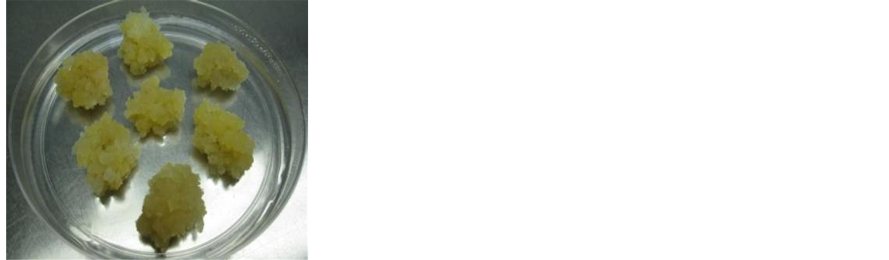 (c)
(c)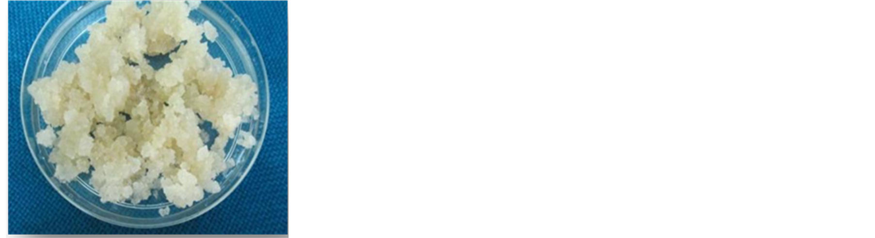 (d)
(d)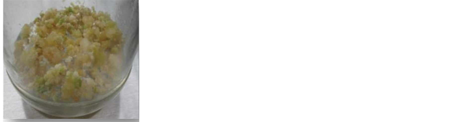 (e)
(e)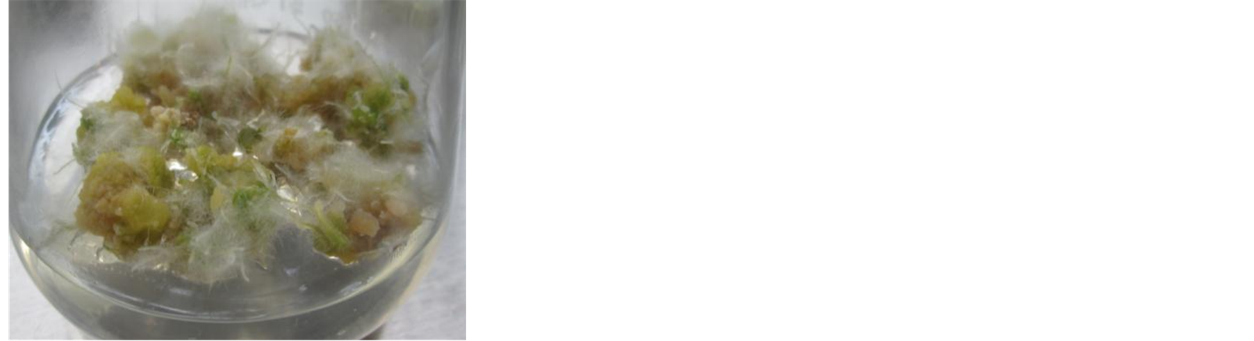 (f)
(f)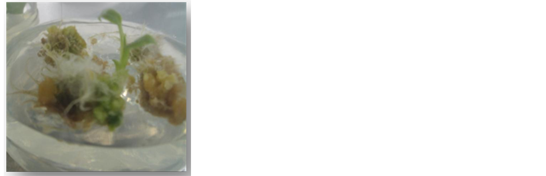 (g)
(g)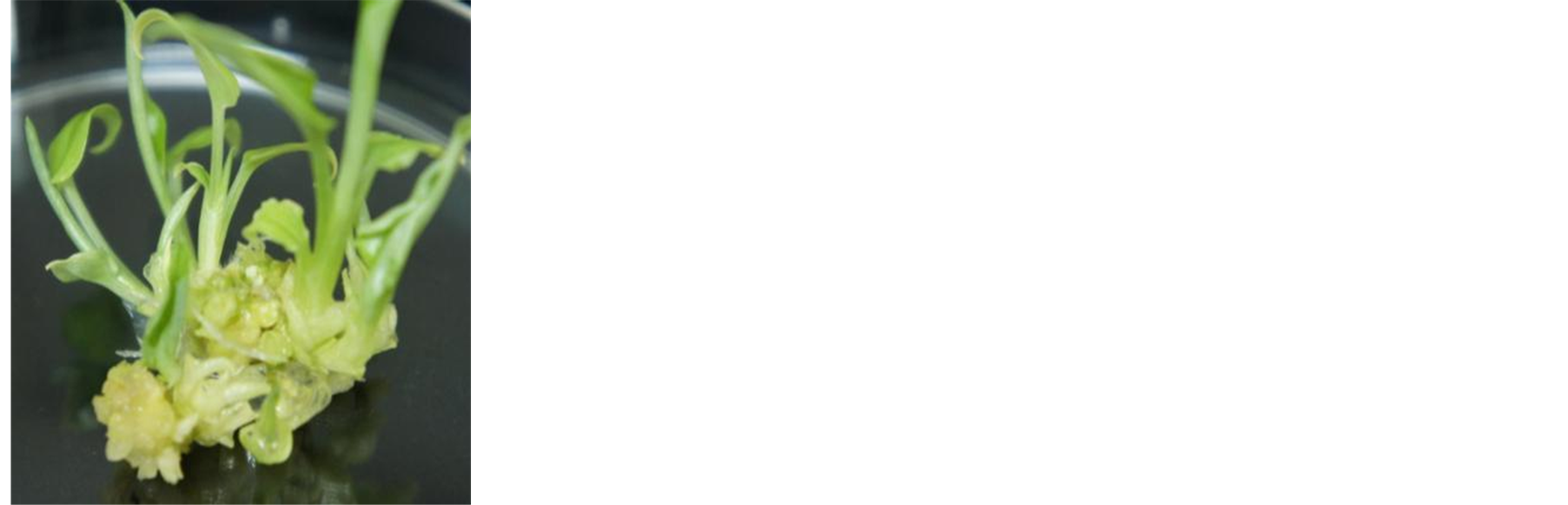 (h)
(h)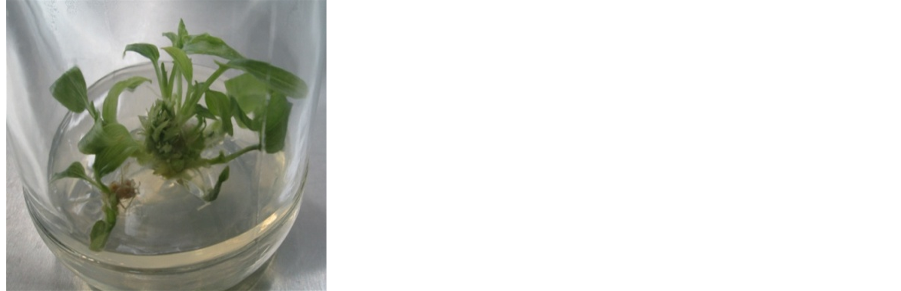 (i)
(i)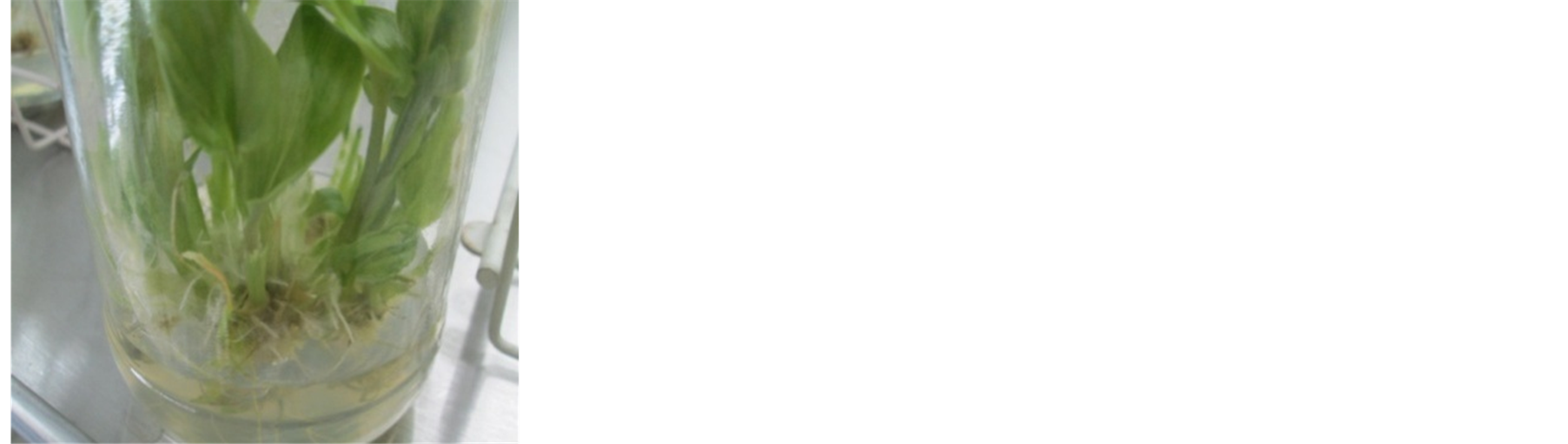 (j)
(j)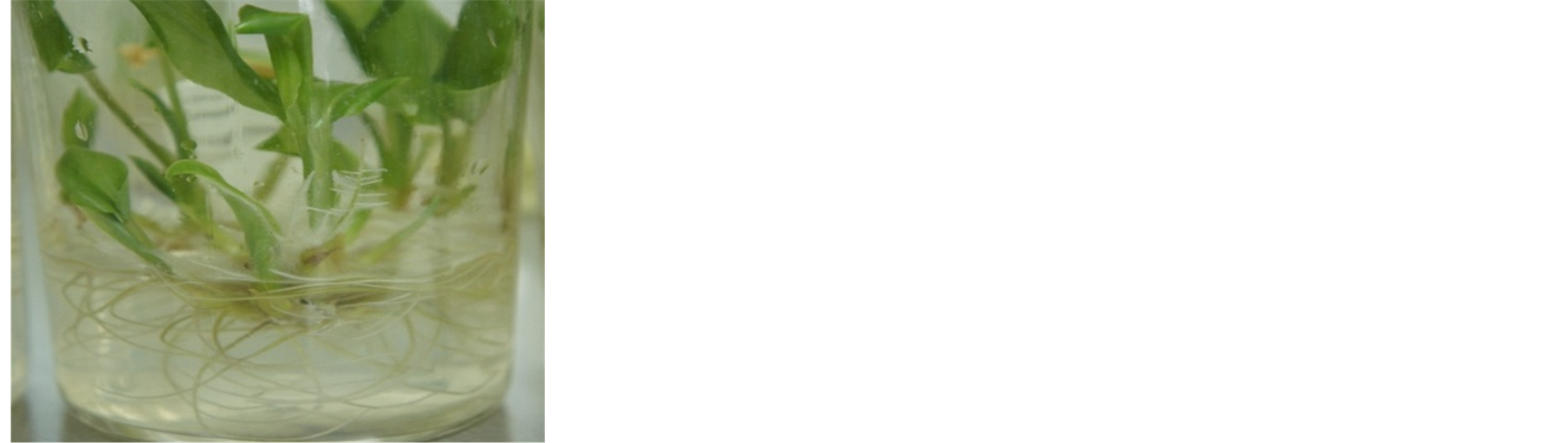 (k)
(k)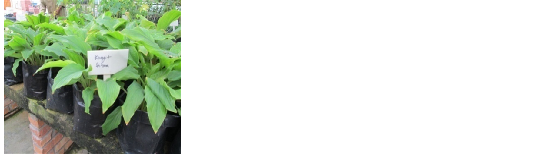 (l)
(l)
Figure 1. Plant regeneration from callus derived form in vitro culture of Curcuma caesia. (a) Explants derived from the junction between the basal stem and the root used for callus induction; (b) Callus proliferation on WPM medium with 2,4-D and BAP; (c) Greenish callus after three sub-cultures; (d) Proliferated embryogenic callus used for plantlet regeneration; (e) and (f) Formation of somatic embryos; (g) - (i) Shoots regeneration; (j) and (k) Plantlet multiplication; (l) Successful plantlet growth after transfer into the net house.

Table 1. Effect of agar concentration and plant growth regulators on the efficiency of somatic embryo production and the number of regenerated plantlets.
Results represent mean ± standard error mean (SEM) of three replicated experiments.
containing 5 mg/L BAP for shoot multiplication, and finally to MS medium that was free of plant growth regulators for leaf elongation and root induction.
2.4. Culture Conditions
The WPM basal media with different PGR in culture jars containing sucrose were adjusted to pH 5.8 before they were solidified with gelrite agar, autoclaved at 121˚C and 104 kPa for 15 min. The cultures were incubated in the culture room under white fluorescent light with a light intensity of 3000 lux and a photoperiod of 16 h at 25˚C ± 2˚C. For callus induction, the culture jars were also kept in the light. Rooted explants with shoots about 4 - 5 cmin length were removed from the culture bottles and the roots were washed under running tap water to remove the agar. The plantlets were individually transplanted in soil contained in polybags and kept under controlled conditions in a net house with 75% shading. To maintain humidity, the plants were watered periodically twice a day. The survival rate of the plantlets was recorded after 6 weeks.
2.5. Statistical Analysis
The data (25 replicates per treatment) were subjected to one way analysis of variance (ANOVA) to assess treatment differences and interaction using the SPSS version 11.0. Significance of differences between means was tested by DMRT’s Test (p ≤ 0.05).
3. Results and Discussion
3.1. Callus Induction
In order to establish the most suitable concentration of plant growth regulator for plantlet regeneration via somatic embryogenesis, we tried various concentrations and combinations of the plant hormones BAP (0.5 - 5.0 mg/L) and 2,4-D (0.5 - 5 mg/L) under light (1200 lux) conditions. Table 2 shows the percentage of callus induction on the surface of the explants after 4 months in culture. Calli were initiated on medium containing 1 - 5
Table 2. Effect of 2,4-D and BAP on the induction of callus in Curcuma caesia.
Results represent mean ± standard error mean (SEM) of three replicated experiments.
mg/L 2,4-D in combination with 1 - 5 mg/L BAP. MS medium supplemented with 2 mg/L 2,4-D combined with 5 mg/L BAP was the most effective in inducing callus (20%). Callus growth was slow and cellular dedifferentiation could take more than 70 days, with maximum callus production up to 95 days.
Medium compositions affected callus induction considerably. [11] state that the success of in vitro cultures largely depends on the nutrition media, growth regulators, variety, and on the interaction between the variety and the medium. The findings in this study indicated that explants from basal part of C. caesia cultured on medium containing 2 mg/L 2,4-D and 5 mg/L BAP performed better in inducing callus compared to the other treatments. Similar results were reported by [12] in their studies on the rice variety Bas-370. They observed that 2 mg/L 2,4-D combined with 5.0 mg/L BAP was the best for callus induction. In the work of [9] on Curcuma kwangsiensi, on the other hand, the combination of 2,4-D and BA gave rise to a few soft calli; the callus could not proliferate and wilted gradually. In addition, the authors also found that neither the auxins NAA and 2,4-D nor the cytokinin BAP were effective when used alone or together in C. kwangsienesis embryogenic callus cultures. The requirement of BA for somatic embryogenesis may vary according to the explant source [13] .
The application of cytokinins in combination with auxins to induce somatic embryogenesis in callus culture has been reported for various plant species [13] -[15] . In the present study, application of 2,4-D alone (0.5 - 5 mg/l) failed to produce any callus. These results were in agreement with the findings of [16] where 2,4-D at all the concentrations tested did not produce any positive response for callus induction of Curcuma zedoaria 50 days after inoculation. Nevertheless, [11] who worked with rice found that callusing in seeds was best when 2 mg/L 2,4-D was supplemented. [17] who similarly worked with rice stated that culture medium supplemented with 2,4-D at concentration of 1 mg/L was most effective for the induction of callusing. [10] observed callus initiation in Curcuma aromatic in all the tested media, but the medium containing 2 mg/l 2,4-D gave the optimum callusing response of 82%. The role of 2,4-D in callus induction has also been reported in Curcuma longa [18] [19] .
3.2. Callus Proliferation
Callus initiated in the growth medium containing 2 mg/L 2,4-D + 5 mg/L BAP were sub-cultured on different media with varying concentrations of BAP (0.5 - 5.0 mg/L) and 2,4-D (0.5 - 5.0 mg/L) to study callus proliferation further (Table 3). Generally, callus growth onmedia with both growth regulators showed faster growth and proliferation than those cultured in medium containing 2,4-D alone. On the other hand, BAP alone at concentrations of 1 - 5 mg/L showed active proliferation. The highest proliferation rate that produced a callus biomass of 921 mg was obtained on a medium containing 5 mg/l BAP. However, the callus was watery and soft rather than friable. Moreover, the dry weight of this callus was low (390 mg after one month of culture). Callus growth on medium containing 2 mg/L 2,4-D combined with 5 mg/L BAP showed faster growth and proliferation than callus cultured on medium containing 2,4-D alone, the former achieving 9.1 g fresh weight and 610 mg dry weight. The texture of the callus surface was friable and yellowish with morphogenic aspects (Figure 1(b)). Faster proliferation of callus occurred when they were sub-cultured to the same fresh medium after 30 days of culture. Proliferation of callus cultured on medium with 5 mg/L 2,4-D alone or in combination with various concentrations of BAP (1 - 5 mg/L BAP) was relatively less active than when lower concentrations of 2,4-D (1 - 2 mg/L) were used.
The faster callus proliferation when transferred to fresh callus induction medium, as noted above, is consistent with the findings of [10] for Curcuma aromatic. In our results also, the best callus induction medium (2 mg/L 2,4-D combined with 5 mg/L BAP) was the best medium for callus proliferation. After two to three subcultures, the calli became friable in texture and greenish in color (Figure 1(c)). These results showed that a combination of cytokinin and of auxin was suitable for callus maintenance or proliferation with the aim of plantlet regeneration, although the lower concentrations BAP were less effective for callus maintenance. Similar results were reported by [20] who found that the best callus proliferation was observed in cauliflower cultured on MS + 3.0 mg/L BAP + 0.1 mg/L 2,4-D. Working with citrus, [21] also observed that green callus proliferation increased when cultured on the same medium (callus induction medium). They also stated that subsequent transfer of callus on medium with 1.0 mg/L 2,4-D and 5.0 mg/L BAP resulted in good proliferation in half of the cultures. In our study, 5 mg/l BAP alone resulted in a high proliferation rate, but this was accompanied by the callus turning watery after 2 to 3 rounds of subculture. [22] found that good callus growth Triticum aestivum was observed on MS medium containing 4 mg/L BAP. These results are consistent with those of [23] who researched on Triticum
Table 3. Effect of 2,4-D and BAP on callus proliferation from callus Curcuma caesia after 4 weeks of culture.
Results represent mean ± standard error mean (SEM) of three replicated experiments.
aestivum. Moreover, sub-culturing on MS medium supplemented with 2.0 mg/l 2, 4-D in combination with 0.5 mg/l BAP improved callus development [22] . In their findings, formation of soft and non-embryogenic callus was observed initially on media with low concentrations of 2,4-D alone, with most of the callus becoming watery after subculture. This callus remained as a cell mass without developing into embryos. Conversely, the induction of embryogeneic callus was usually promoted by a relatively higher concentration of 2, 4-D [24] . [25] notes that the formation of an embryogenic cell is related to nuclear DNA hypermethylation in the presence of high concentrations of 2,4-D.
3.3. Plantlet Regeneration via Embryogenesis
Well-proliferated embryogenic calli cultured on 2 mg/L 2,4-D and 5 mg/L BAP were used for regeneration studies (Figure 1(d)). Approximately 2 g of fresh calli were transferred to a regeneration medium containing different combinations and concentrations of growth regulator and agar as listed in Table 1. The formation of green somatic embryos induced from embryogenic calli was observed in most of the media containing 2,4-D and BAP where agar concentration was between 4.5 - 9.0 g/L (Figures 1(e) and (f)). The presence of both 2,4-D and BAP in the growth medium was more effective than either growth regulator alone. Among the tested media, the highest rate of embryogenesis (53%) was obtained with 5 mg/L BAP and 0.2 mg/l 2,4-D in 6.0 g/L agar (Figures 1(g)-(i)). Fifteen plantlets were obtained with this treatment. While 9.0 g/L agar supported a high rate of embryogenesis (45%), plantlet regeneration on that medium was poor. WPM media containing different concentrations of growth hormones showed different response towards plantlet regeneration. Embryogenic calli that turned green or yellowish in proliferation medium were transferred to regeneration medium for further development of plantlets. In the present study, the combination of 2,4-D and BAP with 6.0 g/L agar was optimal for the production of plantlets. [25] reported on rice, better embryos formation was obtained from calli cultured on pre-regeneration medium supplemented with 6 - 9 g/L agar and 5 - 10 mg/L ABA. According to [21] , somatic embryogenesis in citrus was effectively initiated on media supplemented with 1 mg/L 2,4-D and 1 mg/L BAP.
They stated that embryogenesis increased from 15% to 47% with increasing concentrations of BAP in the medium. Furthermore, [10] found that MS basal medium containing 3 mg/L BA and 0.5 mg/L NAA gave optimum response in inducing an average of 10.13 shoots per culture within 30 - 40 days. The multiplication rate remained unchanged with subsequent subculture. They also reported that the presence of BAP at higher concentrations had an inhibitory effect on shoot regeneration. The important role of BAP in shoot induction from callus of Curcuma aromatic was in agreement with results obtained from other species of Curcuma [27] -[29] . Findings from the present study are in accordance with those of [30] who also used 0.2 mg/L 2,4-D and 2 mg/L BAP for shoot regeneration in cultures initiated from excised embryo wheat explants of cultivars Tajan and Azadi. The same observations were made by [22] who found 2 mg/L BAP and 1.0 mg/L IAA, together with 1.0 mg/L BAP and 0.5 mg/L 2,4-D to be the best combination of growth regulators for plantlet regeneration. [22] stated that plantlet regeneration improved when 0.5 mg/L 2,4-D and BAP at 1.0mg/l were used. These results were similar to those of [31] [32] who observed that the frequency of regeneration was highest in wheat cultured on medium supplemented with 0.01 mg/l 2,4-D and 1.0 mg/l of BAP. WPM medium containing 2 mg/l 2,4-D and 5 mg/l BAP enabled luxuriant regeneration of plantlets. Regenerated plantlets were sub-cultured to WPM media containing 5 mg/L BAP for a month. During sub-culture, the shoot further elongated and multiplied (Figures 1(j) and (k)). These plantlets developed better root systems upon transfer to MS media containing only BAP. The use of BAP in the development of healthy root system in C. aromaticais in agreement with an earlier work on Curcuma [33] [34] . A high percentage of rooted plantlets were successfully transferred to soil and developed to normal plants in the net house with l a survival rate of more than 95% (Figure 1(l)). No morphological variants were observed among these plants.
4. Conclusion
The present work establishes in vitro systems of Curcuma caesia in vitro propagation through direct tuber bud development and through somatic embryogenesis. The system of somatic embryogenesis developed here is reproducible and can be applied to clonal propagation and in vitro conservation. Multiple shoot culture from the standardization of media and hormonal concentrations may provide a viable approach towards securing a good source of pharmacologically active plant constituents.
References
- Sasikumar, B. (2005) Genetic Resource of Curcuma: Diversity, Characterization and Utilization. Plant Genet Resource, 3, 230-251. http://dx.doi.org/10.1079/PGR200574
- Zaman, K., Das, S. and Mondal, P. (2013) Curcuma Caesia Roxb. And It’s Medicinal Uses: A Review. International Journal of Research in Pharmacy and Chemistry, 3, 370-375.
- Tag, H., Das, A.K. and Loyi, H. (2007) Anti-Inflammatory Plants Used by the Khamti Tribe of Lohit District in Eastern Arunachal Pradesh, India. Natural Product Radiance, 6, 334-340.
- Roberts, A.V., Yokoya, K., Walker, S. and Mottley, J. (1995) Somatic Embryogenesis in Rosa spp. In: Jain, S., Gupta, P., Newton, R., Eds., Somatic Embryogenesis in Woody Plants, Kluwer Academic Publishers, Dordrecht, 2, 277-289.
- Merkle, S.A. and Dean, J.F.D. (2000) Forest Biotechnology. Current Opinion in Biotechnology, 11, 298-302. http://dx.doi.org/10.1016/S0958-1669(00)00099-9
- Martinelli, L. and Gribaudo, I. (2001) Somatic Embryogenesis in Grapevine. In: Roubelakis-Angelakis, K.A., Ed., Molecular Biology and Biotechnology of the Grapevine, Kluwer Academic Publishers, Dordrecht, 327-351. http://dx.doi.org/10.1007/978-94-017-2308-4_13
- Gambino, G., Bondaz, J. and Gribaudo, I. (2006) Detection and Elimination of Viruses in Callus, Somatic Embryos and Regenerated Plantlets of Grapevine. European Journal of Plant Pathology, 114, 397-404. http://dx.doi.org/10.1007/s10658-006-0004-6
- Kikkert, J.R., Thomas, M.R. and Reisch, B.I. (2001) Grapevine Genetic Engineering. In: Roubelakis-Angelakis, K.A., Ed., Molecular Biology and Biotechnology of the Grapevine, Kluwer Academic Publishers, Dordrecht, 393-410. http://dx.doi.org/10.1007/978-94-017-2308-4_15
- Zhang, S., Liu, N., Sheng, A., Ma, G. and Wu, G. (2011) In Vitro Plant Regeneration from Organogenic Callus of Curcuma kwangsiensis Lindl. (Zingiberaceae). Plant Growth Regulation, 64, 141-145. http://dx.doi.org/10.1007/s10725-010-9548-8
- Mohanty, S., Panda, M.K., Subudhi, E. and Nayak, S. (2008) Plant Regeneration from Callus Culture Of Curcuma Aromatica and In Vitro Detection of Somaclonal Variation through Cytophotometric Analysis. Biologia Plantarum, 52, 783-786. http://dx.doi.org/10.1007/s10535-008-0153-x
- Pandey, S.K., Ramesh, B. and Gupta, P.K. (1994) Study on Effect on Genotype and Culture Medium on Callus Formation and Plant Regeneration in Rice (Oryza sativa L.). Indian Journal of Genetics, 54, 293-299.
- Hidayat, U., Iltaf, U., Sultan, A.J. and Hamid, R. (2007) Tissue Culture Techniques for Callus Induction in Rice. Sarhad J. Agric. Department of Plant Breeding and Genetics, NWFP Agricultural University, Peshawar-Pakistan. Agricultural Biotechnology Institute, National Agricultural Research Center, Islamabad.
- Bhaskaran, S. and Smith, R.H. (1990) Regeneration in Cereal Tissue Culture: A Review. Crop Science, 30, 1328-1336. http://dx.doi.org/10.2135/cropsci1990.0011183X003000060034x
- Grando, M.F., Franklin, C.I. and Shatters, R.G. (2002) Optimizing Embryogenic Callus Production and Plant Regeneration from “Tifton 9” Bahiagrass (Paspalum notatum Flügge) Seed Explants for Genetic Manipulation. Plant Cell, Tissue and Organ Culture, 71, 213-222. http://dx.doi.org/10.1023/A:1020303522530
- Mol, H. and Von Arnold, S. (1991) Origin and Development of Embryogenic Cultures from Seedling of Norway Spruce (Picea abies). Journal of Plant Physiology, 138, 223-230. http://dx.doi.org/10.1016/S0176-1617(11)80275-0
- Jeanette, I.M., Vera Lúcia, M.R., Antônio Francisco, de C.A., Marcia, O.M., Otto, J.C. and Murilo, M. (2004) Micropropagation and Callogenesis of Curcuma zedoaria Roscoe. Scientia Agricola (Piracicaba, Braz.), 6, 427-432.
- Islam, M.M., Wahed, S.A. and Khan, S.A.K.U. (2004) Studies on Callus Induction and Regeneration from Dehusked Rice (Oryza sativa L.) Seeds. Plant Tissue Culture, 14, 155-160.
- Salvi, N.D., George, L. and Eapen, S. (2001) Plant Regeneration from Leaf Base Callus of Turmeric and Random Amplified Polymorphic DNA Analysis of Regenerated Plants. Plant Cell, Tissue and Organ Culture, 66, 113-119. http://dx.doi.org/10.1023/A:1010638209377
- Sirgurkar, M.V., Naik, V.B. and Von Arnold, S. (2006) An Efficient Protocol for Genetic Transformation and Shoot Regeneration of Turmeric (Curcuma Longa L.) via Particle Bombardment. Plant Cell Reports, 25, 112-116. http://dx.doi.org/10.1007/s00299-005-0033-1
- Hasan, M.M. (2006) In Vitro Regeneration in Cauliflower (Brassica oleracea var Botrytis). An Unpublished Thesis of Master of Science, Department of Biotechnology, Bangladesh Agricultural University, Mymensingh, 1-3.
- Zubeda, C., Hamid, R. and Azra, Q. (1994) Somatic Embryogenesis in Citrus. Pakistan Journal of Agricultural Research, 15.
- Ihsan Shah, M., Jabeen, M. and Ilahi, I. (2003) In Vitro Callus Induction, Its Proliferation and Regeneration in Seed Explants of Wheat (Triticum aestivum L.) var. LU-26S. Pakistan Journal of Botany, 35, 209-217.
- Tanzarella, O.A. and Greco, B. (1985) Clonal Propagation of Triticum durum Desf. from Immature Embryos and Shoot Base Explants. Euphytica, Plant Breeding Institute, University of Bari, Bari, 34, 273-277.
- Ammirato, P.V. (1987) Organizational Events during Somatic Embryogenesis. Plant Tissue and Cell Culture Plant Biology, Green.
- Loschiavo, F., Pitto, L., Giuliano, G., Torti, G., Nuit-Ronchi, V., Marazziti, D., Vergara, R., Orselli, S. and Terzi, M. (1989) DNA Methylation of Embryogenic Carrot Cell Culture and Its Variation as Caused by Mutation Differentiation Hormones and Hypomethyalating. Theoretical and Applied Genetics, 77, 325-331. http://dx.doi.org/10.1007/BF00305823
- Zuraida, A.R., Roowi, S., Wan Zaliha, W.S. and Sreeramanan, S. (2010) Regeneration of Malaysian Indica Rice (Oryzasativa) Variety MR232 via Optimised Somatic Embryogenesis System. Journal of Phytology, 2, 30-38.
- Balachandran, S.M., Bhat, S.R. and Chandel, K.P.S. (1990) In Vitro Clonal Multiplication of Turmeric (Curcuma spp.) and Ginger (Zingiber officinale Rosc.). Plant Cell Reports, 8, 521-524. http://dx.doi.org/10.1007/BF00820200
- Rout, G.R. and Das, G. (2002) In Vitro Studies of Ginger: A Review of Recent Progress. In: Goril, J.N., Anandkumar, P. and Singh, V.K., Eds., Recent Progress in Medicinal Plants, Science Technology Publication, Studium Press, Houston, 4, 307-326.
- Tyagi, R.K., Yusuf, A., Dua, P. and Agrawal, A. (2004) In Vitro Plant Regeneration and Genotype Conservation of Eight Wild ssp. of Curcuma. Biology Plant, 48, 129-132. http://dx.doi.org/10.1023/B:BIOP.0000024289.68669.ef
- Alizadeh, H., Naghavi, M.R., Omidi, M. and Saatian, B. (2004) Effect of Plant Growth Regulators on Direct Shoot Regeneration of Wheat (Triticum aestivum). New Directions for a Diverse Planet: Proceedings of the 4th International Crop Science Congress, 26th September-1st October 2004, Brisbane.
- Lu, W.L. (1992) Study on the Direct Regeneration of Spikelets and Pistil-Like Structures from Callus Derived from Glumella and Lemma Explants of Wheat. Acta Biologiae Experimentalis Sinica, 25, 9-10.
- Mohmand, A.S. (1994) Induced Variability for Some Agronomic and Morphological Characters in Wheat. Pakistan Journal of Agricultural Research, 15, 100-107.
- Nayak, S. (2000) In Vitro Multiplication and Microrhizome Induction in Curcuma aromatica Salisb. Plant Growth Regulation, 32, 41-47. http://dx.doi.org/10.1023/A:1006307316393
NOTES

*Corresponding author.