Advances in Nanoparticles
Vol.05 No.01(2016), Article ID:63457,7 pages
10.4236/anp.2016.51010
Effect of Co Doping on Structural and Optical Properties of Zinc Oxide Nanoparticles Synthesized by Sol-Gel Method
Aravapalli Vanaja1, Karumuri Srinivisa Rao2
1Department of Physics, Lingaya’s University, Old Faridabad, India
2Department of ECE, KL University, Andhra Pradesh, India

Copyright © 2016 by authors and Scientific Research Publishing Inc.
This work is licensed under the Creative Commons Attribution International License (CC BY).
http://creativecommons.org/licenses/by/4.0/


Received 27 July 2015; accepted 13 February 2016; published 16 February 2016
ABSTRACT
In the present work Pure and Cobalt (Co) doped Zinc Oxide (ZnO) Nanoparticles were synthesized by Sol-gel method. The effect of Co doping on structural and optical properties of Nanoparticles were investigated using X-Ray Diffraction (XRD), Scanning Electron Microscopy (SEM) and Fourier Transform Infrared (FTIR) spectroscopic characterization techniques. The XRD spectrum revealed the formation of Wurtzite structure of ZnO. The structural properties of nanoparticles including particle size and lattice strain were calculated from XRD data. SEM shows the morphology of Nanoparticles. The presence of functional groups was analyzed from FTIR spectra. The properties of Cobalt doped zinc oxide nanoparticles prepared through simple cost effective, Sol-gel method can be used in optoelectronics and thermally stable devices.
Keywords:
Nanoparticles, Structural Properties, Optical Properties, X-Ray Diffraction, Scanning Electron Microscopy, ZnO

1. Introduction
In the recent years, researchers have been looking intensively at the use of nanomaterials due to their outstanding chemical and physical properties compared to bulk materials. Nanoparticles can contribute to stronger, lighter, cleaner and smarter surfaces and systems [1] . Today nano-structured materials have already been used in the manufacture of scratch proof eyeglasses, crack-resistant paints and anti-graffiti coatings for walls, transparent sunscreens, stain-repellent fabrics, self-cleaning windows and ceramic coatings for solar cells [2] .
The development of innovative materials is the essential part of research [3] . Zinc oxide is an inorganic compound which has its own importance due to wide range of applications e.g., gas sensor, chemical sensor, bio- sensor, cosmetics, storage, optical and electrical devices, window materials for displays, solar cells, and drug- delivery [4] . ZnO is an attractive material for short-wavelength optoelectronic applications owing to its wide band gap 3.37 eV, large bond strength, and large exciton binding energy (60 meV) at room temperature and the missing absorbance of visible light makes this material one of the best transition metal oxide nanoparicles so far [5] . By using ZnO nanoparticles, it is possible to tune the optical and electrical properties in a wide range [6] . As a wide band gap material, ZnO is used in solid state blue to ultraviolet (UV) opto-electronics, including laser developments [7] . In addition, due to its non-centrosymmetric crystallographic phase, ZnO shows the piezoelectric property, which is highly useful for the fabrication of devices such as electromagnetic coupled sensors and actuators [8] .
The structural, morphological and optical properties of ZnO nanoparticles are investigated regarding doping [9] . The intensity behind doping is to modify the properties of nanoparticles. Further, to enhance the magnetization and their utilization in DMS and spintronics, doping of transition metal ions such as Ni, Co, Mn, and Ni are carried out by the researchers [10] . A number of studies have reported on Co doped ZnO nanoparticles [11] . Talaat 2013 synthesized Co doped ZnO nanoparticles with an average size of 5 - 11 nm using Sol gel method. H. J. Sugahara in his studies reported an increase in photo luminescence properties by Co doping. Shalendra Kumar reported room temperature ferromagnetism of Co doped ZnO. Talaat M. reported the increase in the band gap by Co doping. From theoretical and experimental point of view the transition metal ions doped-ZnO nanoparticles still continue to be an open topic [12] .
In this work we report an investigation on structural and optical properties of ZnO particles doped with Cobalt ions [13] . The advantages of using sol-gel processing instead of high temperature processing methods are: low synthesis temperature, high purity, novel materials, and low capital costs [14] . The effect of cobalt incorporation on structural and optical properties was investigated using X-ray diffraction (XRD), Scanning electron microscopy (SEM) and Fourier transform infrared spectroscopy (FTIR) techniques [15] .
2. Experimental Procedure
2.1. Chemicals Required
Zinc Acetate (ZnCl2), Potassium Hydroxide (KOH), Cobalt Acetate (Co (C2H3O2)2) and Ethanol (C2H6O) were purchased and used without any purification. The chemicals used are of analytical grade purity.
2.2. Apparatus Required
Magnetic stirrer (REMI, 1, MLH), Glass jars, Centrifuge Mission (REMI), Micro oven (VSE 230 A/C) and High Precision Balance (INFRA).
2.3. Procedure for Synthesis of Pure and Cobalt Doped ZnO Nanoparticles
To synthesize Pure ZnO Nanoparticles, Aqueous Ethanol solution of 0.5 M Zinc Acetate was kept under constant stiring using magnetic stirrer to dissolve completely Zinc Acetate for one hour and 0.5 M aqueous ethanol solution of Potassium Hydroxide 0.5 M was also prepared in the same way with stirring of one hour. After complete dissolution of Zinc acetate, aqueous ethanol solution of potassium hydroxide was added under high speed constant stirring drop by drop (slowly for 45 min) touching the walls of the vessel. The reaction was allowed to continue for 2 hours after the completion of reaction the solution sealed and settled over night. Further the supernatant solution separated carefully. The remaining solution centrifuged and precipitate removed. Thus, precipitated ZnO NPs cleaned three times with demonized water and ethanol to remove the byproducts which were bound with the nanoparticles. The solution then dried in an oven at about 60˚C. After drying Zn(OH)2 is completely converted to into ZnO [16] .
For the synthesis of Co-doped ZnO nanopowder 0.5 M concentration of cobalt acetate was added into the zinc solution before potassium hydroxide KOH solution and the same process repeated to obtain the Co doped ZnO nanoparticles. The samples were further characterized to analyze the effect of cobalt doping on properties nanoparicles.
3. Characterization
3.1. X-Ray Diffraction (XRD)
X-ray diffraction is now a common technique for the study of crystal structures and atomic spacing. The patterns for the ZnO nanoparticles were recorded using an X-ray diffractometer (PANLYTICAL) using secondary monochromatic CuKa radiation of wavelength l = 0.1541 nm at 40 Kv/50 mA in the scan range 2q = 20˚ to 90˚. Samples were supported on a glass slide.
3.2. Scanning Electron Microscopy (SEM)
Morphology of the samples was investigated using scanning electron microscope (SEM with EDXA, Sirion) which also has been used for compositional analysis of the prepared ZnO nanoparticles. A drop of nanoparticles dissolved in methanol was placed on a copper grid.
3.3. Fourier Transform Infrared (FTIR) Spectroscopy
The optical characterization of nanopowder is performed using Fourier Transform Infrared spectroscopy (FT- IR). The presence of certain functional groups in a molecule can be obtained from FTIR spectra.
4. Results and Discussions
4.1. X-Ray Diffraction (XRD)
Figure 1 and Figure 2 represents XRD pattern of pure and Co doped ZnO Nanopowders. A definite line broadening of the XRD peaks indicate the synthesized material consists of particles in nanoscale range. The Diffraction analysis provides detailed information on the structural properties like peak intensity, peak position and full width half maximum (FWHM) data. XRD pattern of Pure ZnO nanoparticles consist of diffraction peaks for 2θ = 31.92˚, 34.54˚, 36.398˚, 47.59˚, 56.59˚ and 68.87˚ respectively. XRD pattern of Co doped ZnO Nanoparticles consists of diffraction peaks for 2θ = 18.44˚, 19.15˚, 23.73˚, 23.723˚, 24.07˚, 25.19˚, 27.09˚, 28.7˚, 29.52˚, 32.526˚, 33.04˚, 33.945˚, 34.62˚, 38.45˚, 41.31˚, 41.942˚, 43.76˚, 47.76˚, 48.49˚, 49.78˚, 58.34˚, 68.00˚ and 69.47˚ respectively. The peaks clearly indexed the formation of Hexagonal wurtzite structure from JCPDS powder diffraction card file [17] .
The crystallite size D is calculated using Scherer’s formula

Figure 1. XRD spectrum of pure ZnO nanoparticles.
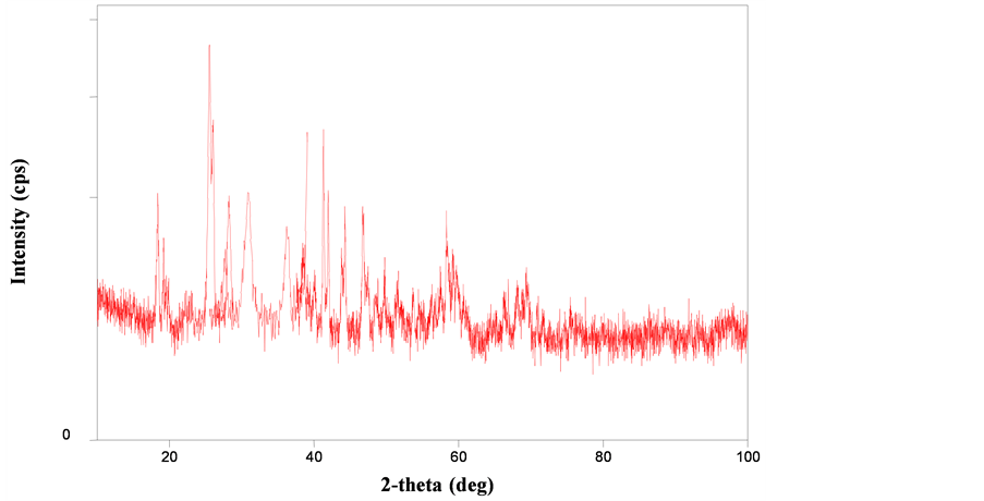
Figure 2. XRD spectrum of cobalt doped ZnO nanoparticles.

The Lattice strain induced in particles calculated using formula

where D is the Average crystalline size, λ represents wavelength of incident beam (1.5406 Å), β is the FWHM (Full Width Half Maxima) of the diffraction peak corresponding to <101>, k is the shape factor (it was set to 0.9 in our work), Ɛ is the lattice strain induced in nanoparticles and θ represents scattering angle of reflection.
The average crystalline size calculated from full width half maximum of high intense peak <101> is found to be 10.34 nm for pure ZnO nanoparticles and 23.58 nm for Co doped ZnO Particles and corresponding lattice strain in the nanoparticles is found to be 0.0221 in the case of undoped ZnO and 0.0071 in the case of doped ZnO Nanoparticles. From spectrum it was clear that significant increase in the crystalline size is observed by doping with cobalt. This show snanoparticles possess good crystallinity. The reduction in the lattice strain explains the grain can grew much easier due to the incorporation of Co dopant.
4.2. Scanning Electron Microscopy (SEM)
SEM has opened doors in fields ranging from chemistry to engineering, allowing scientists to access new, useful information about microscopic processes with macroscopic implications. Figure 3 and Figure 4 represent SEM micrographs of Pure and Cobalt doped ZnO Nanoparticles observed at different magnifications. From micrographs, it is clear that the cauliflower like ZnO comprising of nanoclusters observed on the surface of the ZnO Nanoparticles and almost spherical shape morphology in doped ZnO. The morphological observation of SEM results indicates particles with less aggregation can be obtained from this method [18] .
4.3. Fourier Transform Infrared Spectroscopy (FTIR)
Infrared spectroscopy is an important technique in organic chemistry. FTIR is one of the most widely used tools for the detection of functional groups in pure compounds and mixtures for compound comparison Formation of pure and doped ZnO. Thespectra was recorded in the range 1000 - 3500 cm−1 as shown in Figure 5 and Figure 6. From spectra one can observe the shift in the vibration frequencies of ZnO Nanoparticles corresponding to 3443.46 cm−1, 1632.66 cm−1, 1362.17 cm−1 due to the incorporation of Cobalt ions in to the ZnO Wurtzite structure. The spectrum shows broad absorption peak between 3600 - 3200 Cm−1 due to the O-H stretching vibration of Water molecule. The absorption peak around 1632 cm−1 corresponds to C-H stretching vibration and

Figure 3. SEM Micrographs Pure ZnO Nanoparticles.

Figure 4. SEM Micrographs Co Doped ZnO Nanoparticles.
around 1300 cm−1 corresponds to C-O symmetric stretching vibration. From spectra the main absorption between 600 cm−1 - 1000 cm−1 corresponding to the stretching mode of Zn-O in pure ZnO and characteristic absorption band due to the influence of Co is observed between 600 - 1000−1 in the spectra of doped ZnO Nanoparticles. These results further confirm the formation of Wurtzite structure in nanoparticles synthesized at room temperature.
5. Conclusion
In the present study Pure and Cobalt doped ZnO nanoparticles were synthesized via Sol Gel route and further analyzed through X-ray diffraction, scanning electron microscopy, Fourier transform infrared spectroscopyto analyzing the influence of Co doping on structural and optical properties of ZnO Nanoparticles. The XRD spectrum demonstrates that nanoparticles have the hexagonal wurtzite structure. The crystalline sizes of 10.34 nm and 23.58 nm are obtained from pure and Co doped ZnO. SEM images showed the formation of Cauli flower like ZnO composed of nanoclusters in the case of pure ZnO and almost spherical images due to the influence of Co doping. The vibration frequencies in the FTIR spectra further confirm the formation of wurtzite

Figure 5. FTIR Spectrum of Pure ZnO Nanoparticles.

Figure 6.FTIR Spectrum of Co Doped ZnO Nanoparticles.
structure in ZnO. Thus, the current doping method can be considered as another effective technique to modulate the properties of ZnO nanoparticles. The Co doped ZnO Nanoparticles synthesized by simple cost. Solgel route can be considered as an effective method to modify the properties of ZnO nanoparticles. The particles can be further utililized in the optoelcetonics and thermally stable devices.
Acknowledgements
The authors would like to thank SAIF IIT Chennai and Center for Nanoscience and Technology, Satyabhama University Chennai for proving the facilities for characterization.
Cite this paper
AravapalliVanaja,Karumuri SrinivisaRao, (2016) Effect of Co Doping on Structural and Optical Properties of Zinc Oxide Nanoparticles Synthesized by Sol-Gel Method. Advances in Nanoparticles,05,83-89. doi: 10.4236/anp.2016.51010
References
- 1. Ali, M., Moazzen, M., Borghei, S.M. and Taleshi, F. (2013) Change in the Morphology of ZnO Nanoparticles upon Changing the Reactant Concentration. Applied Nanoscience, 3, 295-302.
- 2. Arora, A.K., Devi, S., Jaswal, V.S., Singh, J., Kinger, M. and Gupta, V.D. (2014) Synthesis and Characterization of ZnO Nanoparticles. Oriental Journal of Chemistry, 30, 1671-1679.
- 3. Prasad, K. and Jha, A.K. (2009) ZnO Nanoparticles: Synthesis and Adsorption Study. Natural Science, 1, 129-135.
http://dx.doi.org/10.4236/ns.2009.12016 - 4. Wang, Z.L. (2004) Zinc Oxide Nanostructures: Growth, Properties and Applications. Journal of Physics: Condensed Matter, 16, R829-R858.
- 5. Kulkarn, S.S. (2015) Optical and Structural Properties of Zinc Oxide Nanoparticles. International Journal of Advanced Research in Physical Science, 2, 14-18.
- 6. Khan, Z.R., Arif, M. and Singh, A. (2012) Development and Study of the Structural and Optical Properties of Hexagonal ZnO Nanocrystals. International Nano Letters, 2, 22.
http://www.inl-journal.com/content/2/1/22 - 7. Bindu, P. and Thomas, S. (2014) Estimation of Lattice Strain in ZnO Nanoparticles: X-Ray Peak Pro?le Analysis. Journal of Theoretical and Applied Physics, 8, 123-134.
- 8. Chen, K.J., Fang, T.H., et al. (2008) The Crystallization and Physical Properties of Al-Doped ZnO Nanoparticles. Applied Surface Science, 254, 5791-5795.
- 9. Ghosh, A., Kumari, N. and Bhattacharjee, A. (2014) Investigations on Structural and Optical Properties of Cu Doped ZnO. Journal of Nano Science and Nano Technology: Spring Edition, 2, 485-489.
- 10. Mukhtar, M., Munisa, L. and Saleh, R. (2012) Co-Precipitation Synthesis and Characterization of Nanocrystalline Zinc Oxide Particles Doped with Cu2+ Ions. Materials Sciences and Applications, 3, 543-551.
- 11. Djaja, N.F., Montja, D.A. and Saleh, R. (2013) The Effect of Co Incorporation into ZnO Nanoparticles. Advances in Materials Physics and Chemistry, 3, 33-41.
- 12. Kshirsagar, S.D., Inamdar, D., Gopalakrishnan, I.K., Kulshreshtha, S.K. and Mahamuni, S. (2007) Formation of Room-Temperature Ferromagnetic Zn1—XCoXO Nanocrystals. Solid State Communications, 143, 457-460.
http://dx.doi.org/10.1016/j.ssc.2007.06.025 - 13. Nair, M.G. (2011) Structural, Optical, Photo Catalytic and Antibacterial Activity of ZnO and Co-Doped ZnO Nanoparticles. Materials Letters, 65, 1797-1800.
- 14. Iqbal, J., Janjua, R.A. and Jan, T. (2014) Structural, Optical and Magnetic Properties of Co-Doped ZnO Nanoparticles Prepared via Wet Chemical Route. International Journal of Modern Physics B, 28, Article ID: 1450158.
- 15. Hays, J., Reddy, K.M., Graces, N.Y., Engelhard, M.H., Shutthanandan, V., Luo, M., Xu, C., Giles, N.C., Wang, C., Thevuthasan, S. and Punnoose, A. (2007) Effect of Co-Doping on the Structural, Optical and Magnetic Properties of ZnO Nanoparticles. Journal of Physics: Condensed Matter, 19, Article ID: 266203.
http://dx.doi.org/10.1088/0953-8984/19/26/266203 - 16. Udayakumarl, S., Renuka, V. and Kavitha, K. (2012) Structural, Optical and Thermal Studies of Cobalt Doped Hexagonal ZnO by Simple Chemical Precipitation Method. Journal of Chemical and Pharmaceutical Research, 4, 1271-1280.
- 17. Sugaharal, H.J., de Melo, E.F., de Melo, C.P. and Alvesl, K.G.B. (1994) Influence of Cobalt Doping on the Luminescence and Magnetic Properties of ZnO Nanoparticles Prepared by Co-Precipitation.
- 18. Rath, C. (2009) Effect of Cobalt Substitution on Microstructure and Magnetic Properties in ZnO Nanoparticles. Indian Journal of Physics, 83, 415-421.