Open Journal of Anesthesiology
Vol.3 No.1(2013), Article ID:27631,3 pages DOI:10.4236/ojanes.2013.31010
Airway Management of an Infant Presenting with Syngnathia for Surgical Correction
![]()
1French Medical Institute for Children, Kabul, Afghanistan; 2Department of Anesthesia and Intensive Care, St. Olavs Hospital, Trondheim, Norway.
Email: hlonnee@gmail.com
Received October 25th, 2012; revised November 27th, 2012; accepted December 14th, 2012
Keywords: Syngnathia; Pediatric Airway; Anesthesia
ABSTRACT
Syngnathia, congenital fusion of the mandible and maxilla, is a rare anomaly. We describe fiberoptic intubation of the trachea whilst controlling the airway with a nasal tube. A simple modification of this technique, previously described in the management of the difficult pediatric airway [1], simplifies the procedure with less potential for complications.
1. Introduction
Reported airway management in case of surgery for syngnathia varies from observation or sedation only, up to full control of the airway via various techniques [2-4]. One of those described is with the aid of a fiberoptic bronchoscope (FOB). Holm-Knudsen et al. recommends the use of a naso-pharyngeal airway (small sized endotracheal tube (ETT)) connected to the anesthetic circuit, and performing FOB guided intubation via the contralateral nasal opening [1]. By introducing a rather larger sized ETT into the vestibulum nasi only, as illustrated by this case report, this technique becomes easier to perform, with less potential for laryngospasm and/or nasal bleeding (Figure 1).
2. Case Report
A 6-month-old female (2.8 kg) was presented to the surgical department of the French Medical Institute for Children (FMIC) in Kabul, Afghanistan [2]. The child was born by normal vaginal delivery and with a congenital inability to open the mouth. There was no other similar anomaly known within the consanguinity-positive family. The patient was awake and alert and otherwise morphologically normal. On examination of the mouth, the gingiva of the mandible and maxilla was fused at the occlusal level except for a central aperture just large enough to let the tip of a sucking bottle pass through (Figure 2). The buccal mucosa was not involved. A computerized tomography scan (CT) and skull X-ray revealed non-bony fusion of the alveolar ridge of the mandible and maxilla, and a hypoplastic mandible (Figure 3). There was no indication of tempero-mandibular fusion or palatal abnormalities. A surgical separation was planned. Anesthesia was induced by facemask with sevoflurane in oxygen whilst allowing the infant to breath spontaneously. Monitoring consisted of ECG, intermittent bloodpressure, pulse-oximetry, rectal temperature, end-tidal CO2 and anesthetic gas concentration. Soon after induction of anesthesia, maintaining a patent airway became more difficult and was not improved by re-adjusting the facemask, jaw thrust or chin lift. Subsequently as planned, a size 4.0 cuffless Ivory ETT was advanced 1 cm into the left nostril and connected to the breathing circuit. An assistant closed the contralateral nostril and the mouth in order to prevent air leakage, obtaining a perfect open airway with unobstructed spontaneous ventilation that was improved by applying continuous positive airway pressure (CPAP). After deepening the level of anesthesia, intravenous access was established, and an antisialogue was given. Meanwhile a size 3.0 Portex cuffed ETT was advanced 1 cm into the right nostril and a fiberoptic bronchoscope (Olympus LF-P 2) passed through its lumen. At this stage anesthesia was maintained by manual ventilation with positive end expiratory pressure (PEEP) of 5 cm H2O. Fiberoptic visualisation of the glottic opening was obtained at the first attempt, and propofol 10 mg was administered intravenously. The FBO was subsequently passed endotracheally, uneventfully followed by the Portex ETT. A nasogastric tube was inserted to deflate any possible gastric air insufflation. No muscle relaxants were used. Intraoperative analgesia provided by fentanyl 20 microgram intravenously. Peroperative oxygen saturation over 95%. Inspection of
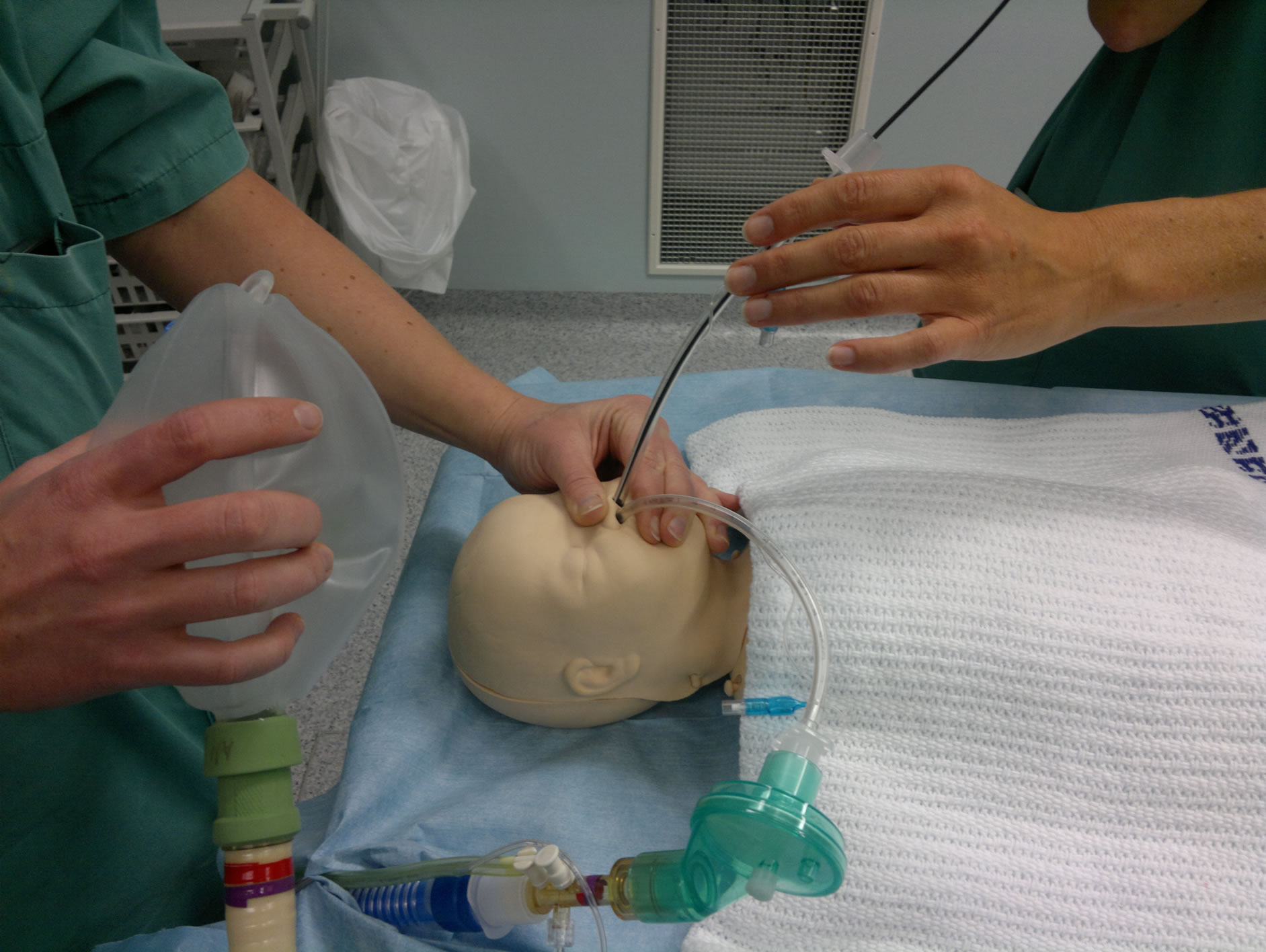
Figure 1. One person controls airway and ventilation by connecting an anaesthetic circuit to an ETT which is placed in the one nasal opening and seals the mouth manually whilst the other person manages FOB via the other nasal opening (dummy scenario).
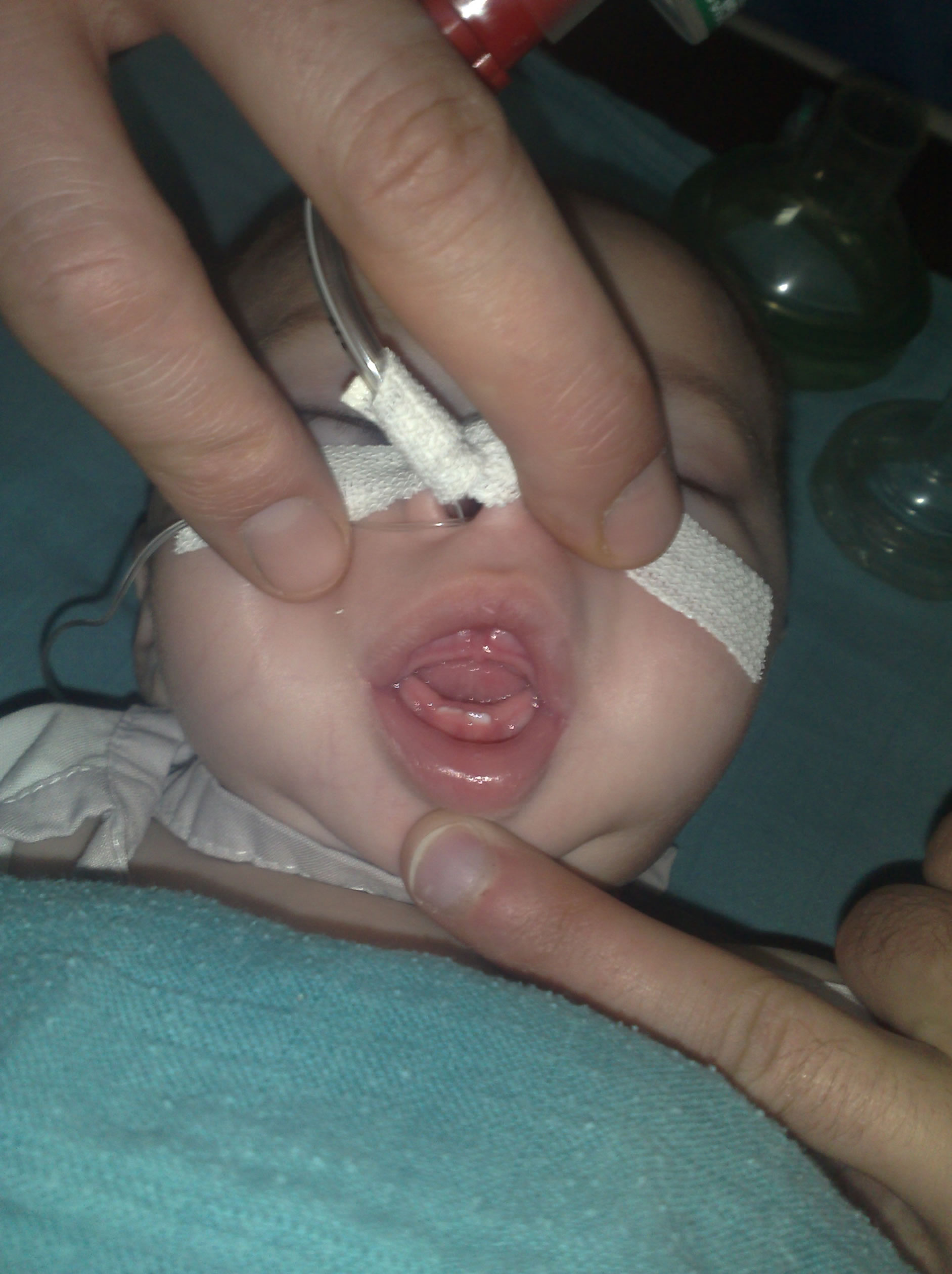
Figure 2. Visualizing the fixed mouth aperture just large enough to pass the tip of a sucking bottle.
the mouth confirmed the preoperative radiological findings and the adhesions appeared not to be exceeding the posterior margin of the alveolar ridge. The surgeon performed an instrumental syngnathia release followed by manual dilatation of the maxillar-manibular opening. There was no bloodloss of note. Laryngoscopy with a
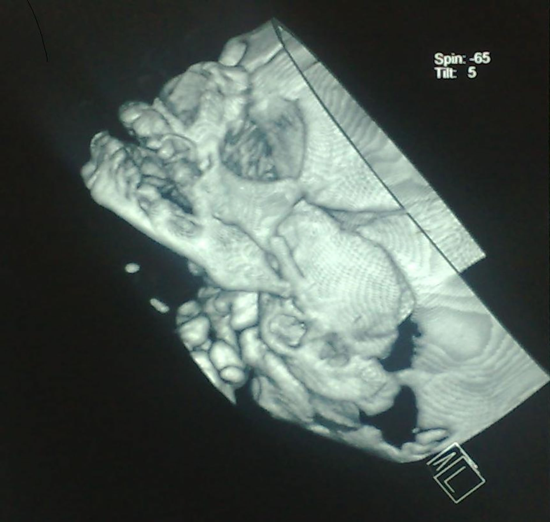
Figure 3. CT scan revealing confirming the non-bony fusion between the hypoplastic mandible and the maxilla.
Miller blade was a grade 4 using the Cormack and Lehane classification. After six hours of ventilation the child was successfully extubated. On day 4 the child had another manual dilatation of the maxillary mandibular opening under ketamine anesthesia. Unfortunately the patient was lost to further follow-up.
3. Discussion
Syngnathia, bony or fibrous, is rare and can occur as an isolated anomaly or in association with other oral abnormalities (cleft palate) or as part of a popliteal pterygium syndrome or Van der Woude syndrome. The adhesion can be complete or incomplete; unilateral or bilateral with an anterior opening. Etiology is unclear although several mechanisms have been proposed. Although surgery has been reported as being relatively simple in case of uncomplicated fibrous strands, airway management is a main challenge and various invasive and non-invasive techniques have been described [3-5].
Loss of muscle tone with increasing level of anesthesia can rapidly lead to a difficult mask ventilation scenario in children with or without expected airway problems. Meier et al. (2002) showed, as visualized by FOB, that CPAP in addition to chin lift and jaw thrust nearly doubled the size of the glottic opening by working as a pneumatic splint [6]. An elegant way of providing CPAP/ PEEP whilst simultaneously handling a difficult airway with a FOB via one nostril was described by HolmKnudsen et al. by inserting an endotracheal tube (3.0 - 3.5) as a nasopharyngeal airway approximately 8 cm into the contralateral nostril attached to an anesthetic circuit [1]. In managing the difficult pediatric airway either with or without FOB we suggest, in contrast to the original technique described, using a larger sized (un)-cuffed endotracheal tube (for optimal sealing) in the nostril dedicated for CPAP/PEEP prior to intubation and advancing it only a centimetre past the margin of the nostril (Figure 1). This nasal, instead of nasopharyngeal airway, will reduce the risk of laryngeal spasm and nose bleeding whilst providing an easily obtainable open airway with option of ventilating during FOB management. In our opinion this method has proven to be useful in several other pediatric patients with a difficult airway; however one should be prepared, in case of difficulties, to proceed with a smaller sized nasopharyngeal ETT dedicated for ventilation.
4. Conclusion
Management of the difficult pediatric airway via a nasal fiberoptic bronchoscope can be facilitated by using the other nostril for ventilation. This can be simply achieved by using an endotracheal tube, connected to an anaesthetic circuit, just one centimetre passed the vestibulum nasi as illustrated by this case report.
REFERENCES
- R. Holm-Knudsen, K. Eriksen and L. S. Rasmussen, “Using a Nasopharyngeal Airway during Fiberoptic Intubation in Small Children with a Difficult Airway,” Pediatric Anesthesia, Vol. 15, No. 10, 2005, pp. 839-845. doi:10.1111/j.1460-9592.2004.01566.x
- http//:www.chainedelespoir.org
- V. Patel, M. C. Theroux and J. Reilly, “Popliteal Pterygium Syndrome with Syngnathia,” Pediatric Anesthesia, Vol. 13, No. 1, 2003, pp. 80-82. doi:10.1046/j.1460-9592.2003.00981.x
- C. Gahm, R. Kuylenstierna and G. Papatziamos, “Popliteal Pterygium Syndrome (PPS) with Intra-Alveolar Syngnathia: A Discussion of Anesthetic and Surgical Considerations,” International Journal of Pediatric Otorhinolaryngology, Vol. 71, No. 10, 2007, pp. 1613-1616. doi:10.1016/j.ijporl.2007.05.024
- A. Jansen and G. Johnston, “The Shikani Optical Stylet: A Useful Adjunct to Airway Management in a Neonate with Popliteal Pterygium Syndrome,” Pediatric Anesthesia, Vol. 18, No. 2, 2008, pp. 188-190. doi:10.1111/j.1460-9592.2007.02401.x
- S. Meier, J. Geiduschek, R. Paganoni, F. Fuehrmeyer and A. Reber, “The Effect of Chin Lift, Jaw Thrust, and Continuous Positive Airway Pressure on the Size of the Glottic Opening and on Stridor Score in Anesthetized, Spontaneously Breathing Children,” Anesthesia & Analgesia, Vol. 94, No. 3, 2002, pp. 494-499. doi:10.1097/00000539-200203000-00004