Journal of Biosciences and Medicines
Vol.05 No.07(2017), Article ID:77589,6 pages
10.4236/jbm.2017.57001
Analysis of the Antimicrobial Properties of Thanaka, a Burmese Powder Used to Treat Acne
Elizabeth V. Seiverling, Jordan P. Trubiano, Jacqueline C. Williams, Hadjh T. Ahrns, David W. Craft, Matthew R. England
Departments of Dermatology, Family and Community Medicine, and Pathology, Pennsylvania State University - Milton S. Hershey Medical Center, Hershey, PA, USA
![]()
Copyright © 2017 by authors and Scientific Research Publishing Inc.
This work is licensed under the Creative Commons Attribution International License (CC BY 4.0).
http://creativecommons.org/licenses/by/4.0/


Received: May 17, 2017; Accepted: July 9, 2017; Published: July 13, 2017
ABSTRACT
Thanaka, powder from Hesperethusa crenulata tree bark, has been used in Burmese culture for acne treatment and prevention for over 2000 years. The purpose of this study was to evaluate the antimicrobial properties of thanaka against Staphylococcus aureus, Escherichia coli, and Propionibacterium acnes. Kirby-Bauer disk diffusion revealed no zones of inhibition for thanaka against the tested microorganisms. Disk diffusion may not be the best modality for definitive analysis of the antimicrobial activity of thanaka. Furthermore, the utility of thanaka in acne treatment may be related to anti-inflammatory, rather than antimicrobial properties.
Keywords:
Thanaka, Myanmar, Disk Diffusion, Propionibacterium acnes

1. Introduction
Thanaka (also spelled thanakha) is a powder produced from the bark of Hesperethusa crenulata or Naringi crenulata trees (Figure 1(a)). Thanaka is used for a variety of cosmetic and dermatologic purposes including photo-protection, acne treatment and prevention, skin cooling, skin lightening, pruritus relief, scar reduction, mosquito repellant, and odor prevention (Figure 1(b)) [1] [2] [3] [4] . Acne treatment and prevention were the most commonly reported reason for applying thanaka to the skin in persons 12 - 16 years old [4] . P. acnes is involved in the release of proinflammatory mediators in the pilosebaceous unit and leads to inflammatory papules, pustules, and nodules on the skin as seen in acne vulgaris [5] . Thus, inhibition of P. acnes with topical antibacterial agents such as
 (a)
(a) (b)
(b)
Figure 1. Thanaka powder and application: (a) An example of thanaka powder produced from the bark of Hesperethusa crenulata or Naringi crenulata trees and (b) An example of direct topical application of thanaka on the face of a Burmese woman.
benzoyl peroxide or blockade of its downstream inflammatory effects is key in treatment for acne [6] . One previous study reported that thanaka possesses some anti-microbial properties against E. coli and S. aureus using a minimum inhibitory concentration (MIC) assay [7] . The purpose of our study was to evaluate the antimicrobial properties of thanaka against P. acnes, S. aureus, and E. coli using Kirby-Bauer disk diffusion and Clinical and Laboratory Standards Institute (CLSI) breakpoints [8] .
2. Methods
2.1. Preparation of Antimicrobial Disks
Kirby-Bauer disks impregnated with thanaka are not commercially available. Thus, thanaka-laced antimicrobial disks were first created mimicking how thanaka is typically used in Myanmar: ground into powder, mixed with water, and applied directly to the faceas a paste (Figure 1(b)). However, the thanaka was not completely soluble in water. In an attempt to improve solubility, thanaka was dissolved into 95% ethanol: water: tween 20 (5:93.5:1.5), to create 1, 5, and 10 mg/ml solutions, as reported in Wangthong et al. [7] . Sterile 6 mm blank paper disks (Becton Dickinson, Cat #231039) were impregnated by soaking in the thanaka solutions for 30 min.
2.2. Clinical and Laboratory Standards Institute (CLSI) Kirby-Bauer Disk Diffusion
Isolates of E. coli (ATCC 25922), S. aureus (ATCC 25923), and P. acnes (clinical isolates identified by matrix-assisted laser desorption/ionization time-of-flight mass spectrometry) were isolated on 5% sheep-blood agar (Becton Dickinson, Cat# 221261). Mueller-Hinton agar plates were inoculated with 1:10 dilutions of 0.5 McFarland bacterial solutions [8] . The following antimicrobial disks were applied to each plate: 1 each of clindamycin (positive control, Beckton Dickinson, Cat #231213), ethanol: water: tween 20 (5:93.5:1.5) (negative control) and 2 each of the 1, 5, and 10 mg/mL thanaka-impregnated disks (Figure 2). Plates were incubated at 24 h at 35˚C for E. coli and S. aureus and 48 h under anaerobic conditions at 35˚C for P. acnes following CLSI protocol.

Figure 2. Kirby-Bauer disk diffusion results: (a) E. coli and (b) S. aureus on MHA with clindamycin and buffer controls and varying concentrations of thanaka disks (values listed in mg/ml) (c) and (d) P. acnes on Brucella blood agar with the clindamycin and buffer controls and varying concentrations of thanaka disks (values listed in mg/ml, c = control, Cl = clindamycin).
3. Results
The measured zone of inhibition for clindamycin was 33 mm against S. aureus, consistent with CLSI guidelines (Figure 2) [8] . The measured zones of inhibition for clindamycin against E. coli was 0 mm and against P. acnes was 48 mm; however there are no disk diffusion guidelines published for clindamycin against E. coli or P. acnes (Figure 2) [8] . There were no zones of inhibition for the disks of 1, 5, and 10 mg/ml thanaka or the negative control disks against S. aureus, E. coli, or P. acnes (Figure 2).
4. Discussion
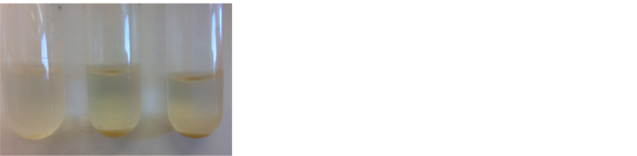
In an attempt to study the antimicrobial properties of thanaka, the Kirby-Bauer disk diffusion protocol was utilized [8] [9] [10] [11] . The lack of a zone of inhibition for the thanaka disks of all three tested concentrations could be attributed to a number of possibilities. First, thanaka is not completely soluble in the sterile, deionized water (Figure 3(a)). In order to create thanaka-impregnated disks, thanaka was first dissolved in sterile, de-ionized water. Mixing thanaka with water mimics how the powder is typically used in Myanmar: the thanaka bark is ground into powder, mixed with water, and applied directly to the face as a paste
 (a) (b)
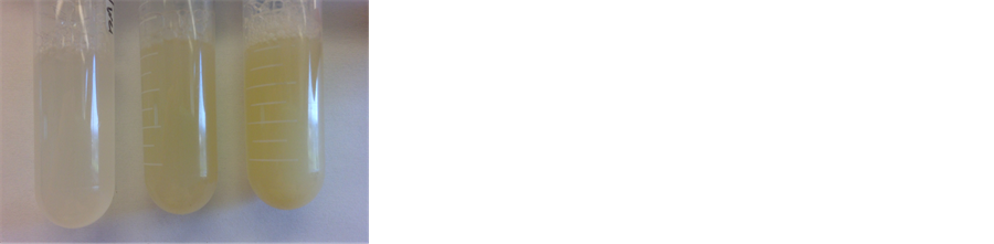
Figure 3. Solubility testing: Solutions of thanaka dissolved in (a) sterile, de-ionized water or (b) 95% Ethanol: Water: Tween 20 (5:93.5:1.5) at 1, 5, and 10 mg/ml.
(Figure 2). In an attempt to improve solubility, the protocol was modified by dissolving the thanaka into 95% ethanol: water: tween 20 (5:93.5:1.5), similar to thatused by Wangthong et al. [7] . Of note, Wangthong et al. utilized extracted compounds from thanaka rather than testing the powder itself. Addition of alcohol and detergent led to improved solubility (Figure 3(b)), but may have impacted the ability to measure antimicrobial activity. Second, the concentrations previously demonstrated by MIC interpretations may not exhibit an equivalent Kirby Bauer disk diffusion in vitro activity. This is not uncommon in the CLSI tables [8] . Third, the thanaka product may not have direct antimicrobial properties against E. coli, S. aureus, or P. acnes. One of the chemical constituents of thanaka is coumarin, which has known anti-oxidant and anti-inflammatory activity [12] [13] . Consequently, the role thanaka plays in acne treatment may be related to anti-inflammatory properties, rather than antimicrobial activity. Fourth, it is possible that the thanaka, or one of its active ingredients, has poor agar diffusion similar to other large, bulky antibiotics, like colistin or other polymyxins [14] . Finally, it is important to note that commercially available thanaka powder is sometimes mixed with other ingredients such as sandalwood or apple wood. Although we attempted to test ‘pure’ unmixed thanaka, we did not have a method to validate its purity. Future research into thanaka would benefit from a standardized product.
5. Conclusion
Thanaka powder is used for acne treatment and prevention, amongst other cosmetic and dermatologic uses. In this study, the antimicrobial properties of thanaka were evaluated according to published CLSI breakpoint interpretation for Kirby-Bauer disk diffusion. No zones of inhibition were observed when testing an alcoholic solution of thanaka against E. coli, S. aureus and P. acnes. Thanaka may have poor agar diffusion or may simply not have antimicrobial activity. Aqueous and alcoholic extracts of thanaka have, however, been shown to inhibit inflammation by blocking free radical release which may be beneficial in acne [7] . Standardizing a system for obtaining extracts of thanaka would benefit future investigations of its anti-acne properties. However, even with a standardized product, disk diffusion may not be the best modality for testing the efficacy of thanaka against P. acnes or skin flora. Ultimately, studies exploring the anti-inflammatory properties of thanaka may provide greater insight into its therapeutic potential.
Acknowledgements
This work was supported by a grant from the James and Joyce Marks Clinician Educator Endowment in the Department of Dermatology at the Pennsylvania State University, Milton S. Hershey Medical Center.
Cite this paper
Seiverling, E.V., Trubiano, J.P., Williams, J.C., Ahrns, H.T., Craft, D.W. and England, M.R. (2017) Analysis of the Antimicrobial Properties of Thanaka, a Burmese Powder Used to Treat Acne. Journal of Biosciences and Medicines, 5, 1-6. https://doi.org/10.4236/jbm.2017.57001
References
- 1. Goldsberry, A., Dinner, A. and Hanke, C.W. (2014) Thanaka: Traditional Burmese Sun Protection. Journal of Drugs and Dermatology, 13, 306-307.
- 2. Lourith, N., Kanlayavattanakul, M. and Pongpunyayuen, S. (2010) Skin Lightening Agent from Naringi Crenulata. World Academy of Science, Engineering and Technology, 46, 1022-1023.
- 3. Lindsay, S.W., Ewald, J.A., Samung, Y., Apiwathnasorn, C. and Nosten, F. (1998) Thanaka (Limonia acidissima) and Deet (dimethyl benzamide) Mixture as a Mosquito Repellent for Use by Karen Women. Medical and Veterinary Entomology, 12, 295-301.
https://doi.org/10.1046/j.1365-2915.1998.00115.x - 4. Seiverling, E.V. and Ahrns, H.T. (2013) Thanakha and Its Dermatologic Uses in Myanmar (Burma) [Abstract]. Journal of Investigative Dermatology, 133, S88-S103.
- 5. Kistowska, M., et al. (2015) Propionibacterium acnes Promotes Th17 and Th17/Th1 Responses in Acne Patients. Journal of Investigative Dermatology, 135, 110-118.
https://doi.org/10.1038/jid.2014.290 - 6. Eichenfield, L.F., et al. (2013) Evidence-Based Recommendations for the Diagnosis and Treatment of Pediatric Acne. Pediatrics, 131, 163-186.
https://doi.org/10.1542/peds.2013-0490B - 7. Wangthong, S., et al. (2010) Biological Activities and Safety of Thanaka (Hesperethusa crenulata) Stem Bark. Journal of Ethnopharmacology, 132, 466-472.
https://doi.org/10.1016/j.jep.2010.08.046 - 8. Clinical and Laboratory Standards Institute (CLSI) (2015) Performance Standards for Antimicrobial Disk Susceptibility Tests; Approved Standards - Twelfth Edition, M02-A12.
- 9. Fattah, N.S.A. and Darwish, Y.W. (2013) In Vitro Antibiotic Susceptibility Patterns of Propionibacterium acnes Isolated From Acne Patients: an Egyptian University Hospital-Based Study. Journal of the European Academy of Dermatology and Venereology, 27, 1546-1151.
https://doi.org/10.1111/jdv.12057 - 10. Eng, W. and Norman, R. (2010) Development of an Oregano-based Ointment With Anti-Microbial Activity Including Activity Against Methicillin-Resistant Staphylococcus aureus. Journal of Drugs in Dermatology, 9, 377-380.
- 11. Antonio-Velmonte, M., Gonzaga, A.J. and Darvin, C.U. (1998) Local Production of Low Cost Quality Antibiotic Susceptibility Disks for the Philippines. Journal of Microbiology and Infectious Disease, 17, 66-75.
- 12. Nayar, M.N.S. and Bhan, M.K. (1972) Coumarins and Other Constituents of Hesperethusa crenulata. Phytochemistry, 11, 3331-3333.
https://doi.org/10.1016/S0031-9422(00)86402-X - 13. Fylaktakidou, K.C., Hadjipavlou-Litina, D.J., Litinas, K.E. and Nicolaides, D.N. (2004) Natural and Synthetic Coumarin Derivatives with Anti-Inflammatory/Antioxidant Activities. Current Pharmaceutical Design, 10, 3813-3833.
https://doi.org/10.2174/1381612043382710 - 14. Galani, I., et al. (2008) Colistin Susceptibility Testing by Etest and Disk Diffusion Methods. International Journal of Antimicrobial Agents, 31, 434-439.
https://doi.org/10.1016/j.ijantimicag.2008.01.011