Open Journal of Orthopedics
Vol.09 No.07(2019), Article ID:93796,8 pages
10.4236/ojo.2019.97014
Polyostotic Melorheostosis: Approach to Clinical Evaluation and Management
Chee Chien Teoh1*, Shawn Ru Zye Teoh1, David Chen Hol Chieng2, Xi Yuan Ang1, Mohamad Hafiz Mohmad Hassim3
1Department of Trauma and Orthopedic, Hospital Bintulu, Sarawak, Malaysia
2Department of Trauma and Orthopedic, Hospital Umum Sarawak, Sarawak, Malaysia
3Orthopaedic Department, International Islamic University Malaysia, Kulliyyah of Medicine, Bandar Indera Mahkota Campus, Kuantan, Malaysia

Copyright © 2019 by author(s) and Scientific Research Publishing Inc.
This work is licensed under the Creative Commons Attribution International License (CC BY 4.0).
http://creativecommons.org/licenses/by/4.0/


Received: June 14, 2019; Accepted: July 20, 2019; Published: July 23, 2019
ABSTRACT
Melorheostosis is a rare form of sclerosing bone dysplasia that involves mainly the long bone of the limbs. However, the involvement of more than one long bone, either involving ipsilaterally or contralaterally, is extremely rare. The aetiology still remains unknown despite approximately 400 cases have been reported in the literature up to date. Diagnosis is mainly by conventional radiographic imaging such as plain X-ray, in which the typical melting wax dripping down a candle appearance is seen. The main treatment options are conservative management involving mainly analgesias, physiotherapy, braces, and possible nerve block and symphathectomies if indicated. The aim of this case report is to discuss the approach to a case of melorheostosis involving two long bones of the ipsilateral lower limb (polyostotic) that presented to us in our centre. Appropriate investigations and treatment modalities must be tailored according to the patient’s complaints in order to achieve satisfactory treatment outcomes. With adequate analgesia complimenting with appropriate physiotherapy and rehabilitation, our patient’s symptoms improved dramatically.
Keywords:
Melorheostosis, Melting Wax Syndrome, Candle-Wax Appearance, Leri Disease, Rare

1. Introduction
Melorheostosis (synonyms: candle bone disease, melting wax syndrome, Leri disease) is an atypical, non-hereditary, sclerosing bone dysplasia that affects both cortical bone and adjacent soft tissue structures [1] . It mainly affects the long bone of the limbs, and involvement of more than one long bone is rare. First described in 1922 by Leri and Joanny, it is a benign bone disorder exhibiting patterns of hyperostosis characterised by linear distribution along the major axis of long bones, resembling a “dripping candle wax” appearance [2] . It has equal distribution among men and women [3] , which usually presents after early childhood or adolescence [4] . Although benign in nature, it may be debilitating due to limb deformity, contracture, pain and stiffness. Management involves addressing symptoms presented by the patient and is often a multidisciplinary approach involving orthopaedics, physiotherapist, rehabilitation and physical medicine. Here, we described a polyostotic melorheostosis case of a 26-year-old gentleman with chronic pain and deformity of his left knee and leg. The patient agreed to participate in this case report by means of a free and informed consent form.
2. Case Report
This is a 26-year-old gentleman who presented to our out-patient clinic with a history of left knee deformity for four years associated with gradual pain, swelling and stiffness over left knee and leg progressively worsening for past six months. No recent trauma/fall to suggest a possible traumatic cause. Clinically the left knee has mild valgus deformity and limb length discrepancy of 5 cm longer compared to the contralateral lower limb, with multiple ill-defined non-tender hard swelling over the anteromedial aspect of the left knee (Figure 1). Further examination showed discrepancy of the left femur bone as the causation of the significant limb length difference (Figure 2). Hyperpigmentation was also noted over the entire length of the left leg. Range of motion of the left knee was slightly limited due to pain. All blood parameters including serum calcium, alkaline phosphatase, C-reactive protein and erythrocyte sedimentation rate were within normal limits.


Figure 1. Initial presentation of 5 cm longer limb length discrepancy over the left leg with valgus deformity of the left knee.
Plain radiographs of the affected limb showed typical hyperostotic appearance “dripping candle wax” phenomenon located eccentrically on the inner and outer medial cortex of left femur (Figure 3) and tibia (Figure 4), query possible chip fracture of the distal femur with intra-articular involvement (Figure 5). Similar appearances can be seen within the medulla of the left proximal femur (Figure 6). Calcifications can also be seen over medial aspect of the distal femur. The diagnosis of melorheostosis was raised based on clinical and radiological judgment and we completed the investigations by a CT scan of the left knee to exclude possible intra-articular chip fracture of the left femur.
CT scan findings of the left knee showed undulating cortical hyperostosis at medial aspect of femur and proximal tibia plateau with hyperostosis within medulla of proximal tibia (Figure 7). Linear hyperdensity is seen between vastus medialis and sartorius with irregular calcium deposits seen at the medial aspect of medial femoral condyle, anterior knee joint and posterior knee joint, not suggesting

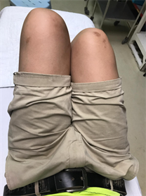
Figure 2. Discrepancy of left femur length can be observed with hyperpigmentation of skin over the left leg.

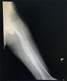
Figure 3. X-ray of the left femur (AP/lateral view) with classical “dripping candle wax” appearance characterised by dense, irregular and eccentric hyperostosis of medial cortex of femur, causing narrowing of medullary cavity.


Figure 4. X-ray of left tibia/fibula (AP/lateral view) showing osteoma-like hyperostosis involving the long axis of tibia. Narrowing of medullary cavity is also observed here.


Figure 5. X-ray of the left knee (AP/lateral view) showing irregular cortical hyperostosis, with discontinuity over medial condyle of femur extending into knee joint suggesting possible chip fracture (red arrow). Calcifications can be observed over medial aspect of femur (yellow arrows).

Figure 6. Flowing candle wax appearance in the proximal left femur on a plain radiograph (white arrow).


Figure 7. CT scan of the left knee showed characteristic candle wax condensation of the distal femur and medial tibia plateau.
of chip fracture in knee joint [5] .
In view of no evidence of fracture involving the knee joint, surgical intervention was not indicated in this case. As such, conservative management was chosen to address his complaints. The patient was started on Paracetamol 1000 mg QID, Tramadol 50 mg TDS and Celecoxib 200 mg PRN dosing for pain control, and further pain control modalities were not needed as his pain is well controlled with regular 2 monthly follow-ups and review. He was also referred to physiotherapy for the range of motion and stretching exercises of the left knee to alleviate his joint stiffness problem. Occupational therapist was also involved for a shoe lift over the right lower limb to optimize ambulation in view of limb length discrepancy of 5 cm. Subsequent follow-ups showed improvement of symptoms in terms of pain alleviation and improved mobility. Patient is able to return to work as well with no worsening of symptoms.
3. Discussion
Melorheostosis is a rare connective tissue disease characterized by sclerosing bone dysplasia. The incident is approximately 0.9 cases per million inhabitants [1] [6] . With about 400 cases of melorheostosis being reported in the literature up to date [7] , the aetiology of the disease itself remains unknown. Several postulations such as genetic factors, metabolic predisposition or malformation of vessels have been made and proposed, but the exact cause is yet to be proven. Loss of function mutation in LEMD3 gene (12q12-12q14.3) [7] was initially proposed to be the cause of loss of functional protein in the inner nuclear membrane, which is crucial in bone morphogenic protein and tumor growth factor-beta signaling. However, further studies demonstrated that these mutations were not associated with the idiopathic melorheostosis. Hellemans et al. showed that LEMD3 gene mutation was not noted in isolated and sporadic cases of melorheostosis, suggesting that the genetic basis postulation remains questionable [8] . Kim et al. proposed that down-regulation of adhesion proteins involved in osteoblastic regulation, specifically of the TGF-B-induced gene product, which may lead to the occurrence of hyperostosis and associated soft tissue abnormalities [9] .
Clinical presentation of melorheostosis can be highly variable ranging from asymptomatic to debilitating symptoms such as chronic limb pain, deformity, joint stiffness and swelling, which may involve one or more limbs. Skin lesions such as circumscribed hyperpigmentation, edema, fibrosis, and circumscribed scleroderma have also been reported. Bruno et al. reported a case of a 24-year-old patient with melorheostosis involving both right upper and lower limb who presented with swelling, decreased range of motion and hyperpigmentation in the affected limb [6] , similar to our patient’s presentation who has left lower limb deformity, chronic pain, skin pigmentation and limited mobility of the affected knee joint. Rakesh et al. also described a case of 35-year-old woman with melorheostosis of left tibia bone who presented with similar non-tender bony swelling, hyperpigmentation and restriction of knee movement [3] .
Conventional radiographic imaging such as plain X-ray is used to diagnosed melorheostosis, in which the typical melting wax dripping down a candle appearance may be seen, showing extensive, dense, irregular eccentric hyperostosis of both periosteal and endosteal surfaces of cortex resulting in deformity over bones and narrowing of marrow space. Both patients reported by Bruno et al. and Rakesh et al. has similar pathognomonic findings of “dripping candle wax” appearance in plain X-ray suggesting the diagnosis of melorheostosis [3] [6] . Freyschmidt described three additional patterns of radiological patterns:
1) “osteoma-like” hyperostosis involving only the endosteal surface, which orientates in the long axis of involved bone. Lesions may be 5 cm or larger in diameter, may involve more than one bone and eccentrically located. If only one bone involved, circumscribed scleroderma or subcutaneous fibrosis above the lesion may be seen [7] .
2) “osteopathia striata-like” hyperostosis, showing unilateral, long and dense hyperostosis striations near the inner surface of the cortex in two or more bones [7] .
3) “myositis ossificans-like” pattern in two or more unilateral regions, with or without intraosseous hyperostosis [7] .
Computer tomography (CT) can be helpful in showing osseous sclerosis and reduction of medullary space at higher resolution than plain radiology but is often not needed in diagnosing melorheostosis [8] .
In our case study, plain imaging shows “osteoma-like” hyperostosis involving the endosteal surface along the long axis of femur and tibia. Calcium deposits can be observed over medial aspect of the femur. Proceeded with CT, which reported as undulating cortical hyperostosis at medial aspect of the femur and proximal tibia plateau with hyperostosis within medulla of proximal tibia and linear hyperdensity is seen between vastus medialis and sartorius with irregular calcium deposits seen at the medial aspect of medial femoral condyle, anterior knee joint and posterior knee joint.
Blood investigations for melorheostosis are unremarkable, with no rise in tumour markers level and normal level of baseline investigation amid the severity of the disease. Histopathology results are non-specific [10] and often show a mixture of mature and immature bone in dense formation with increased trabecular bone. As such, it can only be used as an exclusion criterion against osteosarcoma. Thus with clinical presentation, physical examination and plain radiograph images, these are sufficient to diagnose melorheostosis.
Different modalities of treatment had been used to treat the pain and deformities associated with melorheostosis. Conservative management includes use of oral medications such as bisphosphonates and NSAIDs, physiotherapy, manipulation, braces, serial casting, nerve block and symphathectomies [11] . Good response to treatment was seen in the patient presented by Bruno et al. with physical therapy program and Ibuprofen as analgesia [6] . Bisphosphonate can also be used to reduce inflammatory bone pain as it inhibits osteoclast-mediated bone resorption by direct and indirect actions on osteoblast and macrophages and bone vascularity [12] . In the case reported by Rakesh et al., the patient showed good response with Pamidronate 30 mg daily for 6 days as well as analgesia and physiotherapy [3] . Surgical options are offered after failed conservative therapy, limbs deformity or patient preferences, which includes tendon lengthening, limb lengthening, excision of fibrous tissue, fasciotomies, capsulotomies, osteotomies, excision of hyperostosis, arthrodesis, contralateral epiphysiodesis and amputation [4] . Freyschmidt noted success in relieving bone pain in 2 patient performing cortical fenestration of large hyperostosis, based on the idea that elevated intraosseous pressure leads to pain in melortheostosis patient. However, it is important to note that surgical therapy often results in recurrence [13] . To sum up, treatment must be tailored according to the patient’s presentation and preference in order to achieve good prognosis.
4. Conclusion
Melorheostosis is a rare benign sclerosing bone dysplasia with an indistinct etiology. It usually remains asymptomatic and undetected until early childhood or adolescence, where cortical hyperostosis and surrounding soft tissues involvement causes pain, joint stiffness, contractures, and limb deformity. Diagnosis is achieved by a combination of clinical assessment and radiological features of extensive, dense, irregular eccentric hyperostosis of the bone cortex which is classically described as “dripping candle wax” appearance. Blood investigations will be within normal limits, and histopathology results are non-specific and only indicated in the exclusion criterion against osteosarcoma. Treatment wise is mainly symptomatic, with care emphasizing on pain and functional limitations management. Thus, a multimodal approach is essential which commonly revolves around orthopedics, physical medicine, rehabilitation, and pain management. Surgery may have a role in large deformations, contractures and joint damage, but with great care and planning.
Conflicts of Interest
The authors declare no conflicts of interest regarding the publication of this paper.
Cite this paper
Teoh, C.C., Teoh, S.R.Z., Chieng, D.C.H., Ang, X.Y. and Hassim, M.H.M. (2019) Polyostotic Melorheostosis: Approach to Clinical Evaluation and Management. Open Journal of Orthopedics, 9, 137-144. https://doi.org/10.4236/ojo.2019.97014
References
- 1. Clifford, P.D. and Jose, J. (2009) Melorheostosis. The American Journal of Orthopedics (Belle Mead NJ), 38 360-361. https://doi.org/10.1093/ageing/afp071
- 2. Leri, A. and Joanny, J. (1992) Une Affection non des os hyperostose “en coulee” sur toute la longeur d’un member “oumelorheostoses”. Bulletins et Mémoires de la Société Médicale des H?pitaux de Paris, 46, 1141-1145.
- 3. Kumar, R., Sankhala, S.S., Bijarna, I. (2014) Melorheostosis—Case Report of Rare Disease. Journal of Orthopedic Case Report, 4, 25-27.
- 4. Rozencwaig, R., Wilson, M.R. and McFarland Jr., G.B. (1997) Melorheostosis. The American Journal of Orthopedics, 26, 83-86.
- 5. Hamli, Q.I.B. and Jin, M.W. (2018) Report CT Bilateral Knee Joints LID 999904265763.
- 6. Alpoim, B.P., Rodrigues, M.E.G.R., Felix, A.J.M., Marques, P.M.D.C., Sá, P.M.G. and Silva, L.F.N.P. (2013) Melorheostosis: A Case Report. Revista Brasileira de Ortopedia (English Edition), 48, 282-285. https://doi.org/10.1016/j.rboe.2012.07.007
- 7. Kotwal, A. and Clarke, B.L. (2017) Melorheostosis: A Rare Sclerosing Bone Dysplasia. Current Osteoporosis Reports, 15, 335-342. https://doi.org/10.1007/s11914-017-0375-y
- 8. Suresh, S., Muthukumar, T. and Saifuddin, A. (2010) Classical and Unusual Imaging Appearances of Melorheostosis. Elsevier Clinical Radiology, 65, 593-600. https://doi.org/10.1016/j.crad.2010.02.004
- 9. Kim, J.-E., Kim, E.-H., Han, E.-H., et al. (2000) A TGF-β-Inducible Cell Adhesion Molecules, βig-h3, Is a Downregulated in Melorheostosis and Involved in Osteogenesis. Journal of Cellular Biochemistry, 77, 169-178. https://doi.org/10.1002/(SICI)1097-4644(20000501)77:2<169::AID-JCB1>3.0.CO;2-L
- 10. Freyschmidt, J. (2001) Melorheostosis: A Review of 23 Cases. Springer-Verlag 2001. European Radiology, 11, 474-479. https://doi.org/10.1007/s003300000562
- 11. Gagliardi, G.G. and Mahan, K.T. (2010) Melorheostosis: A Literature Review and Case Report with Surgical Consideration. The Journal of Foot & Ankle Surgery, 49, 80-85. https://doi.org/10.1053/j.jfas.2009.08.004
- 12. Wood, J., Bonjean, K., Ruetz, S., et al. (2002) Novel Antiangiogenic Effects of the Bisphosphomnate Compound Zolendronic Acid. Journal of Pharmacology and Experimental Therapeutics, 302, 1055-1061. https://doi.org/10.1124/jpet.102.035295
- 13. Goldman, A.B., Schneider, R., Huvos, A.S. and Lane, J. (1993) Case Report 778. Melorheostosis Presenting as Two Soft-Tissue Masses with Osseous Changes Limited to the Axial Skeleton. Skeletal Radiology, 22, 206-210. https://doi.org/10.1007/BF00206157