Modern Plastic Surgery
Vol.2 No.3(2012), Article ID:21199,3 pages DOI:10.4236/mps.2012.23011
Repair of the Median Microform Cleft Lip Using Z-Plasty
![]()
Section of Plastic and Reconstructive Surgery, Department of Surgery, Yale University School of Medicine, New Haven, USA.
Email: derek.steinbacher@yale.edu
Received January 14th, 2012; revised February 15th, 2012; accepted March 7th, 2012
Keywords: Median Cleft Lip; Midline Cleft Lip; Mucosal Z-plasty
ABSTRACT
A median cleft lip is a rare central midline deficiency of the upper lip. Multiple surgical techniques are described in the literature to address this defect, though there is little consensus on the preferred surgical technique. We describe an intra-oral approach for repair of the median upper lip cleft using mucosal Z-plasty. This technique provides excellent access to the attenuated orbicularis oris muscle and the frenulum fibrosed to the labial margin. The tethered lip can be mobilized and the notch converted with appropriate mucosal length, lip height, and vermillion fullness. The contour of the free labial border immediately improves, all while avoiding a cutaneous scar. The midline cleft lip notch can be effectively treated by adhering to 3 major principles: 1) excision of the tight, constrictive labial band; 2) achieving midline orbicularis oris muscle approximation; and 3) establishing mucosal lengthening using a Z-plasty.
1. Background
A median or midline cleft lip is a vertical defect in the labial tissue. This physical anomaly is a rare variant that represents less than 1% of all cleft patients [1]. Phenotypically, median clefts can range from a small central vermillion notch to a wide midline cleft in combination with a bifid nose, bony defect, and hypertelorism. The median cleft lip defect has been found to occur both sporadically and as a part of a larger constellation of inherited anomalies.
Midline clefts can be grouped into two major categories: 1) false clefts, or those thought to be embryologically-related to tissue agenesis; and 2) true clefts, or those that result when fusion of the medial nasal processes fails. False clefts are further characterized by forebrain abnormalities and are thought to be a subtype of holoprosencephaly [2,3]. Flattening, or clefting, of the midline structures and widening of the lateral facial elements may also be appreciated in patients with false midline clefts. In the most severe cases, false clefts are not compatible with life, obviating the need for surgical correction.
Multiple terms have been used to describe the constellations of abnormalities observed with true median clefts, including median cleft face syndrome [4], frontonasal dysplasia [5], and Tessier #0 clefts [6]. True median clefts can be distinguished by a lack of forebrain abnormalities. However, hypertelorbitism, midline craniofacial osseous defects, hairline abnormalities, and a midline upper lip cleft may be present to varying degrees in these cases. The mildest form includes a small notch in the soft tissue of the upper lip that does not cross the vermillion border. The majority of these cases are sporadic, but familial aggregation has been documented [7, 8].
Multiple techniques have been described to repair the median cleft lip deformity. We describe a mucosal Zplasty and midline orbicularis oris muscle unification to repair a median cleft lip notch.
2. Methods
2.1. Clinical Presentation
A 13-month-old male presented to the Yale Craniofacial Center with a midline cleft lip. The defect appeared as a notch in the upper lip wet and dry mucosa crossing the vermillion border (Figure 1(A)). Intraorally, the maxillary labial frenulum was comprised of several bands which were attached abnormally close to the free vermillion border. The maxillary alveolus was notched at the midline, but no osseous cleft was present (Figure 1(B)). Other pertinent facial findings included a shortened nasal length, maxillary hypoplasia, and delayed eruption of the maxillary anterior teeth. The anterior nasal spine was palpable and present.
2.2. Surgical Procedure and Outcome
A Z-plasty was designed on either side of the elongated frenulum and the constriction band was excised (Figure 1(C)). Raising the mucosal flaps exposed the orbicularis
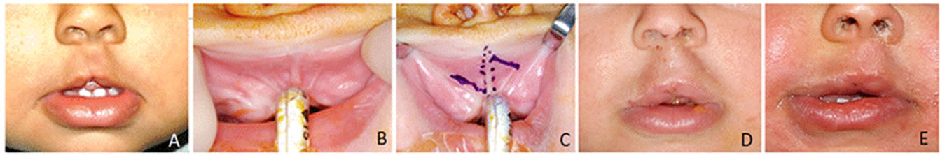
Figure 1. (A) Median cleft lip with mild phenotype; (B) Notched maxillary alveolus, with multiple labial frenum; (C) Design of intra-oral Z-plasty; (D) Immediate intra-operative result following release of fibrous bands; (E) Results at 2 months postoperatively.
oris muscle, which was discontinuous and underlying the median cleft notch. The midline fibrotic band was excised and the orbicularis oris muscle was dissected on both sides and united in the midline using horizontal mattress sutures. The Z-limbs were then transposed and sutured into position, recruiting additional mucosal length. The contour of the free labial border immediately improved. The notch was corrected and appropriate lip height and fullness were achieved and maintained postoperatively (Figures 1(D), 1(E)).
This case report and technique description is exempt from IRB approval and thus was not submitted to this body for review prior to data collection and reporting.
3. Discussion
Surgical correction of the true median cleft lip has been described for both minor and severe defects. The findings of a duplicated frenulum, notched lip free border, notched alveolus, attenuated orbicularis oris muscle, and a fibrous band are the characteristic findings in these microform cases [9].
For the treatment of a mild deformity Urata & Kawamoto [9] described a V-Y flap based in the maxillary vestibule and apex at the lip free border; their report also detailed the excision of a fibrous band and re-approximation of the orbicularis oris muscle. Weimer [10] described the excision of a diamond-shaped piece of skin and mucosa, as well as a fibrotic band with overlap of the orbicularis oris muscle and a zig-zag closure of the mucosa. Frietas and colleagues [11] described a similar technique involving excision of abnormal tissue, muscle approximation, and mucosal Z-transposition in patients with true microform median clefts. For more severe defects, including those involving the philtrum, Millard [12] recommended a combination of an inverted V-excision as well as a 90-degree angle in the excision 2 mm above the white roll on each side of the cleft. Francesconi [7] described local flaps which were rotated inferiorly at the midline to reconstruct a central defect that extended into the lip skin.
Since such wide variation exists within this deformity, each case must be individually considered. We agree with Urata & Kawamoto [9] that a hidden intraoral incision is most appropriate to correct this mild deformity, which does not cross the vermillion border. We have found that the intraoral incision allows access to the fibrous band and orbicularis oris muscle and provides the surgeon with the ability to augment the tubercle with mucosa through a Z-plasty.
4. Conclusions
The midline cleft lip notch can be effectively treated by adhering to 3 major principles:
1) Excision of the tight, constrictive labial band;
2) Achieving midline orbicularis oris muscle approximation;
3) Establishing mucosal lengthening using a Z-plasty.
The technique described successfully releases the tethered lip, provides mucosal length and vermillion fullness, and avoids a cutaneous scar.
REFERENCES
- C. J. Pinto and K. S. Goleria, “Transactions of the Fifth International Congress of Plastic and Reconstructive Surgery,” Butterworths, Melbourne, 1971.
- W. Demyer, W. Zeman and C. G. Palmer, “The Face Depicts the Brain: Diagnosis and Significance of Median Facial Anomalies for Holoprosencephaly with Median Cleft Lip and Palate,” Pediatrics, Vol. 11, 1964, pp. 256- 263.
- M. M. Cohen, “An Update on the Holoprosencephalic Disorders,” The Journal of Pediatrics, Vol. 101, No. 5, 1982, pp. 865-869. doi:10.1016/S0022-3476(82)80349-1
- W. Demyer, W. Zeman and C. G. Palmer, “Familial Alobar Holoprosencephaly,” Neurology, Vol. 13, 1963, pp. 913-918. doi:10.1212/WNL.13.11.913
- H. O. Sedano, M. M. Cohen, J. Jirasek, et al., “Frontonasal dysplasia,” The Journal of Pediatrics, Vol. 76, No. 6, 1970, pp. 906-913. doi:10.1016/S0022-3476(70)80374-2
- P. Tessier, “Anatomical Classification of Facial, Craniofacial, and Latero-Facial Clefts,” Journal of Maxillofacial Surgery, Vol. 4, 1976, pp. 69-92. doi:10.1016/S0301-0503(76)80013-6
- G. Francesconi and G. Fortunato, “Median Dysraphia of the Face,” Plastic and Reconstructive Surgery, Vol. 43, No. 5, 1969, pp. 481-491.
- K. Boo-Chai, “The Bifid Nose with a Report of 3 Cases of Siblings,” Plastic and Reconstructive Surgery, Vol. 36, 1965, pp. 626-628. doi:10.1097/00006534-196512000-00007
- M. M. Urata and H. Kawamoto, “Median Clefts of the Upper Lip: A Review and Surgical Management of a Minor Manifestation,” The Cleft Palate-Craniofacial Journal, Vol. 47, No. 5, 2010, pp. 104-106.
- D. R. Wiemer, S. B. Hardy and M. Spira, “Anatomical Findings in Median Cleft of Upper Lip,” Plastic and Reconstructive Surgery, Vol. 62, No. 6, 1978, pp. 866-869. doi:10.1097/00006534-197812000-00006
- R. da Silva Freitas, N. Alonso, J. H. Shin, L. Busato, M. C. Ono and G. A. Cruz, “Surgical Correction of Tessier Number 0 Cleft,” Journal of Craniofacial Surgery, Vol. 15, No. 9, 2008, pp. 1348-1352. doi:10.1097/SCS.0b013e318184326e
- D. R. Millard and S. Williams, “Median Clefts of the Upper Lip,” Plastic and Reconstructive Surgery, Vol. 42, 1968, pp. 4-14. doi:10.1097/00006534-196807000-00002