New Journal of Glass and Ceramics
Vol.4 No.3(2014), Article ID:47661,6 pages
DOI:10.4236/njgc.2014.43007
Crystal Growth of ZnO Microneedles in Water Containing Microbubbles
Yomei Tokuda1*, Hiroaki Matsuki1, Yoshikatsu Ueda2, Hirokazu Masai1, Toshinobu Yoko1
1Institute for Chemical Research, Kyoto University, Kyoto, Japan
2Research Institute for Sustainable Humanosphere, Kyoto University, Kyoto, Japan
Email: *tokuda@noncry.kuicr.kyoto-u.ac.jp
Copyright © 2014 by authors and Scientific Research Publishing Inc.
This work is licensed under the Creative Commons Attribution International License (CC BY).
http://creativecommons.org/licenses/by/4.0/


Received 20 May 2014; revised 15 June 2014; accepted 10 July 2014
ABSTRACT
Microbubble technology is now available in a wide range of industrial fields. The liquid containing microbubbles possesses a large number of air-liquid interfaces, and also generates radicals during bubble collapse. Here, we synthesized ZnO powder to explore the potential of microbubbles as starting materials for the formation of crystalline microor nanoparticles. The bubbles facilitated the growth of ZnO microneedles in high yields, and enhanced the reaction by radicals generated on bubble collapsing.
Keywords:Microbubble, Microcrystal, ZnO

1. Introduction
Microbubbles are defined as having diameters less than 50 mm, and have important technical applications in industrial fields [1] -[3] . Although the characteristics of water that contains microbubbles are not completely understood [3] -[5] , such water is currently used in semiconductor plants to remove oil [6] and on Japanese highways to remove de-icing salts [7] .
Microbubbles can be generated in water by various methods. One of the most efficient methods for producing fine bubbles, such as nanoor microbubbles, involves centrifugal gas-liquid separation [8] , in which a mixture of gas and liquid is rotated at a high rotational velocity. The gas bubbles in the liquid are fractured because of the centrifugal shearing force during rotation. Advantages of this method include the generation of bubbles of ~100 nm in diameters and the long-term stability [9] . Additionally, the gas-liquid shearing method is versatile because any gas and liquid can be used as starting materials.
Microbubbles tend to decrease in size due to the dissolution of their interior gas into the surrounding water, and to collapse due to the high interior gas pressure on collapsing. Free radicals are generated from the bubbles even in the absence of ultrasound waves. These free radicals are known to accelerate chemical reactions. Indeed, the radicals generated by cavitation are used in sonochemistry [10] . Moreover, such radicals have affected the crystal growth of ZnO [11] , in which ZnO nanorods were aligned on a substrate. On the other hand, it was expected that microbubbles could act as crystal nuclei for the heterogeneous nucleation because of the high interfacial surface area between the bubbles and the liquid. From the view point of crystal growth, the advantage of using microbubble nucleation would be the absence of residue in the final product.
A preparative approach employing microbubbles would constitute a novel preparation method for nanomaterials. However, no reports are presently available. Here, we synthesize ZnO microcrystals using water containing microbubbles (MB water), in order to demonstrate its potential in the field of nanomaterials.
2. Experimental Section
MB water was prepared by the centrifugal gas-liquid separation method (BUVITAS HYK-32-D, Ligaric, Japan [7] . Distilled water (11 L) at a flow rate of 45 L/min and gas at a rate of 0.7 L/min were subjected to centrifugal rotation for 90 min to afford MB water. The gases used in this study were N2 and O3; the latter was generated by UV irradiation of oxygen gas (EO-OG-R4, Ecodesign-labo, Japan). It should be noted that O3 MB water contains both O2 and O3 gases.
The concentration of ozone in O3 MB water was determined by the difference in the UV absorption between the purified and ozonated water (ozone densitometer OZM 5000G, Okitrotec Co. Ltd., Japan).
The size distributions of the gas bubbles in the purified and MB waters were characterized using a laser-illuminated optical microscopic technique based on particle-tracking analysis (NanoSight LM10, NanoSight, Ltd.) [12] . The detection limit ranged from 50 to 1000 nm, depending on the particles and solvent.
All chemical reagents were used without further purification. Solutions of Zn(NO3)2 (25 mM) and of hexamethylenetetramine (HMT, 25 mM) were prepared using different water medium (purified water without bubbles for comparison, N2 MB water, or O3 MB water), Zn(NO3)2 (Wako Pure Chemicals Industries, Ltd., Japan), and HMT (Wako Pure Chemicals Industries, Ltd., Japan). A mixture of Zn(NO3)2 solution (50 mL) and HMT solution (50 mL) was stirred for 90 min at 90˚C to produce a precipitate. Microparticles were collected on a Si substrate by dipcoating.
Scanning electron microscopy (SEM) images were obtained for the microparticles using a field-emission scanning electron microscope (JSM6500F, JEOL, Japan). X-ray diffraction (XRD) patterns were also obtained for the precipitate (RINT-2000, Rigaku, Japan).
3. Results and Discussion
Figure 1 and Figure 2 show the time dependence of the mode values of the diameter and the bubble concentration (108 particles/mL) at the mode value of the diameter, respectively. The error bars indicate the standard dev-

Figure 1. Mode value of the N2 bubble diameter versus centrifugal gas-liquid separation time. The error bars indicate the standard deviation over seven experiments.

Figure 2. Bubble concentration (108 particles/mL) at the mode values of the diameter for N2 MB water versus centrifugal gas-liquid separation time. The error bars indicate the standard deviation for over seven experiments.
iation over seven repetitions. The bubbles in the water were determined to be microbubbles of ~100 nm in diameter after 10 min operation. The standard deviation of the concentration before 30 min was rather large, but became small after 30 min. Moreover, over 50 min, the concentration decreased due to the heat generated by the centrifugal apparatus. Therefore, because of the stability of both the diameter and concentration, N2 microbubble (MB) water centrifugally rotated for 50 min was used for the preparation of ZnO.
In the case of O3 MB water, the bubble diameters showed similar behavior to the N2 MB water. However, the ozone concentration reached a maximum and then decreased after ~30 min (Figure 3). This was also due to the heat generated by the centrifugal apparatus, because the ozone spontaneously decomposed above room temperature. Therefore, because of the stability of the concentration, O3 MB water operated for 50 min was used for the preparation of ZnO.
Table 1 shows the reaction yields of ZnO together with the bubble concentrations, the maximum bubble diameter values, and the ozone concentration. The ZnO yields obtained from purified water, N2 MB water, and O3 MB water were 0.11, 0.35, and 0.60, respectively. Thus, the yield was increased by using the MB waters instead of pure water, and the O3 MB water produced a much higher yield than the N2 MB water.
The increase in the yield through the use of MB water can be interpreted based on the growth mechanism of ZnO nanorods as follows [13] -[15] ,
 (1)
(1)
 (2)
(2)
 (3)
(3)
In the case of MB water, Reaction (3) was expected to be enhanced due to the increased interfacial surface area between the bubbles and water.
In the case of O3 MB water, the increment can be also interpreted based on the generation of radicals on O3 decomposition. That is, ozone decomposes to generate free radicals as follows [16] ,
 (4)
(4)
 (5)
(5)
where the symbol “·” indicates the radical. The generated radical reacts with Zn2+ to produce ZnO as follows [11] ,
 (6)
(6)
XRD measurements were carried out to examine whether the microbubbles affected the ZnO crystal structure or not. Figure 4 shows XRD patterns of ZnO prepared using purified water, N2 MB water, and O3 MB water,

Figure 3. Aqueous ozone concentration in water plotted against the operation time. The error bars indicate the standard deviation for over seven experiments.
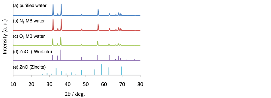
Figure 4. XRD patterns of ZnO prepared with (a) purified water, (b) N2 MB water, or (c) O3 MB water, together with XRD patterns derived from the JCPDS database.

Table 1. Bubble concentration, mode value of the diameter, ozone concentration, and reaction yields.
together with the powder diffraction patterns of wurtzite and zincite from the JCPDS database. The results clearly showed that there were no differences among the samples. Hence, neither the interfacial surfaces nor the radicals had distinct effect on the crystal structure of ZnO.
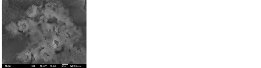
Finally, SEM observations were carried out to determine the effects of the microbubbles on the sizes or shapes of ZnO crystals (Figure 5). From purified water, the island-shaped ZnO crystals were grown, while in the cases of water with microbubbles, the ZnO crystals in needle-like structures of ~1 mm in length were grown. Note that purified water has no interfacial surfaces in the reaction system. As a result, the mechanism of crystal growth is mainly due to homogeneous nucleation. The number of initial crystallites is small, which results in large crystals and a low reaction yield. On the other hand, in the MB water, a lot of interfacial surfaces are present in the reac-
 (a)
(a)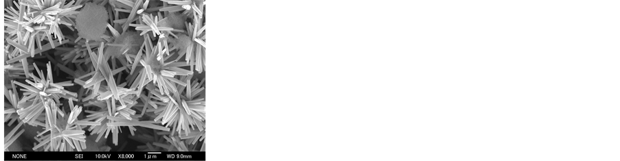 (b)
(b)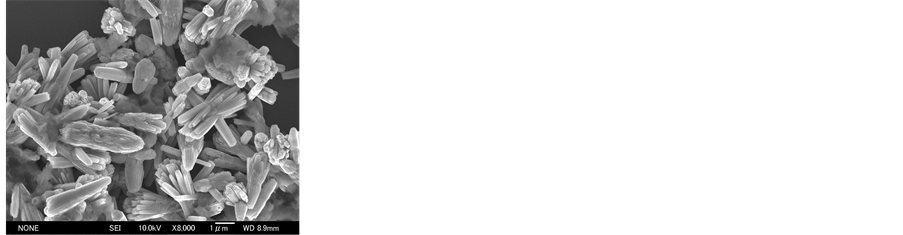 (c)
(c)
Figure 5. SEM images of ZnO prepared with (a) purified water, (b) N2 MB water, and (c) O3 MB water.
tion system, which acts as nuclei for heterogeneous nucleation. The number of ZnO crystallites is expected to be large. Accordingly, the concentration of Zn2+ decreased rapidly, resulting in small crystals and high yields. In addition, from O3 MB water, ZnO crystals with low aspect ratio were grown, while in the case of N2 MB water, that with high aspect ratio. We believe that this result is due to the enhancement of the oxidation reaction by O3.
4. Conclusions
ZnO crystals were grown from purified water, N2 MB water, and O3 MB water mediums. On the basis of the yield, crystal structure, and crystal shape, we reviewed whether the presence of the bubbles and radicals affected crystal growth. The yields in the experiments that used MB water were larger than in those that used purified water. We believe that this increment by means of MB water was due to the presence of interfacial surfaces between the bubbles and water, which acted as nuclei for heterogeneous nucleation. O3 MB water provided a higher yield than N2 MB water. This increment was likely due to the radicals generated by O3. The use of MB water resulted in the formation of ZnO with needle-like structures of ~1 mm in length, whereas that of purified water produced islandshaped ZnO. This result was also interpreted as being due to heterogeneous nucleation: the concentration of Zn2+ decreased rapidly, resulting in small crystals. On the other hand, the crystal structures were identical and independent of the presence of MB in the initial water. Thus, MB water is a potential and novel starting material for the facile preparation of microcrystals.
We have presented a simple and facile method for producing ZnO microneedles using water containing microbubbles. In the reaction system, the interfaces between the bubbles and water are expected to promote heterogeneous nucleation. In addition, the radicals generated on the collapse of O3 bubbles can be used as oxidizing agents. We expect that the present process can also be applied to the synthesis of other types of oxide nanoor microparticles because we can select any gas as inert, oxidative or reductive gas for preparing bubbles.
Acknowledgements
This work was supported by the Center for Exploratory Research on Humanosphere of the Research Institute for Humanosphere, Kyoto University. This work was also financially supported by a Grant-in-Aid for Scientific Research, No. 20613007 and Mazda Foundation.
References
- Burns, S.E., Yiacoumi, S. and Tsouris, C. (1997) Microbubble Generation for Environmental and Industrial Separations. Separation and Purification Technology, 11, 221-232. http://dx.doi.org/10.1016/S1383-5866(97)00024-5
- Kodama, Y., Kakugawa, A., Takahashi, T. and Kawashima, H. (2000) Experimental Study on Microbubbles and Their Applicability to Ships for Skin Friction Reduction. International Journal of Heat and Fluid Flow, 21, 582-588. http://dx.doi.org/10.1016/S0142-727X(00)00048-5
- Agarwal, A., Ng, W.J. and Liu, Y. (2011) Principle and Applications of Microbubble and Nanobubble Technology for Water Treatment. Chemosphere, 84, 1175-1180. http://dx.doi.org/10.1016/j.chemosphere.2011.05.054
- Takahashi, M. (2005) ζ Potential of Microbubbles in Aqueous Solutions: Electrical Properties of the Gas-Water Interface. The Journal of Physical Chemistry B, 109, 21858-21864. http://dx.doi.org/10.1021/jp0445270
- Takahashi, M., Izawa, E., Etou, J. and Ohtani, T. (2002) Kinetic Characteristic of Bubble Nucleation in Superheated Water Using Fluid Inclusions. Journal of the Physical Society of Japan, 71, 2174-2177. http://dx.doi.org/10.1143/JPSJ.71.2174
- Miyamoto, T.U.M. (2010) Japanese Patent, 4595764.
- Yamazaki, Y. (2012) Japanese Patent, 4916526.
- Tsuji, H. (2008) Japanese Patent, 4118939.
- Ueda, Y., Tokuda, Y., Shigeto, F., Nihei, N. and Oka, T. (2013) Removal of Radioactive Cs from Gravel Conglomerate Using Water Containing Air Bubbles. Water Science and Technology, 67, 996-999. http://dx.doi.org/10.2166/wst.2013.650
- Bang, J.H. and Suslick, K.S. (2007) Sonochemical Synthesis of Nanosized Hollow Hematite. Journal of the American Chemical Society, 129, 2242.
- Jung, S.H., Oh, E., Lee, K.H., Park, W. and Jeong, S.H. (2007) A Sonochemical Method for Fabricating Aligned ZnO Nanorods. Advanced Materials, 19, 749.
- Gallego-Urrea, J.A., Tuoriniemi, J. and Hassellov, M. (2011) Applications of Particle-Tracking Analysis to the Determination of Size Distributions and Concentrations of Nanoparticles in Environmental, Biological and Food Samples. TrAC Trends in Analytical Chemistry, 30, 473-483. http://dx.doi.org/10.1016/j.trac.2011.01.005
- Hu, X.L., Zhu, Y.J. and Wang, S.W. (2004) Sonochemical and Microwave-Assisted Synthesis of Linked Single-Crystalline ZnO Rods. Materials Chemistry and Physics, 88, 421-426. http://dx.doi.org/10.1016/j.matchemphys.2004.08.010
- Li, W.J., Shi, E.W., Zhong, W.Z. and Yin, Z.W. (1999) Growth Mechanism and Growth Habit of Oxide Crystals. Journal of Crystal Growth, 203, 186-196. http://dx.doi.org/10.1016/S0022-0248(99)00076-7
- Chaparro, A.M., Maffiotte, C., Gutierrez, M.T. and Herrero, J. (2003) Study of the Spontaneous Growth of ZnO Thin Films from Aqueous Solutions. Thin Solid Films, 431, 373-377. http://dx.doi.org/10.1016/S0040-6090(03)00248-7
- Gurol, M.D. and Akata, A. (1996) Kinetics of Ozone Photolysis in Aqueous Solution. AIChE Journal, 42, 3283-3292. http://dx.doi.org/10.1002/aic.690421128
NOTES
![]()
*Corresponding author.