Natural Science
Vol.5 No.6(2013), Article ID:33090,11 pages DOI:10.4236/ns.2013.56090
Essential oil of Thymus vulgaris L. and Rosmarinus officinalis L.: Gas chromatography-mass spectrometry analysis, cytotoxicity and antioxidant properties and antibacterial activities against foodborne pathogens
![]()
1Laboratory of Analysis, Treatment and Valorisation of Environment Polluants and Products, Faculty of Pharmacy, Monastir, Tunisia; *Corresponding Author: miladi_h@yahoo.fr
2UR Study & Management of Urban and Coastal Environments, LARSEN—National Engineering School, Sfax, Tunisia
3Laboratory of Biochemistry, Faculty of Medicine, Monastir, Tunisia
4Range Ecology Laboratory, Arid Land Institute of Medenine, Medenine, Tunisia
Copyright © 2013 Hanene Miladi et al. This is an open access article distributed under the Creative Commons Attribution License, which permits unrestricted use, distribution, and reproduction in any medium, provided the original work is properly cited.
Received 20 March 2013; revised 22 April 2013; accepted 30 April 2013
Keywords: Thymus vulgaris L.; Rosmarinus officinalis L.; GC-MS; Cytotoxicity; Antioxidant Activity; Antimicrobial Activity
ABSTRACT
The essential oil composition of Thymus vulgaris L. and Rosmarinus officinalis L. endemic to France were determined by GC and GC-MS. Oils were assessed for their cytotoxic, antioxidant and antimicrobial activity. 31 and 37 different compounds were identified representing 99.64% and 99.38% of the thyme and rosemary oils respectively, where oxygenated monoterpenes constituted the main chemical class. Thymol (41.33%) and 1.8-cineole (24.10%) were identified as the main constituents of T. vulgaris L. and R. officinalis L., respectively. Essential oils (EOs) of selected plant species were evaluated for their in vitro cytotoxicity against the human lung adenocarcinoma epithelial cell line (A549). Cytotoxicity was measured using MTT [3-(4,5-dimethylthiazol-2-yl)-2,5-diphynyltetra- zolium bromide] colorimetric assay. Dose-dependent studies revealed IC50 of 8.50 ± 0.01 µg/mL and 10.50 ± 0.01 µg/mL after 72 h on the A549 cells for R. officinalis L. and T. vulgaris L., respectively. Antioxidant activity was determined using a quantitative DPPH (1,1-diphenyl- 2-picryl hydrazyl) assay. Thymus and rosemary EOs exhibited effective radical scavenging capacity with 50% inhibitory concentration (IC50) of 437 ± 5.46 µg/mL and 189 ± 2.38 µg/mL respectively and therefore acts as a natural antioxidant agent. The antimicrobial activity of these species has also been studied against several foodborne pathogens and food isolated Salmonella spp. including S. enteritidis of significant importance. According to the results, T. vulgaris L. showed higher bactericidal effect than those from R. officinalis L. These results suggest that the essential oil from T. vulgaris L. and R. officinalis L. have potential to be used as a natural cytotoxic, antioxidant and antimicrobial agent in food processing.
1. INTRODUCTION
Microbial contamination is an important factor promoting food deterioration and contributing to food-borne disease incidence. The presence and growth of microorganisms in food may cause spoilage and result in a reduction of quality and quantity [1,2]. Food-borne illnesses associated with Listeria monocytogenes, Staphylococcus aureus, Escherichia coli O157:H7 and Salmonella enteritidis present a major public health concern [2,3]. It has been reported that the incidence of foodborne diseases caused by microbial contamination and environmental pollution will globally increase [4,5]. Furthermore, the consumption of foods contaminated with pathogenic microorganisms, such as bacteria, mould, viruses and parasites active the growth of a pathogen. Moreover, new examinations of antimicrobial activity on a wider spectrum of microorganisms, including some new multiresistant strains of bacteria and fungi were necessary [6,7]. In addition, to the increasing demand for safe and natural food in recent years, the great economic costs of deterioration and poisoning of food products by food pathogens have motivated many researchers to explore new alternatives to traditional food preservation practices [8]. In this context, food safety has become a complex problem related to food products frequently introduced into the market and posed crucial public health and economic concerns for the human society [2, 9].
With the growing incidence of infections resistant to antibiotics, an arsenal of either new agents of the supplementation of current antibiotics was needed. According to Daferera et al. (2003) [10], the use of essential oils as antimicrobial agents assume important role in the combat to the development of microbial resistance [11]. So, essential oils and their components are widely used in medicine as constituents of different medical products, in the food industry as flavoring additives and also in cosmetics as fragrances [12]. In addition, demand is growing for natural, high-quality, preservative-free products that at the same time are safe and stable. Among these natural products, essential oils (EOs) from aromatic and medicinal plants have received particular attention as potential natural agents for food preservation to improve the flavor and organoleptic properties [13,14]. Also, they have great potential in the emerging nutritious industry, because these materials are often considered as food and medicines, as well, and are used in prevention and curative treatments throughout the world [7]. Their use in phytotherapy is mostly related to different biological activities such as antiviral, antimicrobial, antioxidant, anticarcinogenic, antiparasitic, and insecticidal effects [7,14]. The essential oils are considered among the most important antimicrobial agents present in the plants, and may also have cytotoxic and antioxidant activities [2]. They are a rich source of biologically active compounds mainly monoterpenes, sesquiterpenes, and their oxygenated derivatives such as alcohols, aldehydes, esters, ethers, ketones, and phenols which may be involved in its physiological and biological activities [15,16].
Numerous studies have described the chemical composition, the antioxidant and antimicrobial activity of the EOs of several species of the genus Thymus, in the family Lamiaceae [8,17,18] how serves as preservative for foods and as an aromatic ingredient for seasoning various dishes [19]. Regarding Rosmarinus officinalis EOs (Lamiaceae family), it has been previously reported variations in the chemical composition and biological activity of plants growing in different countries [8,20]. This oil was rich in phenolic acids such as rosmarinic acid and carnosic acid with strong antioxidant properties, it has been proposed to be used as preservation for certain foods and nutraceutical products to avoid synthetic antioxidants [21]. However, data regarding this activity of essential oils of thyme and rosemary are not abundant and the methods for determination are different [22,23]. On the contrary, the antimicrobial activity of the thyme and rosemary essential oils is well documented [7,8,24- 26]. But, these investigations are not so often performed with a precisely defined chemical composition of the essential oil in question. Furthermore, new examinations of antimicrobial activity on a wider spectrum of microorganisms, including some new multiresistant strains of bacteria and fungi, could help the pharmaceutical industry in synthesis or semisynthesis of new antibiotics [7].
In this study, the cytotoxicity, the antioxidant activity and antibacterial effects of essential oils of thyme and rosemary (Thymus vulgaris L. and Rosmarinus officinalis L., Lamiaceae) against several foodborne pathogens, especially the most common causative agent of foodborne salmonellosis, were reported. The chemical characterization of the investigated essential oils was performed by gas chromatography—mass spectrometry (GCMS).
2. MATERIALS AND METHODS
2.1. Plant Material and Essential Oil Extraction
Thymus vulgaris L. and Rosmarinus officinalis L. plants were freshly collected in 2011 during the period of full flowering on the mountain in the south of France (Mediterranean climate country and mountainous region). The specimens of collected plants were identified according to the forester flora of France [27]. The seeds were dried at room temperature. Air-dried leaves of thyme and rosemary were submitted to hydrodistillation (HD) for 3 h with 500 ml distilled water using a Clevenger-type apparatus according to the European Pharmacopoeia (1975) [28]. The extracted oil were collected and dried over anhydrous sodium sulfate, then stored in sealed glass vials in a refrigerator at 4˚C prior to analysis. The quantities of the essential oils were determined gravimetrically.
2.2. Essential Oil Analyses
2.2.1. Gas Chromatography (GC)
An Agilent Technologies 6890N GC equipped with HP-5MS capillary column (30 m × 0.25 mm i.d., film thickness 0.25 μm; Hewlett-Packard) and connected to a FID was used. The column temperature was programmed at 50˚C for 1 min, then 7˚C/min to 250˚C, and finally left at 250˚C for 5 min. The injection port temperature was 240˚C; while that of the detector was 250˚C (split ratio: 1/60).
The carrier gas was helium (99.995% purity) with a flow rate of 1.2 ml/min. The analyzed essential oil volume was 2 μl. Percentages of the constituents were calculated by electronic integration of FID peak areas, without the use of response factor correction. Mean percentage of Thymus vulgaris L. and Rosmarinus officenalis L. volatiles compounds represented the average calculated on three individuals. Retention indices (RI) were calculated for separate compounds relative to C9- C16 n-alkanes mixture (Aldrich Library of Chemicals Standards) [29].
2.2.2. Gas Chromatography/Mass Spectrometry (GC/MS)
The volatile compounds isolated by HD were analysed by GC/MS, using an Agilent Technologies 6890N GC. The fused HP-5MS capillary column (the same as that used in the GC/FID analysis) was coupled to an Agilent Technologies 5973B MS (Hewlett-Packard, Palo Alto, CA, USA). The oven temperature was programmed as previously (50˚C for 1 min, then 7˚C/min to 250˚C, and then left at 250˚C for 5 min). The injection port temperature was 250˚C and that of the detector was 280˚C (split ratio: 1/100). The carrier gas was helium (99.995% purity) with a flow rate of 1.2 ml/min. The MS conditions were as follow: ionization voltage, 70 eV; ion source temperature, 150˚C; electron ionization mass spectra were acquired over the mass range 50 to 550 m/z.
2.2.3. Volatile Compounds Identification
The volatile compounds of Thymus vulgaris L. and Rosmarinus officinalis L. leaves were identified by comparing the mass spectra data with spectra available from the Wiley 275 mass spectra libraries (software, D.03.00). Further identification confirmations were made referring to RI data generated from a series of known standards of n-alkanes mixture (C8 to C26) [29] and to those previously reported in the literature [30-32].
2.4. Antioxidant Activity
DPPH radical method. The free-radical scavenging activity of Thymus vulgaris L. and Rosmarinus officinalis L. EOs were measured by 2,2-diphenyl-2-picrylhydrazyl (DPPH, Sigma-Aldrich, France) using the method described by Hanato et al. (1988) [33]. One milliliter of the essential oil at known concentration was added to 0.25 ml of a DPPH methanolic solution. The mixture was shaken vigorously and left standing at room temperature for 30 min in the dark. The absorbance of the resulting solution was then measured at 517 nm and corresponded to the ability of the essential oil to reduce the stable radical DPPH to the yellow-colored diphenylpicrylhydrazine. The antiradical activity was expressed as IC50 (µg/ml), the extract dose required to cause a 50% inhibition. Absorption of a blank sample containing the same amount of methanol and DPPH solution acted as negative control. All determinations were performed in triplicate. The ability to scavenge the DPPH radical was calculated using the following equation:
 (1)
(1)
where A0 was the absorbance of the control at 30 min, and A1 was the absorbance of the sample at 30 min. All samples were analyzed in triplicate.
2.4. Cytotoxic Activity
Thymus vulgaris L. and Rosmarinus officinalis L. EOs were screened for cytotoxic activities using the MTT [3-(4,5-dimethylthiazol-2-yl)-2,5-diphynyltetrazolium bromide] colorimetric assay against the human lung adenocarcinoma epithelial cell line (A549) as described previously [34]. Briefly, cells were treated with concentrations of EOs ranging from 12.5 to 800 µg/ml and seeded in 96-well micro plates. The essential oil was first dissolved in DMSO and then in RPMI 1640 supplemented with 2% foetal calf serum (FBS). The final DMSO concentrations in the test medium and controls were 1% (v/v). Each concentration was tested in quadruplicate together with the control and repeated two times in separate experiments.
After incubation for 24, 48 and 72 hours, the medium in each well was collected and the cytotoxic effect was measured with the MTT colorimetric assay. To determine the cell viability, 20 µl of MTT (5 mg/ml) were added to each well and cells were cultured in additional incubation for 4 h. After washing the supernatant out, the insoluble formazan product was dissolved in acidified isopropanol. Then, optical density (OD) of 96-well culture plates was measured using an enzyme-linked immunosorbent assay (ELISA) reader at 540 nm. The OD of formazan formed in untreated control cells was taken as 100% of viability.
2.5. Antimicrobial Activity
2.5.1. Microorganisms
The tested microorganisms included the following Gram-positive bacteria: Staphylococcus aureus ATCC 25923, Staphylococcus epidermidis CIP 106510, Micrococcus luteus NCIMB 8166, Bacillus cereus ATCC 11778, Bacillus cereus ATCC 14579, Listeria monocytogenes ATCC 19115 and Gram negative bacteria: Escherichia coli ATCC 35218, Pseudomonas aeruginosa ATCC 27853, Enterococcus feacalis ATCC 29212, Vibrio alginolyticus ATCC 17749, Vibrio alginolyticus ATCC 33787, Salmonella typhimurium ATCC 1408, Salmonella typhimurium LT2 DT104. The antibacterial effect was also tested against 31 strains belonging to Salmonella genus, including 12 species of enteritidis responsible for collective food intoxication isolated in hospital Fatouma Bouguiba Monastir (Tunisia) in June 2000. These microorganisms were kindly provided by Prof. Rhim Amel from the Regional Laboratory of Public Health of Monastir (Tunisia) and the serotyping of the strains was performed at the Pasteur institute, Tunisia.
2.5.2. Disc-Diffusion Assay
Antimicrobial activity testing was done according to the Clinical and Laboratory Standards Institute (2006) guidelines [35]. For the experiments, a loopful of the microorganisms working stocks were enriched on a tube containing 9 ml of Mueller-Hinton (MH) broth, then incubated at 37˚C for 18 h - 24 h. The overnight cultures were used for Thymus vulgaris L. and Rosmarinus officinalis L. EOs antimicrobial activities test and the optical density was adjusted at 0.5 McFarland turbidity standards with a DENSIMAT (Biomérieux). The inoculums were streaked onto MH agar plates at 37˚C.
A sterile filter discs (diameter 6 mm, Whatman Paper No. 3) were impregnated with 10 µl of EO placed on the MH agar mediums. The treated Petri dishes were placed at 4˚C for 1 h - 2 h and then incubated at 37˚C for 18 h - 24 h. The antibacterial activity was evaluated by measuring the growth inhibition diameter zone around the disk. Standard disks of the antibiotic ciprofloxacin (5 µg), served as the positive antibacterial controls according to the Comité de la Société Française de l’Antibiogramme for all strains except L. monocytogenes which standard disks of the antibiotic gentamycin (10 µg/disc), served as the positive antibacterial controls [36]. Each experiment was carried out in triplicate and the mean diameter of the inhibition zone was recorded.
2.5.3. Micro-Well Determination of MIC and MBC
The minimal inhibition concentration (MIC) and the minimal bactericidal concentration (MBC) values were determined for all bacterial strains used in this study as described by Güllüce et al. (2007) [37]. The inoculums of the bacterial strains were prepared from 12 h broth cultures and suspensions were adjusted to 0.5 McFarland standard turbidity. Thymus vulgaris L. and Rosmarinus officinalis L. EOs were dissolved in 10% dimethylsulfoxide (DMSO) and then serial twofold dilutions were made in a concentration range from 0.0488 to 50 mg/ml and from 0.1953 to 200 mg/ml for Thymus vulgaris L. and Rosmarinus officinalis L. respectively, in 5 ml sterile test tubes containing nutrient broth f. The 96-well plates were prepared by dispensing into each well 95 µl of nutrient broth and 5 µl of the inoculum. A 100 µl aliquot from the stock solutions of each essential oil was added into the first wells. Then, 100 µl from the serial dilutions were transferred into 100 µl consecutive wells. The last well containing 195 µl of nutrient broth without essential oil and 5 µl of the inoculum on each strip was used as the negative control. The final volume in each well was 200 µl. The plates were incubated at 37˚C for 18 h - 24 h.
After incubation, bacterial growth was evaluated by the presence of turbidity and a pellet on the well bottom. The MIC was defined as the lowest concentration of the compounds to inhibit the microorganism growth. The MBC values were interpreted as the highest dilution (lowest concentration) of the sample, which showed clear fluid with no turbidity development and without visible growth. All tests were performed in triplicate.
3. STATISTICAL ANALYSIS
Values were expressed as means ± standard deviation. Analysis of variance was conducted and differences between variables were tested for significance by one-way ANOVA with a SPSS 11 (Statistical Package for the Social Sciences) programme. Differences at p < 0.05 were considered statistically significant.
4. RESULTS AND DISCUSSION
4.1. Chemical Essential Oil Composition
GC-MS analyses of the thyme and rosemary oils led to the identification of 31 and 37 different components, representing 99.64% and 99.38% of the total oil respectively. The identified compounds of the volatile constituents of the essential oils (percentage content of each compound, retention index (RI), and structural subclass) are listed in Table 1 according to their elution order on a HP-5MS column. Both the oil contained a complex mixture consisting of mainly oxygenated monoand sesqueterpenes, and monoand sesqueterpene hydrocarbons. The basic components of the studied thyme oil were thymol (41.33%), p-cymene (18.08%), and γ-terpinene (13.12%). According to Burt (2004) [13], thyme oil consists of 10% - 64% thymol and 10% - 56% p-cymene. Also the rosemary oil used in this study mostly consisted of monoterpenes: 1,8-cineole, camphor, and α-pinene, constituting 24.1%, 19.87% and 19.49% of the essential oil, respectively. Flamini et al., (2002) [38] classified rosemary oil into two chemotypes: the α- pinene chemotype with the main compounds being α-pinene (20.6%) and 1,8 cineole (6.6%) and the 1,8-cineole chemotype with the major components being 1,8 cineole (40.2%) and α-pinene (13.2%). The monotepenes hydrocarbons (42.03%), represented mainly
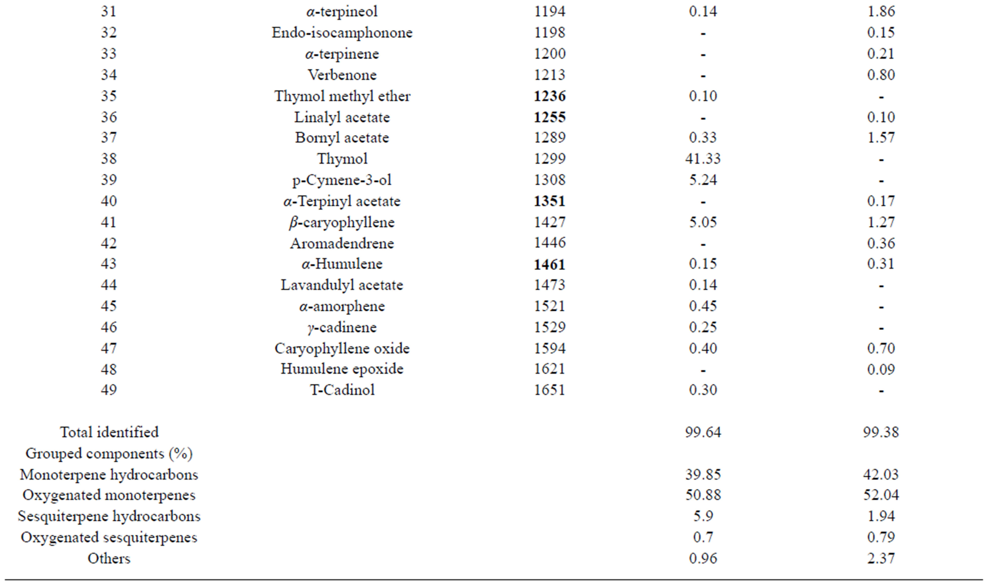
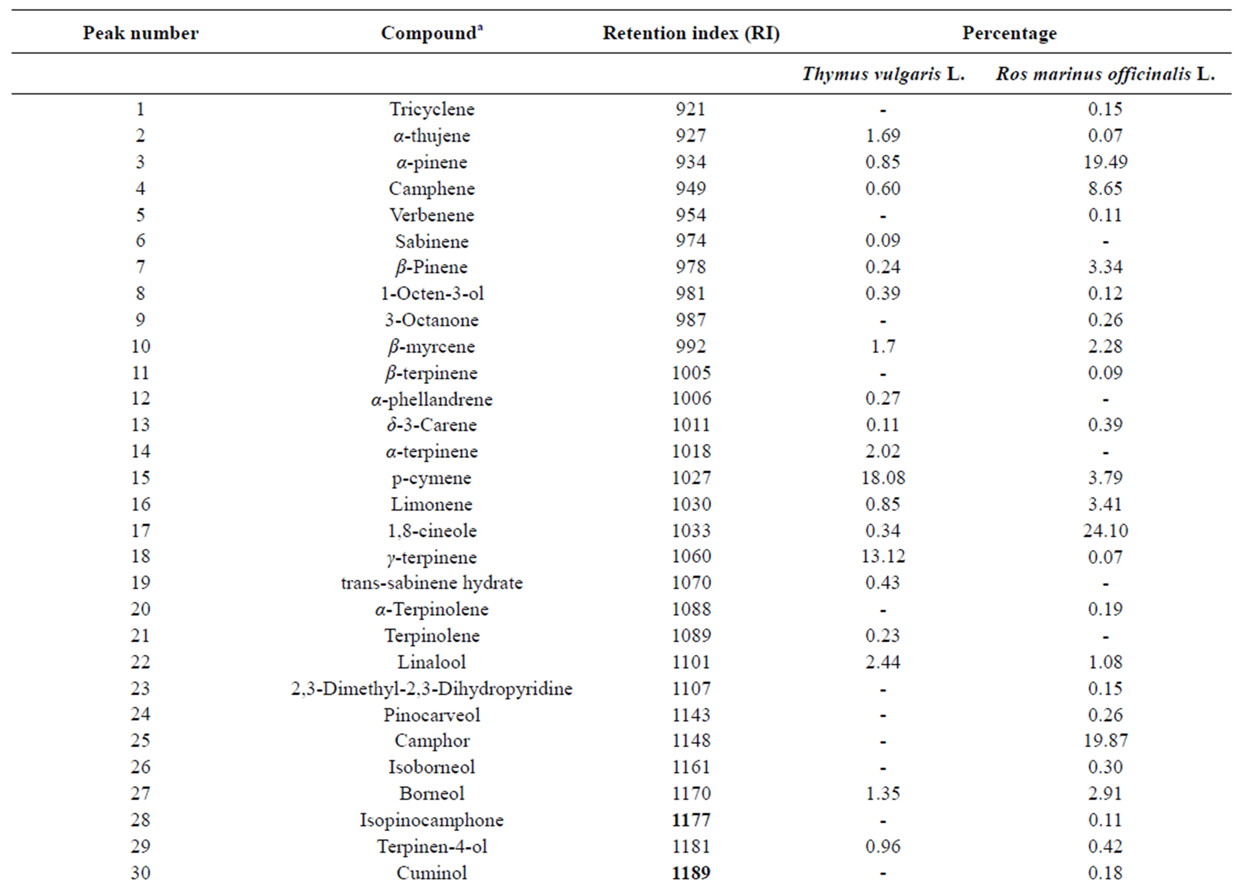
Table 1. Chemical composition of Thymus vulgaris L. and Rosmarinus officinalis L. essential oils.
by 1,8-cineole, α-pinene, camphene, formed the major group. Ketones constitute 20.67% and camphor was the major compound of this class (Table 1). At the species level, our results on the composition of french R. officinalis oils were in accordance with those previously re ported for other Mediterranean Rosemary samples [39- 41].
4.2. Antioxidant Activity
The antioxidant activity of Thymus vulgaris L. and Rosmarinus officinalis L. EOs was assessed by DPPH assay, evaluating the H-donating or radical scavenging ability of the oils using the stable radical 2,2-diphenyl-1- picrylhydrazyl (DPPH) as a reagent. The concentrations that led to 50% inhibition (IC50) for thyme and rosemary oil are 437 ± 5.46 µg/mL and 189 ± 2.38 µg/mL respectively. In this study, IC50 of both used oil were less potent than the reference antioxidants butylated hydroxytoluene (BHT) and quercetin (IC50 values of 4.21 ± 0.08 µg/mL and 1.07 ± 0.01 µg/mL respectively) [26]. However a significant correlation was observed across the oils between their antioxidant activity and the content of oxygenated monoterpenes (p = 0.001). It seems to be a general trend that the essential oils which contain oxygenated monoterpenes and/or sesquiterpenes have greater antioxidative properties [9]. Ruberto and Barratta (2000) [42], who tested the antioxidant activity of about 100 pure components of essential oils, pointed out that the phenolic compounds such as thymol, carvacrol and camphor showed the highest activity. Thus, many aromatic plants are today considered as the most important sources for the extraction of compounds with strong antioxidant activity. Rosemary (R. officinalis L.) and Thyme (T. vulgaris L.) are two spices widely used in folk medicine, cosmetics, phytopharmacy, and the flavoring of food products [12]. Furthermore, rosemary is the only spice commercially available for use as an antioxidant in Europe and the United States [23,43].
4.3. Cytotoxic Activity
Cell viability was performed after 24, 48 and 72 h exposure to R. officinalis L. and T. vulgaris L. for their anticancer activity using the MTT colorimetric assay.
The EOs were prepared and screened for their in vitro cytotoxic effects against human respiratory epithelial cell line (A549). A concentration and time dependent inhibitory effect on A549 cell were observed. After 24 h of incubation, rosemary and thymol oil cytotoxicity were considered whenever cell survival percent were less than 50. The extracts were not cytotoxic towards A549 cell line in all tested concentrations. But after 48 and 72 h essential oil exposure, R. officinalis L. was strongly inhibited the proliferation of the A549 cells and IC50 is 80.00 ± 0.02 µg/mL and 8.50 ± 0.01 µg/mL respectively (Figure 1). A low toxicity was observec for T. vulgaris L., which IC50 is 390.00 ± 0.03 µg/mL after 48 H EO exposure and it is 10.50 ± 0.01 µg/mL after 72 h (Figure 2). It is interesting to note that rosemary and thyme EOs exhibited varying cytotoxicity against the A549 cells. The anticarcinogenic activity of rosemary is due to the major bioactive compounds such as 1,8-cineole, camphor, and α-pinene [44]. In a similar study where compounds extracted from R. officinalis were tested on various cancer cell lines, such as NCI-H82 (small lung carcinoma), DU- 145 (prostate carcinoma), Hep3D (liver carcinoma), K- 562 (chronic myelois carcinoma), MCF-7, (breast adenocarcinoma), PC-3 (prostate adenocarcinoma) and MDAMB-231 (breast adenocarcinoma) the IC50 values ranged from 8.82 μg/mL to over 100 μg/mL [45].
4.4. Antimicrobial Activity
The in vitro antimicrobial activity of T. vulgaris L. and R. officinalis L. EOs estimated by the diameter of inhibittion varied according to essentials oils and bacteria strains were summarised in Table 2. In fact, the data obtained of zones of growth inhibition (mm) scored in Mueller-Hinton agar demonstrated that Gram-positive bacteria exhibited the highest diameters of growth inhibition between 26 and 41 mm recorded to thyme oil and between 8 and 17 mm recorded to rosemary oil. T. vulgaris L. EO was particularly effective against M. luteus NCIMB 8166 and L. monocytogenes ATCC 19115 with inhibition diameter exceeding those of the tested antibiotics. On the other hand, Gram-negative bacteria were less sensitive to T. vulgaris L. essential oil with a diameter of growth inhibition ranging from 20 (S. typhimurium LT2 DT104) to 32 mm (P. aeruginosa ATCC 27853 and V. alginolyticus ATCC 33787 ). The oil of T. vulgaris L. was generally active against the majority of food intoxitation isolated S. enteritidis and the diameters of growth inhibition were ranging from 16 mm to 45 mm. Concerning 19 food isolated strains Salmonella spp., R. officinalis L. EO (8.6 mg/disc) was not active showing a clear zone of inhibition ranging from 0 to 15 mm.
The bacteriostatic and bactericidal effectiveness of the thyme and Rosemary EOs estimated by minimum inhibitory concentration (MIC) and minimum bactericidal concentration (MBC) respectively are shown in Table 2 against 13 reference strains and 31 strains belonging to Salmonella genus, of them 12 belong to the species enteritidis and responsible for collective food intoxication in June 2000 in hospital Fatouma Bouguiba Monastir (Tunisia). Both essential oils showed a significant antibacterial activity against Gram positive as well as Gram negative bacteria that confirms previous findings [17, 41,46]. Thyme essential oil exhibited a higher anti-microbial activity than that of rosemary essential oil, which was similar to the results of the sensitivity test
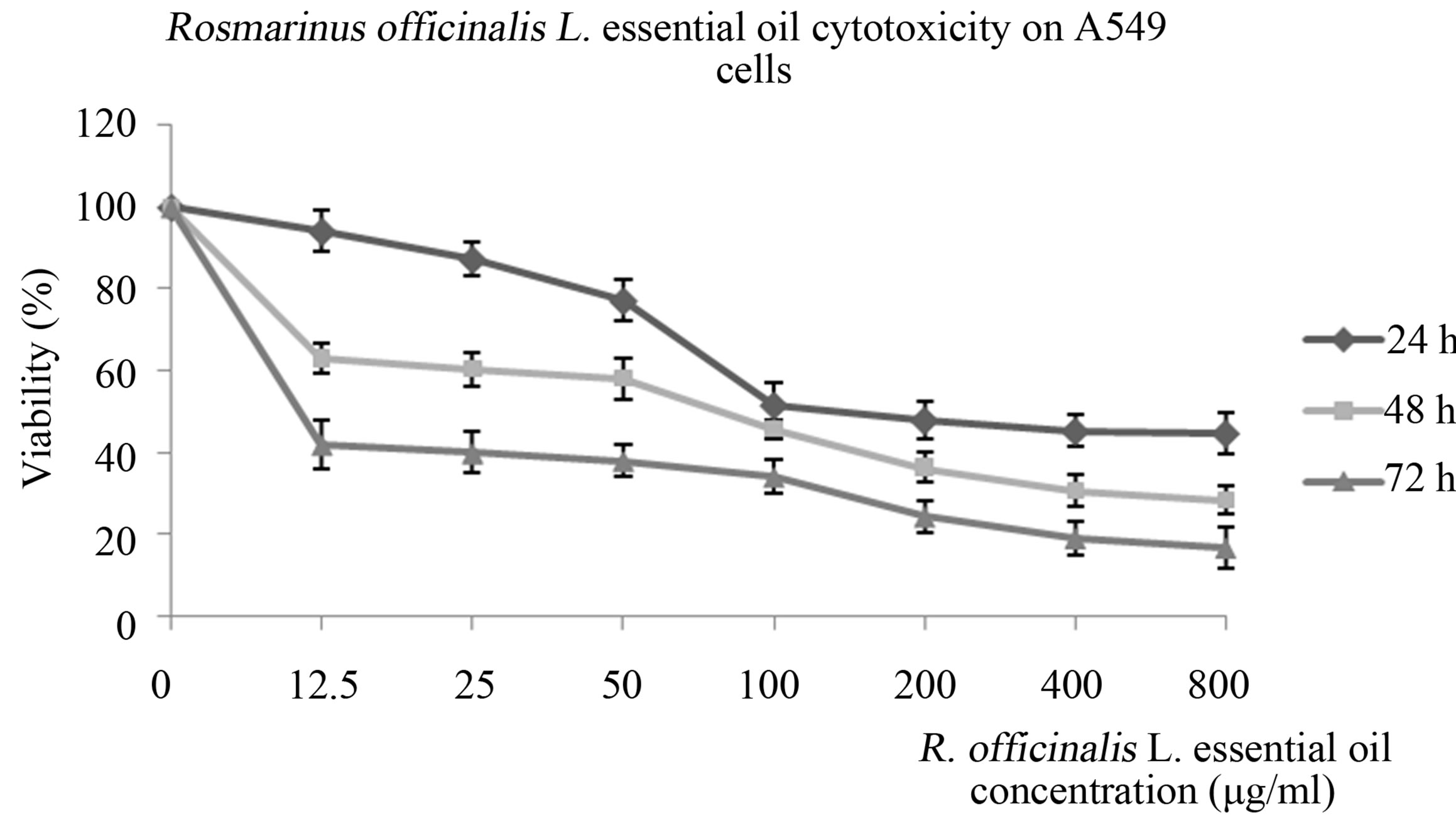
Figure 1. Cytotoxicity of Rosmarinus officinalis L. essential oil on A549 cells.
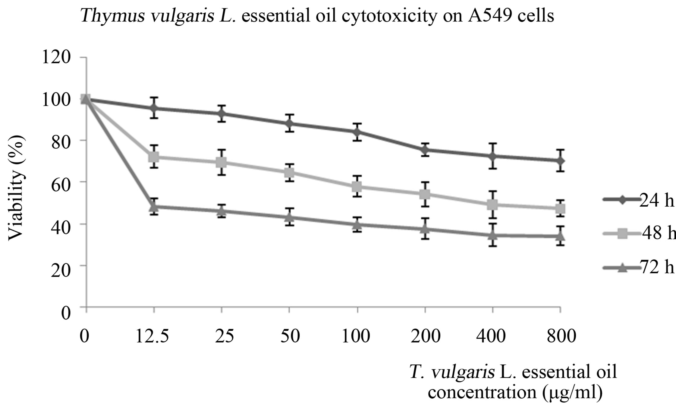
Figure 2. Cytotoxicity of Thymus vulgaris L. essential oil on A549 cells.
(Table 2). The antimicrobial activity of rosemary essential oil against S. typhimurium ATCC 1408 (50 mg/mL) was less than against the other reference bacteria. The MICs for the thyme essential oil ranged from 0.78 to 3.12 mg/mL for all test microorganisms, while MICs for rosemary oil ranged from 6.25 to 50 mg/mL, MBC values of the two oils were similar or even higher than the corresponding MIC values.
In the antimicrobial action of essential oil componentsis as follows: phenols > aldehydes > ketones > alcohols > ethers > hydrocarbons [47]. The main component of investigated thyme oil was thymol, a monoterpene with phenolic ring. In the case of rosemary oil, the main component was 1,8-cineole belonging to ethers group. Based on composition of investigated oils we expected that the antimicrobial activity of thyme oil will be higher than those of rosemary oil. It was confirmed during study, the MIC of thyme oil against tested strains was significantly (p < 0.05) 16-fold lower than the MIC of rosemary oil. Results found in this study are in accordance with reports that have shown MIC of thyme oil against B. thermosphacta was 10-fold lower than the MIC of rosemary oil [47]. The antimicrobial activity of the EO of R. officinalis L. studied in this work may also be attributed to the dominant presence of 1,8-cineole, which has been found to have relatively strong antimicrobial properties against many important pathogens [48]; however, there are some contradictory reports on the role of 1,8-cineole and the compounds camphor, α-pinene, and p-cymene, also found in reasonably high content in the R. officinalis L. EO assayed in this survey. It is difficult to attribute the activity of a complex mixture to a single or particular constituent; thus, a higher concentration of the major component does not necessarily mean the best antimicrobial effects for most of the strains assayed [49], and possible synergistic and/or antagonistic effects of compounds in the oil should also be given consideration. In addition, R. officinalis EO analysed in this study, even with a high content of 1,8-cineole, showed less antibacterial activity than T. vulgaris EO. In fact, some of the Salmonella spp. microorganisms tested R. officinalis showed no inhibition (Table 2). The MIC and MBC values (≥50 mg/mL) also confirmed this lower antimicrobial activity. Actually, the EO from R. officinalis has been previously reported to possess moderate antibacterial activity [50].
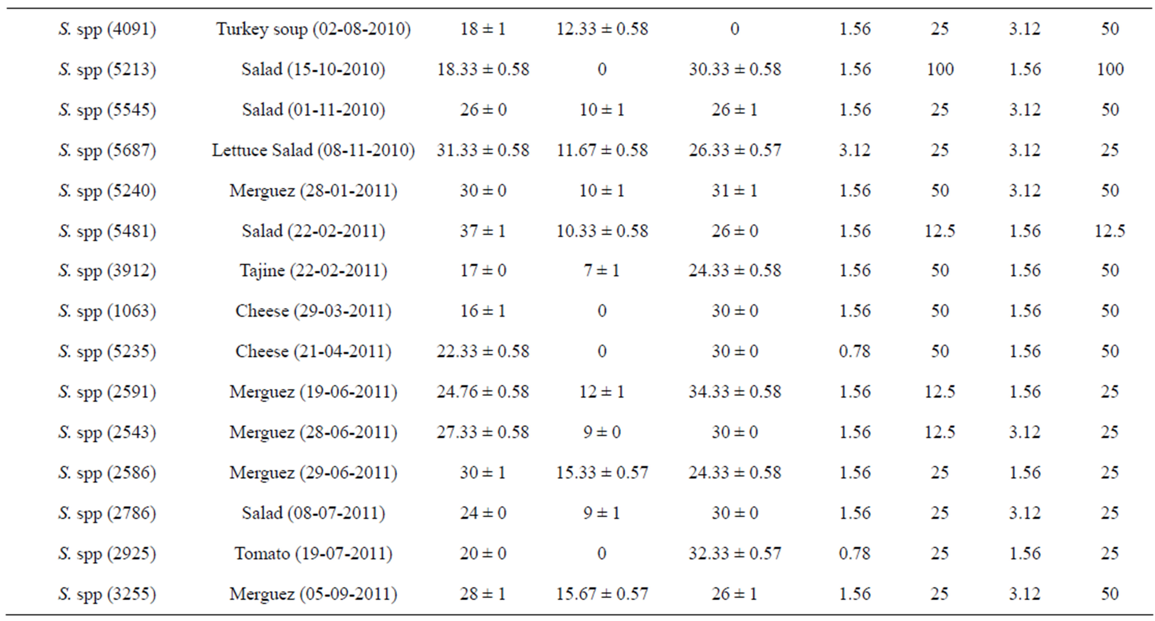
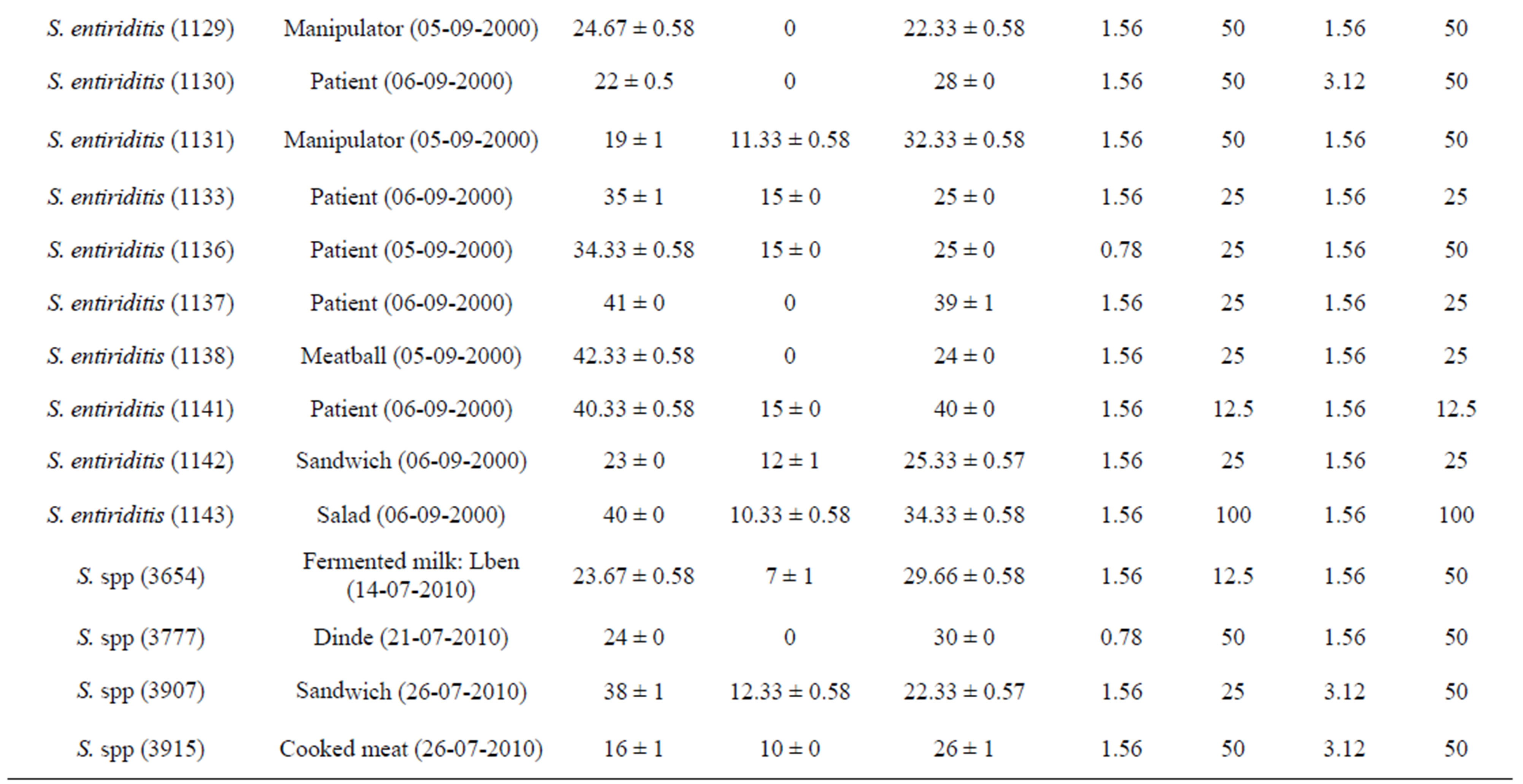
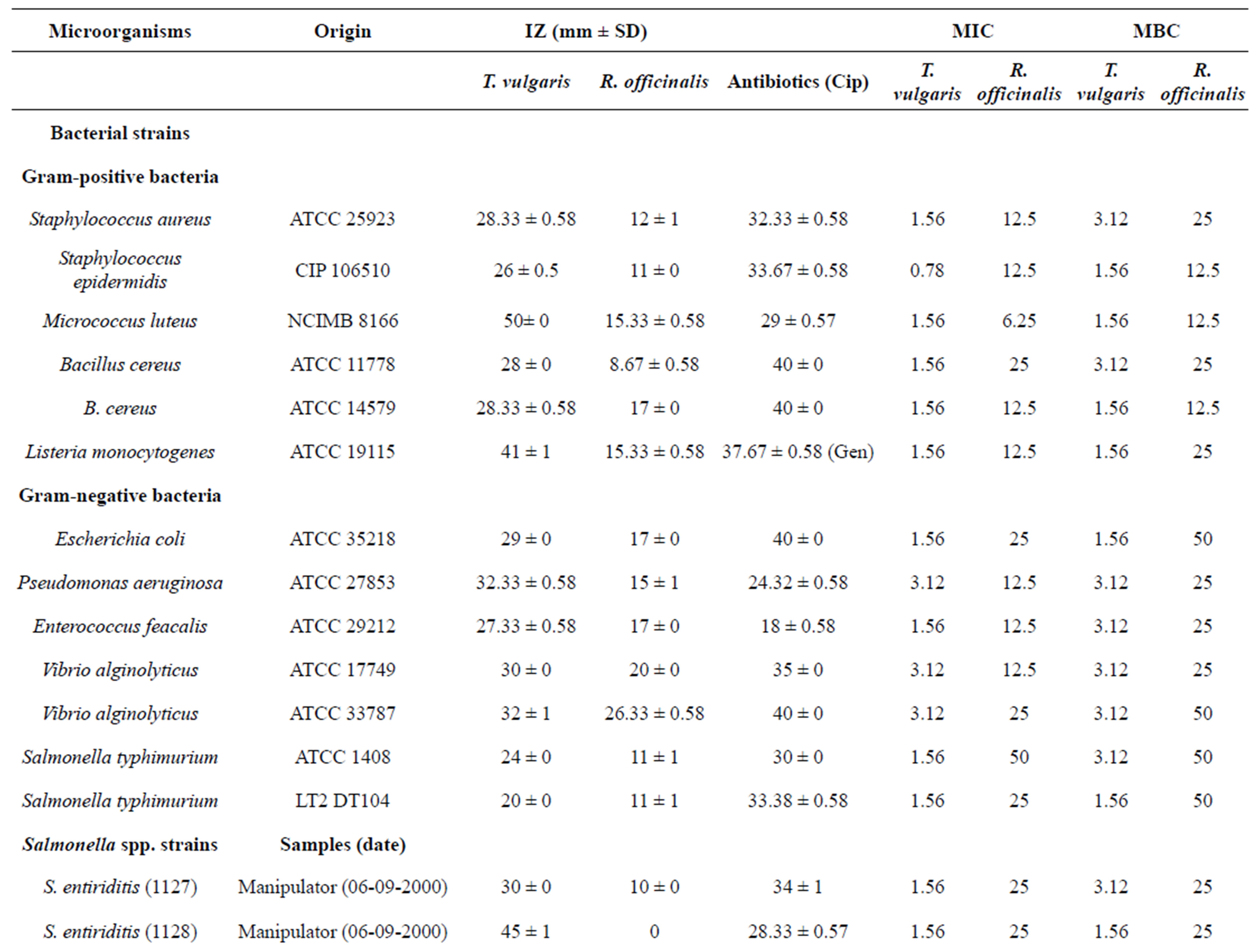
Table 2. Antibacterial activity of Thymus vulgaris L. and Rosmarinus officinalis L. essential oils against human pathogenic bacteria using agar disc diffusion method and determination of MIC (mg/ml) and MBC (mg/ml) values.
Regarding the susceptibility of different bacteria to the EOs tested, it was verified that Gram-negative bacterial strain is known to have a high level of intrinsic resistance to many antimicrobials and antibiotics because of a very restrictive outer membrane barrier, and it is highly resistant even to synthetic drugs [51].
5. CONCLUSION
In summary, the results presented here contribute to the knowledge of antimicrobial activities and chemical composition of the tested EOs obtained from aromatic plants growing in the mountain in the south of France. Our data also support the possible use of EOs of T. vulgaris and R. officinalis, in particular the EO of T. vulgaris, as potential natural agents for food preservation. Despite the moderate activity of R. officinalis, the data presented in this study are also significant given that this is the first time its bacteriostatic and bactericidal effects against the bacteria strains assayed have been reported. The case of L. monocytogenes, which has shown in most cases a significant sensitivity to the both EOs tested, is also noteworthy. L. monocytogenes is able to multiply under refrigerated conditions and so is a pathogen of great concern to the food industry. Because of that, the use of EOs as an additional barrier of food preservation should be considered. In general, the use of food presservation methods conjointly with the use of EOs could enhance the antimicrobial activity of these EOs; therefore, more research into the biological activities of these EOs, alone or combined with food preservation techniques, is recommended.
6. ACKNOWLEDGEMENTS
We are grateful to Prof. Rhim Amel from the Regional Laboratory of Public Health of Monastir (Tunisia) for her help to collect the microorganisms.
REFERENCES
- Soliman, K.M. and Badeaa, R.I. (2002) Effect of oil extracted from some medicinal plants on different mycotoxigenic fungi. Food Chemical Toxicology, 40, 1669- 1675. doi:10.1016/S0278-6915(02)00120-5
- Vivek, K.B., Atiqur, R. and Sun C.K. (2008) Chemical composition and inhibitory parameters of essential oil and extracts of Nandina domestica Thunb. to control foodborne pathogenic and spoilage bacteria. International Journal of Food Microbiology, 125, 117-122. doi:10.1016/j.ijfoodmicro.2008.03.011
- Hall, R.L. (1997) Food-borne illness: Implications for the future. Emerging Infectious Diseases, 3, 555-559. doi:10.3201/eid0304.970421
- Loizzo, M.R., Tundis, R., Chandrika, U.G., Abeysekera, A.M., Menichini, F. and Frega, N.G. (2010) Antioxidant and antibacterial activities on foodborne pathogens of Artocarpus heterophyllus Lam. (Moraceae) leaves extracts. Journal of Food Science, 75, 291-295.
- Zeng, W.C., Zhang, Z., Gao, H., Jia, L.R. and He, Q. (2012) Chemical composition, antioxidant, and antimicrobial activities of essential oil from pine needle (Cedrus deodara). Journal of Food Science, 77, 824-829. doi:10.1111/j.1750-3841.2012.02767.x
- Cowan, M.M. (1999) Plant products as antimicrobial agents. Clinical Microbiology Reviews, 12, 564-582.
- Bozin, B., Mimica-Dukic, N., Samojlik, I. and Jovin, E. (2007) Antimicrobial and antioxidant properties of rosemary and sage (Rosmarinus officinalis L. and Salvia officinalis L., Lamiaceae) essential oils. Journal of Agricultural Food Chemistry, 55, 7879-7885. doi:10.1021/jf0715323
- Ait-Ouazzou, A., Loran, S., Bakkali, M., Laglaoui, A., Rota, C., Herrera, A., Pagana, R. and Conchello, P. (2011) Chemical composition and antimicrobial activity of essential oils of Thymus algeriensis, Eucalyptus globulus and Rosmarinus officinalis from Morocco. Journal of the Science of Food and Agriculture, 91, 2643-2651. doi:10.1002/jsfa.4505
- Tepe, B., Donmez, E., Unlu, M., Candan, F., Daferera, D., Vardar-Unlu, G., Polissiou, M. and Sokmen, A. (2004) Antibacterial and antioxidative activities of the essential oils and methanol extracts of Salvia cryptantha (Montbret et Aucher ex Benth.) and Salvia multicaulis p (Vahl). Food Chemistry, 84, 519-525. doi:10.1016/S0308-8146(03)00267-X
- Daferera, D.J., Ziogas, B.N. and Polissiou, M.G. (2003) The effectiveness of plant essential oils on the growth of Botrytis cinerea, Fusarium sp. and Clavibacter michiganensis subsp. michiganensis. Crop Protection, 22, 39-44. doi:10.1016/S0261-2194(02)00095-9
- Helenicy, N.H., Veras Fabíola, F.G.R., Colares, A.V., Menezes, I.R.A., Coutinho Henrique, D.M., Botelho, M.A. and Costa, J.G.M. (2012) Synergistic antibiotic activity of volatile compounds from the essential oil of Lippia sidoides and thymol. Fitoterapia, 83, 508-512. doi:10.1016/j.fitote.2011.12.024
- Schelz, Z., Molnar, J. and Hohmann, J. (2006) Antimicrobial and antiplasmid activities of essential oils. Fitoterapia, 77, 279-285. doi:10.1016/j.fitote.2006.03.013
- Burt, S. (2004) Essential oils: Their antibacterial properties and potential application in foods e a review. International Journal of Food Microbiology, 94, 223-253. doi:10.1016/j.ijfoodmicro.2004.03.022
- Bakkali, F., Averbeck, S., Averbeck, D. and Idaomar, M. (2008) Biological effects of essential oils—A review. Food Chemistry and Toxicology, 46, 446-475. doi:10.1016/j.fct.2007.09.106
- Zhang, C., Li, H., Yun, T., Fu, Y., Liu, C., Gong, B. and Neng, B. (2008) Chemical composition, antimicrobial and antioxidant activities of the essential oil of Tibetan herbal medicine Dracocephalum heterophyllum Benth. Natural Product Research, 22, 1-11. doi:10.1080/14786410701619076
- Jeong-Ho, L., Byung-Kyu, L., Jong-Hee, K., Sang Hee, L. and Soon-Kwang, H. (2009) Comparison of chemical compositions and antimicrobial activities of essential oils from three conifer trees; Pinus densiflora, Cryptomeria japonica, and Chamaecyparis obtuse. Journal of Microbiology and Biotechnology, 19, 391-396. doi:10.4014/jmb.0803.191
- Rota, C., Carrami Nana, J.J., Burillo, J. and Herrera, A. (2004) In vitro antimicrobial activity of essential oils from aromatic plants against selected foodborne pathogens. Journal of Food Protection, 67, 1252-1256.
- Hazzit, M., Baaliouamer, A., Verissimo, A.R., Faleiro, M.L. and Miguel, M.G. (2009) Chemical composition and biological activities of Algerian Thymus oils. Food Chemistry, 16, 714-721. doi:10.1016/j.foodchem.2009.03.018
- Zarzuelo, A. and Crespo, E. (2002) The medicinal and nonmedicinal uses of thyme. In thyme. The genus Thymus. In: Stahl-Biskup, E. and Saez, F., Eds. Medicinal and Aromatic Plants Industrial Profiles, New York, Taylor and Francis, 263-292.
- Gachkar, L., Yadegari, D., Rezaei, M.B., Taghizadeh, M., Astaneh, S.A. and Rasooli, I. (2007) Chemical and biological characteristics of Cuminum cyminum and Rosmarinus officinalis essential oils. Food Chemistry, 102, 898- 904. doi:10.1016/j.foodchem.2006.06.035
- Chizzola, R., Michitsch, H. and Franz C. (2008). Antioxidative properties of Thymus vulgaris leaves: Comparison of different extracts and essential oil chemotypes. Journal of Agricultural Food Chemistry, 56, 6897-6904. doi:10.1021/jf800617g
- Lee, S.E., Lee, H.S. and Ahn, Y.J. (1999) Scavenging Effect of plant-derived materials on free radicals and active oxygen species. Agricultural Chemistry and Biotechnology, 42, 40-44.
- Bicchi, C., Binello, A. and Rubiolo, P. (2000) Determination of phenolic diterpene antioxidants in rosemary (Rosmarinus officinalis L.) with different methods of extraction and analysis. Phytochemical Analysis, 11, 236-242. doi:10.1002/1099-1565(200007/08)11:4<236::AID-PCA503>3.0.CO;2-B
- Yesil Celiktas, O., Hames Kocabas, E.E., Bedir, E., Vardar Sukan, F., Ozek, T. and Baser, K.H.C. (2007) Antimicrobial activities of methanol extracts and essential oils of Rosmarinus officinalis, depending on location and seasonal variations. Food Chemistry, 100, 553-559. doi:10.1016/j.foodchem.2005.10.011
- Barbosa, L.N., Mores Rall, V.L., Henrique Fernandes, A.A., Ushimaru, P.I., da Silva Probst, I. and Ary Fernandes, J. (2009) Essential oils against foodborne pathogens and spoilage bacteria in minced meat. Foodborne Pathogens and Disease, 6, 725-728. doi:10.1089/fpd.2009.0282
- Alaoui Jamalia, C., El Bouzidia, L., Bekkouchea, K., Lahcenb, H., Markouka, M., Wohlmuthc, H., Leachc, D. and Abbad A. (2012) Chemical composition and antioxidant and anticandidal activities of essential oils from different wild moroccan thymus species. Chemistry and Biodiversity, 9, 1188-1197.
- Rameau, J.C., Mansion, D., Dumé, G., Gauberville, C., Bardat, J., Bruno, E. and Keller, R. (2008) Flore forestière Française: Guide ecologique illustré. Tome 3, région méditerranéenne. Institut Pour le Developpement Forestier, Paris & Ministere de l’Agriculture et de la Peche, Paris.
- European Pharmacopoeia (1975) Volume 3. Maisonneuve, Sainte-Ruffine.
- Kovàts, E. (1958) Characterization of organic compounds by gas chromatography. Part 1. Retention indices of aliphatic halides, alcohols, aldehydes and ketones. Helvetica Chimica Acta, 41, 1915-1932.
- Adams, R.P. (2001) Identification of essential oil components by gas chromatography/quadrupole mass spectrometry. Allured Publishing Corporation, Carol Stream.
- Sibanda, S., Chigwada, G., Poole, M., Gwebu, E.T., Noletto, J.A., Schmidt, J.M., Rea, A.I. and Setzer, W.N. (2004) Composition and bioactivity of the leaf essential oil of Heteropyxis dehniae from Zimbabwe. Journal of Ethnopharmacology, 92, 107-111. doi:10.1016/j.jep.2004.02.010
- Zouari, S., Zouari, N., Fakhfakh, N., Bougatef, A., Ayadi, M.A. and Neffati, M. (2010) Chemical composition and biological activities of a new essential oil chemotype of Tunisian Artemisia herba alba Asso. Journal of Medicinal Plants Research, 4, 871-880.
- Hanato, H., Kagawa, T., Yasuhara, J. and Okuda, T. (1988) Two new flavonoids and other constituents in licorice root: Their relative astringency and radical scavenging effect. Chemical and Pharmaceutical Bulletin, 36, 1090- 1097.
- Mossman, B.T., Jean, L. and Landesman, J.M. (1983) Studies using lectins to determine mineral interactions with cellular membranes. Environmental Health Perspectives, 51, 23-25. doi:10.1289/ehp.835123
- Clinical and Laboratory Standards Institute (2006) Performance standards for antimicrobial susceptibility testing. 16th Informational Supplement, M100-S16, Clinical and Laboratory Standards Institute, Wayne.
- Cavallo, J.D., Chardon, H., Chidiac, C., Choutet, P., Courvalin, P., Dabernat, H., Drugeon, H., Dubreuil, L., Goldstein, F., Jarlier, V., Leclerq, R., Nicolas-Chanoine, M.H., Philippon, A., Quentin, C., Rouveix, B., Sirot, J. and Soussy, C.J. (2006) Antibiogram committee of the French microbiology society. Pathology Biology, Paris.
- Güllüce, M., Sökmen, M., Daferera, D., Ağar, G., Ozkan, H., Kartal, N., Polissiou, M., Sökmen, A. and Sahin, F. (2003) In vitro antibacterial, antifungal, and antioxidant activities of the essential oil and methanol extracts of herbal parts and callus cultures of Satureja hortensis L. Journal of Agricultural and Food Chemistry, 51, 3958- 3965. doi:10.1021/jf0340308
- Flamini, G., Cioni, P.L., Morelli, I., Macchia, M. and Ceccarini, L. (2002) Main agronomic productive characteristic of two ecotypes of two Rosmarinus officinalis L. and chemical composition of their essential oils. Journal of Agricultural and Food Chemistry, 50, 3512-3517. doi:10.1021/jf011138j
- Lawrence, B.M. (1997) Progress in essential oils: Rosemary oil. Perfum Flavor, 22, 71.
- Pintore, G., Usai, M., Bradesi, P., Juliano, C., Boatto, G. and Tomi, F. (2002) Chemical composition and antimicrobial activity of Rosmarinus officinalis L. oils from Sardinia and Corsica. Flavor and Fragrance Journal, 17, 15-19. doi:10.1002/ffj.1022
- Zaouali, Y., Bouzaine, T. and Boussaid, M. (2010) Essential oils composition in two Rosmarinus officinalis L. varieties and incidence for antimicrobial and antioxidant activities. Food and Chemical Toxicology, 48, 3144-3152. doi:10.1016/j.fct.2010.08.010
- Ruberto, G. and Baratta, M.T. (2000) Antioxidant activity of selected essential oil components in two lipid model systems. Food Chemistry, 69, 167-174. doi:10.1016/S0308-8146(99)00247-2
- Madsen, H.L. and Bertelsen, G. (1995) Spices as antioxidants. Trends Food Science Technology, 6, 271-277. doi:10.1016/S0924-2244(00)89112-8
- Bai, N., He, K., Roller, M., Lai, C.S., Shao, X., Pan, M.H. and Ho, C.T. (2010) Flavonoids and phenolic compounds from Rosmarinus officinalis. Journal of Agricultural and Food Chemistry, 58, 5363-5367. doi:10.1021/jf100332w
- Yesil-Celiktas, O., Sevimli, C., Bedir, E. and Vardar-Sukan, F. (2010) Inhibitory effects of rosemary extracts, carnosic acid and rosmarinic acid on the growth of various human cancer cell lines. Plant Foods for Human Nutrition, 65, 158-163. doi:10.1007/s11130-010-0166-4
- Lopez, P., Sanchez, C., Batle, R. and Nerin, C. (2005) Solid and vaporphase antimicrobial activities of six essential oils: Susceptibility of selected food borne bacterial and fungal strains. Journal of Agricultural and Food Chemistry, 53, 6939-6946. doi:10.1021/jf050709v
- Nowak, A., Kalemba, D., Krala, L., Piotrowska, M. and Czyzowska, A. (2012) The effects of thyme (Thymus vulgaris) and rosemary (Rosmarinus officinalis) essential oils on Brochothrix thermosphacta and on the shelf life of beef packaged in high-oxygen modified atmosphere. Food Microbiology, 32, 212-216. doi:10.1016/j.fm.2012.05.001
- Rosato, A., Vitali, C., De Laurentis, N., Armenise, D. and Milillo, M. (2007) Antibacterial effect of some essential oils administered alone or in combination with norfloxacin. Phytomedicine, 14, 727-732. doi:10.1016/j.phymed.2007.01.005
- Rota, M.C., Herrera, A., Martinez, R.M., Sotomayor, J.A. and Jordan, M.J. (2008) Antimicrobial activity and chemical composition of Thymus vulgaris, Thymus zygis and Thymus hyemalis essential oils. Food Control, 19, 681- 687. doi:10.1016/j.foodcont.2007.07.007
- Angioni, A., Barra, A., Cereti, E., Barile, D., Cosson, J.D., Arlorio, M., Dessi, S., Coroneo, V. and Cabras, P. (2004) Chemical composition, plant genetic differences, antimicrobial and antifungal activity investigation of the essential oil of Rosmarinus officinalis L. Journal of Agricultural Food Chemistry, 52, 3530-3535. doi:10.1021/jf049913t
- Skocibusic, M., Bezic, N. and Dunkic, V. (2006) Phytochemical composition and antimicrobial activities of essential oils from Satureja subspicata Vis. growing in Croatia. Food Chemistry, 96, 20-28. doi:10.1016/j.foodchem.2005.01.051