Open Journal of Stomatology, 2011, 1, 121-125
doi:10.4236/ojst.2011.14019 Published Online December 2011 (http://www.SciRP.org/journal/ojst/ OJST
).
Published Online December 2011 in SciRes. http://www.scirp.org/journal/OJST
The accuracy of brackets placement in direct bonding
technique: a comparison between the pole-like bracket
positioning gauge and the star-like bracket positioning gauge
Lahcen Ousehal, Laila Lazrak
Department of Orthodontics, Faculty of Dentistry, Casablanca, Morocco.
Email: lahcen2228@yahoo.fr
Received 13 April 2011; revised 24 June 2011; accepted 3 August 2011.
ABSTRACT
The accuracy of brackets placement is a key factor in
successful orthodontic therapy. An in vitro study was
conducted in ten models from a natural maxillary
teeth model in order to compare the accuracy of brac-
kets placement between two direct bonding instru-
ments: the pole-like Bracket Positioning Gauge and
the star-like Bracket Positioning Gauge. Our results
have shown that: the star-like Bracket Positioning
Gauge is more precise in placing brackets vertically,
whereas the pole-like Bracket Positioning Gauge al-
lows a better angulation of the bracket. Considering
each tooth separately, there is no statistically signifi-
cant difference between the two positioning gauges,
except that the star-like gauge gives better results in
bracket’s height for the second premolar and the ca-
nine, whereas the pole-like gauge allows for a better
positioning and a better vertical angulation of the
brackets for the lateral incisor. No statistically signi-
ficant difference was found between the two gauges
on the mesiodistal position. Overall, the star-like gau-
ge showed a better accuracy in positioning brackets.
Keywords: Direct Bonding; Pole-Like Gauge; Star-Like
Gauge
1. INTRODUCTION
Good placement of orthodontic brackets guarantees a suc-
cessful mechanical treatment [1]. Direct bonding is the
technique that is most used by orthodo ntists in their dai-
ly practice [2].
Two positioning instruments are available to the practi-
tioner, the pole like gauge and the star like gauge also
known as Boone gauge.
The question that arises is which of these two instru-
ments is more accurate in terms of positioning orthodon-
tic brackets.
In literature, there are no studies comparing the pole
like gauge and the star like gauge in bonding brackets.
Our work has taken as objective to compare the accuracy of
orthodontic bracket placement between two instruments
of direct bonding: the star like and the pole like gauges.
2. MATERIALS AND METHODS
An experiment with a set of healthy natural teeth taken
from 15 to 25 was conducted using a mold. Ten extra
hard plaster models were made from the original model
using alginate.
The natural teeth model was used for the ideal brack-
ets’ placement.
Eleven sets of Edgwise standard metal brackets 022.
028 were used fo r the stu dy.
The plaster models were mounted on mannequins to be
close to the clinical conditions during the bonding of
brackets.
The right hemi arch was reserved for direct bonding
using the pole like gauge, and the left hemi arch was us-
ed for direct bonding using the star like gauge.
Four experienced orthodontists have realized the bon-
ding of brackets following the rules of correct bonding
which are: th e bracket axis sho uld be parallel to the long
axis of the tooth, the bracket should be centered in the
crown, the height of bonding should be set to 4, 4.5 and
3.5 mm for the central incisors, bicuspids, canines and
lateral incisors respectively.
The bonding material used is 3M TRANSBOND self
cured composite resin.
The natural teeth from the reference model have been
uncovered upwards to see their roots. This facilitates the
alignment of th e vertical axis of th e tooth. Only one pr ac-
titioner positioned the brackets ideally on the reference
model, held by hand, in order to control the bonding in
three dime nsi ons (Figure 1).
L. Ousehal et al. / Open Journal of Stomatology 1 (2011) 121-125
122
Figure 1. Pos i tioni ng t he brac kets on the referen ce mode l.
A metal ruler and a protractor scale 0.5 mm were used
for linear and angular measures.
Standardized photographs of each bonded tooth of ten
plaster models and the reference model have been taken
with an analog camera (III Yascika dental eye.) The
camera was fixed on a tripod allowing for an effective
control of the vertical distance from which the images
were taken.
The expansion used is 1/1 and the photographs were
enlarged ten times to facilitate measurements.
Methods
The contour of the crown, the vertical center line and the
horizontal center line of the bracket are drawn on a trac-
ing paper laid over the photo of each tooth.
Each paper is laid over the picture of the natural tooth
equivalent in the reference model.
Two linear measures and one angular measure were
calculated between the ideal group and the tested group
(Figure 2).
Figure 2. Tracing p aper ov erla id on t he photo of th e dog stu ck
by the direct method showing the different steps.
The height of the bracket: A zero value was assigned
to the bracket that bonded perfectly to the center.
A negative value was assigned when the vertical gap
between the bracket bonded with the gauge and the ideal
bracket is occlusal. Conversely, if the gap is cervical, a
positive value is assigned.
Mesio distal inclination: The horizontal distance be-
tween the center of the ideal bracket and that of the test-
ed bracket was measured to assess the mesio distal gap
of the bracket. A positive value was assigned when the
elevation of the tested bracket is mesial compared to that
of the ideal bracket, and a negative value is assigned
when the gap is distal.
Angulation: It is determined by the angle between the
horizontal center line of the ideal bracket and that of the
tested bracket.
This measure is positive when the center line of the
tested bracket is cervical relative to the ideal bracket and
negative in the oppo site case.
Statistical analysis: A statistical software (EPI-INFO)
was used in order to collect and analyze all the measure-
ments. The Student T test was used for statistical com-
parison of the results. The measurements were repeated
for reproducibility and reliability of results. A paired t
test has been realized and th e t test is less than the calcu -
lated t-test table, we can say there is no significant dif-
ference between the 2 measurements.
3. RESULTS
Tables 1 and 2 show the averages and standard devia-
tion for each group of teeth of the three measures: verti-
cal, mesiodistal, and angular.
Vertical positioning: The comparison between the gr-
oup of teeth bonded with the star-like gauge and the pole-
like gauge shows a very significant difference (P = 0.006)
for the second premolar and a significant difference for
the canine and the lateral incisor ( Table 3).
The star-like gauge is more accurate for the second pre-
molar and the canine. The pole-like gauge is more accu-
rate for the lateral incisor.
Mesiodistal positioning: The comparison between the
group of teeth bonded with the star-like gauge and the
pole-like gauge shows a significant difference for the fir-
st premolar and the lateral incisor (Table 4). Indeed, the
mesiodistal gap are opposed relatively to the center in
the two groups. For the first premolar, we have a mesial
gap of 0.32 mm on average for the group of teeth that
bonded with the pole-like gauge, and a distal gap of 0,
46 mm for the group of teeth that bonded with the star-
like gauge.
Similarly, for the lateral incisor, we have a mesial gap
of 0.42 mm with the pole-like gauge, and a distal gap of
0.38 mm with the star-like gauge.
Both instruments lead to mesiodistal placement errors.
C
opyright © 2011 SciRes. OJST
L. Ousehal et al. / Open Journal of Stomatology 1 (2011) 121-125
Copyright © 2011 SciRes.
123
OJST
Inclination of the bracket: The angular gap of the brac-
ket shows a significant difference only for the lateral in- cisor (Table 5). The pole-like gauge is more accurate in
this case.
Table 1. Mean values and standard deviation (STDV) of the vertical, the mesiodistal and the angular brackets’ position on the left
hemi arch.
25 24 23 22 21
Teeth Mean STDV Mean STDV Mean STDV Mean STDV Mean STDV
Height (mm) –0.43 0.129 –0.46 0.314 –0.29 0.207 –0.38 0.109 –0.31 0.217
M-D (mm) 0.10 0.237 –0.45 0.228 –0.03 0.262 –0.38 0.230 0.01 0.242
Å (degrees) –0.32 0.228 –0.12 0.177 –0.17 0.130 –0.30 0.146 –0.24 0.133
Table 2. Mean values and standard deviation of the vertical, the mesiodistal and the angular brackets’ position on the right hemi arch.
15 14 13 12 11
Teeth Mean SDTV Mean SDTV Mean SDTV Mean SDTV Mean SDTV
Height (mm) –1.02 0.430 –0.38 0.204 –0.47 0.440 0.26 0.214 –0.005 0.281
M-D (mm) –0.04 0.205 0.32 0.155 0.32 0.216 0.42 0.116 0.03 0.155
Å (degrees) –0.23 0.131 –0.17 0.056 –0.21 0.228 0.16 0.168 –0.05 0.215
Table 3. Com parison of the vertical gaps between the star-like and the pole-like gauges.
Pole-like gauge Star-like gaug e
Teeth Mean (mm) SDTV Mean (mm) SDTV P-value
2nd PM 1.020 0.430 0.435 0.129 0.006**(S)
1st PM –0.315 0.204 –0.460 0.228 0.54
Canine –0.470 0.440 –0.030 0.262 0.05*(S)
Late ra l incis o r 0.260 0.214 0.380 0.109 0.04*(S)
Central incisor –0.005 0. 281 –0.31 5 0.217 0.87
Table 4. Comparison of the mesiodistal gap between the star-like and the pole-like gauges.
Pole-like gauge Star-like gauge
Tooth Mean (mm) SDTV Me an (mm) S DTV P-value
2nd PM –0.040 0.205 0.105 0.237 0.66
1st PM 0.320 0.155 0.460 0.428 0.04* (S)
Canine 0.328 0.216 –0.030 0.262 0.43
Lateral incisor 0.425 0.116 0.380 0.230 0.03* (S)
Central incisor –0.030 0.155 0.010 0.242 0.74
Table 5. Comparison of the inclination gaps between the star-like and the pole-like gauges.
Pole-like gauge Star-like gauge
Teeth Mean (degrees) SDTV Mean (degrees) SDTV P-value
2nd PM –0.235 0.131 –0.320 0.258 0.73
1st PM –0.173 0.056 –0.125 0.177 0.84
Canine –0.210 0.228 –0.170 0.130 0.86
Latéral Incisor 0.160 0.168 –0.305 0.146 0.04*(S)
Central Incisor –0.050 0.215 –0.240 0.133 0.05*(S)
L. Ousehal et al. / Open Journal of Stomatology 1 (2011) 121-125
124
4. DISCUSSION
The objective of our work was to compare two instru-
ments of direct bonding of brackets that are commonly
used by orthodontists: the star-like gauge and the pole-
like gauge.
We conducted our study on the maxillary arch because
we believe that it is the arch in which the placement of
brackets in direct bonding is the most difficult.
We have also reproduced (in part) the clinical condi-
tions by mounting the models on mannequins and using
the same adhesive product used in clinical practice.
The choice of experienced orthodontists in our study
reduces errors related to the competence of the practi-
tioner.
Similarly, the use of multiple practitioners is more re-
alistic in that it avo ids ending up with th e same bonding
errors related to a single practitioner.
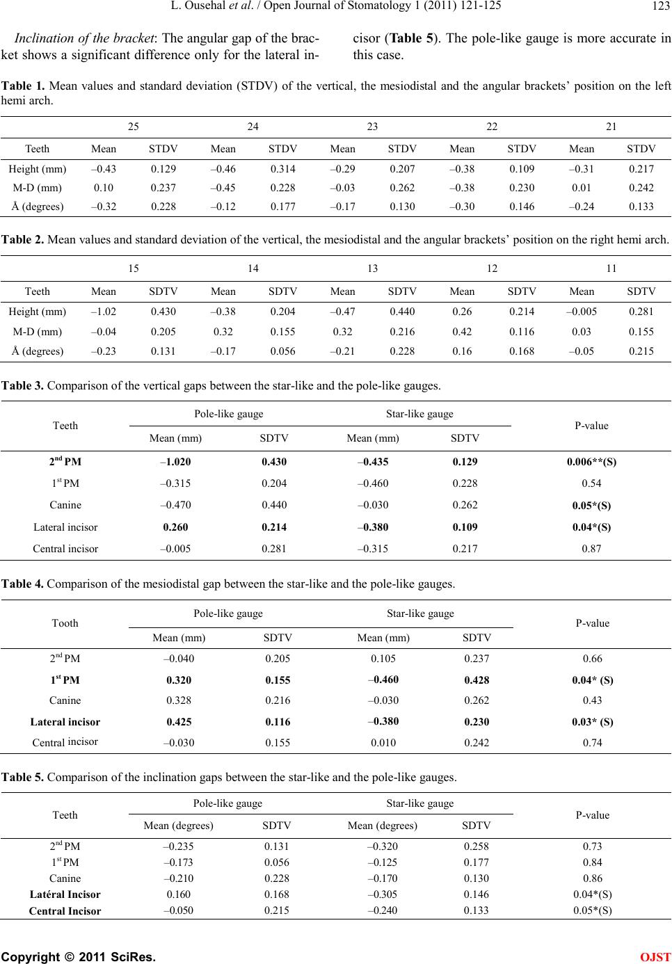
The comparison between the two methods showed
that the two ins truments cau se overall the same p lacemen t
errors, but that we have more accuracy with the star-like
gauge, especially for the vertical placement of brackets.
The photographic assessment is a reliable way to
study the position of the bracket, provided the same pro-
tocol and the same parameters are followed.
In our study, we used a system of rigid fixation and the
same magnification for all pictures. This greatly reduces
errors related to distortions of the phot ographic image.
The enlargement of the photographs was performed
only for conveniently reading the various measures.
The pole-like gauge sho wed less accuracy in the verti-
cal placement of bracket; this can be explained by its
nature that allows it to be inclined too occlusally or too
cervically, depending on the practitioner’s position rela-
tive to the bonding tooth.
Our results showed a greater inclination of the bracket
with the pole-like gauge on the lateral incisor.
The orthodontic literature has focu sed mainly o n com-
paring the accuracy of brackets placement between di-
rect and indirect bonding [3-7]. No study has taken be-
fore this one the objective of comparing bonding with
the pole-like gaug e and the star-like gauge.
BOHN CHAN KOO [8] conducted an in vitro study
comparing the accuracy of bracket placement between
direct and indirect technique.
For this, 19 sets of models of the same occlusion were
divided into two groups, one for direct bonding (9 mod-
els), the second for indirect bonding (9 models).
A model has been set aside for ideal bonding (compa-
rison model). The bonding was done by nine experienc-
ed orthodontists.
Direct bonding has been achieved by the st ar-like gauge
similarly to our work, comparing the accuracy bonding on
the basis of three measurements: vertical, angular, and
mesiodistal using enlarged photographs of each tooth.
The author concluded that there is a gap between in-
direct bonding and ideal bondin g. This is consistent with
our results on the direct bonding.
He also noted that for the maxillary second premolar,
indirect bonding is more accurate, whereas for the max-
illary lateral incisor, direct bonding was better for the in -
clination of the bracket.
In our work we found that the pole-like gauge allows
precise angle of the bracket for the la teral incisor, and we
recorded largest gaps for the maxillary second premolar.
Similar result s have been presented by B ALUT [2] .
In his work, he studied the accuracy of direct bonding
on models having the same malocclusion. He found that
the average inclination was 5.54˚ and 0.34 mm for the
height.
Another study was conducted by Aguirre, KING and
WALDRON [9]. It is a clinical evaluation done to de-
termine the advantages and disadvantages of both tech-
niques.
It focuses on 11 patients, 206 bonded brackets, and
189 measured brackets. The maxillary and mandibular
arches are divided into hemi-arches, randomly bonding
one side according to a technique different to the reverse
side.
In direct technique [10], the brackets are positioned
with the star-like gauge (gauge Boone UNITEK).
To assess the placement of brackets, photographs of
each bonded tooth are taken in similar ways and are
compared with the corresponding tooth on the other he-
mi arch. To get accurate conclusions, the authors analy-
zed in turn:
- Linear measurements.
- Angular values.
Concerning the linear measurements (height place-
ment), the results showed that neither techniques was
100% accurate. If we compare direct and indirect me-
thods, no significant difference in the maxillary arch was
found, except for the canines for which brackets bonded
using the indirect technique are placed near the ideal
height.
The angular measurements have also shown that both
methods fail to place ideally brackets with great vari-
ability. Apparently, the practitioner has more difficulty in
judging the ang les relative to height. In comparing direct
and indirect technique in the upper arch, there is no sig-
nificant difference except for the canines with an advan-
tage with the indirect technique, which led to an angle
closer to the ideal angle of 90. The results of this study
should perhaps be brought into perspective, because all
the brackets are bonded by the same practitioner. Indeed,
these results may reflect the clinical skill of a particular
practitioner rather than the difference between the two
techniques.
C
opyright © 2011 SciRes. OJST
L. Ousehal et al. / Open Journal of Stomatology 1 (2011) 121-125 125
5. CONCLUSIONS
With the advent of straight-wire techniques, th e po sition -
ing of brackets has taken a significant role in the success
of orthodontic treatment.
The placement of orthodontic brackets is a difficult
but crucial, even in straight wire techniques, which use
pre-informed brackets.
In this case, any small placement error leads all the
brackets data to be distorted proportionally to the place-
ment error.
In everyday practice, most orthodontists use two typ es
of gauge for brackets placement: the star-like gauge and
the pole-like gaug e.
Our study revealed by comparing these two types of
gauges that:
- The star-like gauge provides better placement of the
vertical bracket, whereas the pole-like gauge allows for a
better angle of the bracket .
- No statistically significant difference between these
two gauges has been highlighted for the mesiodistal po-
sition.
REFERENCES
[1] Andrews, L.F. (1976) The straight-wire appliance. Ex-
plained and compared. Journal of Clinical Orthodontics,
10, 174-195.
[2] Balut, N., Klapper, L., Sandrik, J. and Bowman, D. (1992)
Variations in bracket placement in the preadjusted or-
thodontic appliances. American Journal of Orthodontics
and Dentofacial Orthopedics, 102, 62-67.
doi:10.1016/0889-5406(92)70015-3
[3] Shpack, N. (2007) Bracket placement in lingual vs labial
systems and direct vs indirect bonding. Angle Orthodon-
tist, 77, 509-517.
doi:10.2319/0003-3219(2007)077[0509:BPILVL]2.0.CO
;2
[4] Hodge, T.M. (2004) A randomized clinical trial compar-
ing the accuracy of direct versus indirect bracket place-
ment. Journal of Orthodontics, 31, 132-137.
doi:10.1179/146531204225020427
[5] Wiechman, D. (2003) Customized brackets and Archwire
for lingual orthodontic treatment. American Journal of
Orthodontics and Dentofacial Orthopedics, 124 , 593-
599. doi:10.1016/j.ajodo.2003.08.008
[6] Joiner, M. (2010) In-house precision bracket placement
with the indirect bonding technique. American Journal of
Orthodontics and Dentofacial Orthopedics, 137, 850-854.
doi:10.1016/j.ajodo.2009.04.023
[7] Thomas, D.S., Salome, N., Hatch, J. and Rugh, J. (2007)
Practice-based comparison of direct and indirect bonding.
American Journal of Orthodontics and Dentofacial Or-
thopedics, 132, 73 8- 74 2.
doi:10.1016/j.ajodo.2006.01.037
[8] Koo, B.C. (1999) Comparison of the accuracy of brack-
ets placement between direct and indirect bonding tech-
niques. American Journal of Orthodontics and Dentofa-
cial Orthopedics, 116, 346-351.
doi:10.1016/S0889-5406(99)70248-9
[9] Aguirre, M., King, G. and Waldron, J. (1982) Assessment
of bracket placement and bond strength when comparing
direct bonding to indirect bonding. American Journal of
Orthodontics and Dentofacial Orthopedics, 82, 269-276.
[10] Nassib, B. and Klapper, L. (1992) Variations in bracket
placement in the preadjusted orthodontic appliance. Ame-
rican Journal of Orthodontics and Dentofacial Orthope-
dics, 102, 62-67. doi:10.1016/0889-5406(92)70015-3
C
opyright © 2011 SciRes. OJST