Surgical Science, 2011, 2, 456-458
doi:10.4236/ss.2011.29100 Published Online November 2011 (http://www.SciRP.org/journal/ss)
Copyright © 2011 SciRes. SS
Gracilis Muscular Flap for Large Urethral Defect
Yuichiro Yoshioka, Shigeaki Moriura, Hiroshi Yuba, Atsushi Hirano, Ryohei Hattori
Department of Surgery and Urol o gy , Yachiyo Hospital, Anjo, Japan
E-mail: moriura@yachiyo-hosp.or.jp
Received July 22, 2011; revi sed Octobe r 13, 2011; accepted O ct o be r 28, 2011
Abstract
We report the use of Gracilis muscle to repair a large urethral defect. A 57-year-old-man with rectal cancer
underwent abdominoperineal resection including part of the prostate and seminal vesicle. Soon after surgery,
he presented with massive urinary leakage from the prostatic urethra. Conservative treatment for one month
failed. The defect of the prostatic urethra, measuring 2.5 cm in diameter, was closed with the right gracilis
muscular flap. About five years and 6 months after surgery, the patient can void spontaneously without in-
continence. Cystoscopy demonstrated good epithelization of the reconstructed urethra without stenosis. The
gracilis muscular flap was easily available and useful for closure of a large urethral defect.
Keywords: Urethral Injury, Reconstruction
1. Introduction
In a case of rectal cancer with a limited invasion to the
prostate, partial resection of the prostate should be tried
to avoid pelvic exenteration. However, there is a risk of
urethral injury, which may cause urethral fistula. We
describe a case of a postoperative urethral rupture in
which a gracilis muscular flap was useful for repair.
2. A Case Report
A 57-year-old man was referred to our hospital with a
diagnosis of lower rectal cancer. Abdominoperineal re-
section was performed. We resected the posterior part of
the prostate and right seminal vesicle because of the tu-
mor invasion to the prostate. There was no apparent in-
jury to the urethra. The resected prostate was round and
measured 2 cm in diameter, 2 - 4 mm in thickness. Soon
after surgery, there was significant urinary discharge
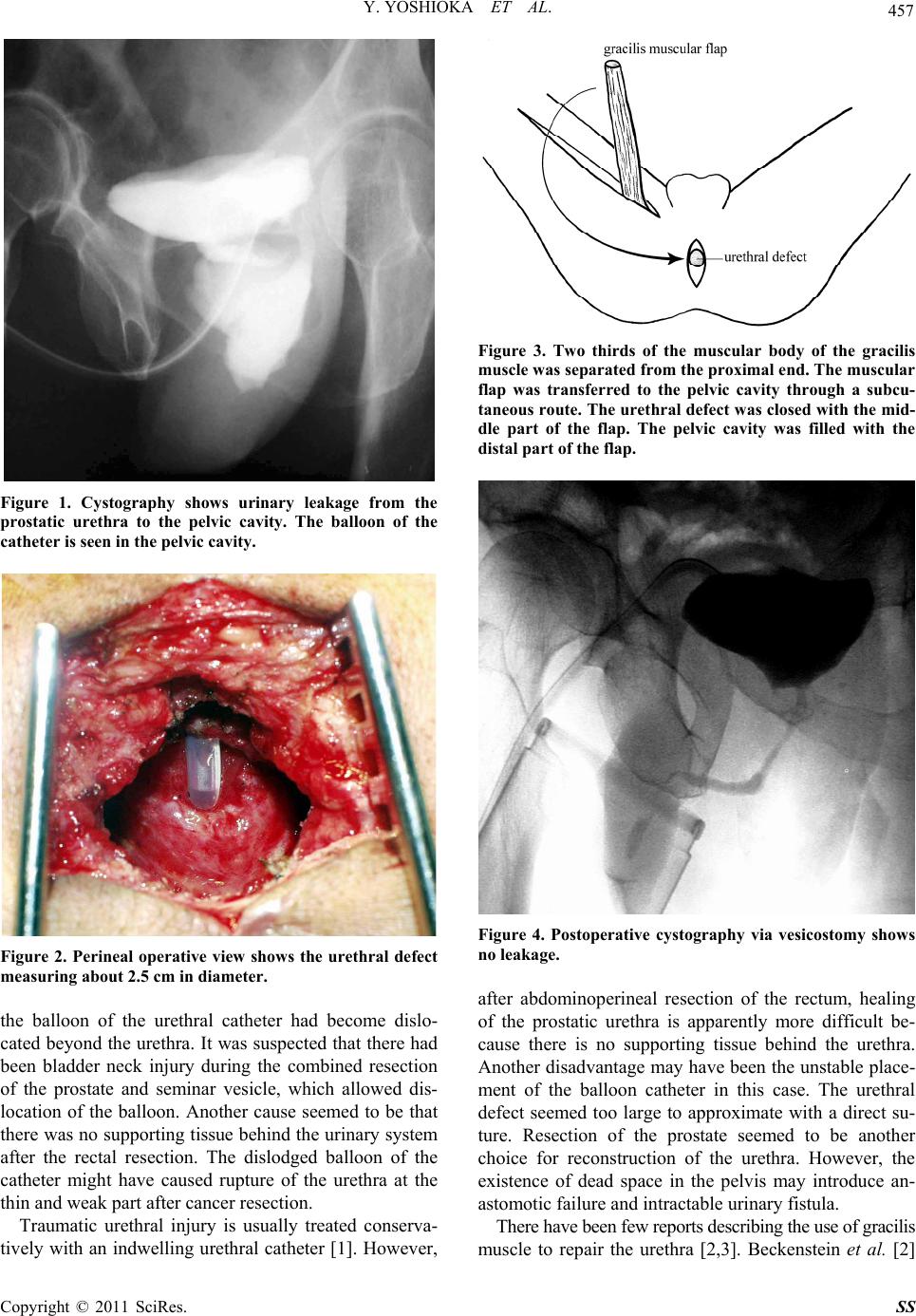
from the pelvic drain. Cystogr aphy demonstrated urinary
leakage from the prostatic urethra to the pelvic cavity
(Figure 1). The balloon of the catheter was seen in the
pelvic cavity. We changed the urethral catheter to that
with a larger balloon and tried to close the fistula con-
servatively. One month later, there was no improvement
of leakage or change in fistula size. We decided to close
the fistula surgically.
The patient was placed in the lithotomy position and
we re-opened the perineal wound. There were no signs of
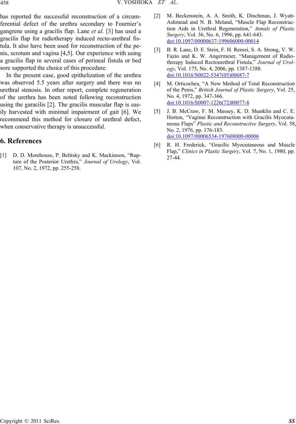
infection in the pelvic cavity. The size of the urethral
defect was about 2.5 cm in diameter (Figure 2). Two
thirds of the muscular body of the gracilis muscle was
separated from the proximal end and the vascular pedicle
located at the proximal part was preserved. The gracilis
muscular flap was transferred to the pelvic cavity
through a subcutaneous route. The urethral defect was
closed using the middle part of the flap with absorbable
sutures as if the muscle were a patch. The pelvic cavity
was filled with the distal p art of the flap (Figure 3) Suc-
tion drains were placed in the pelv ic cavity and th e donor
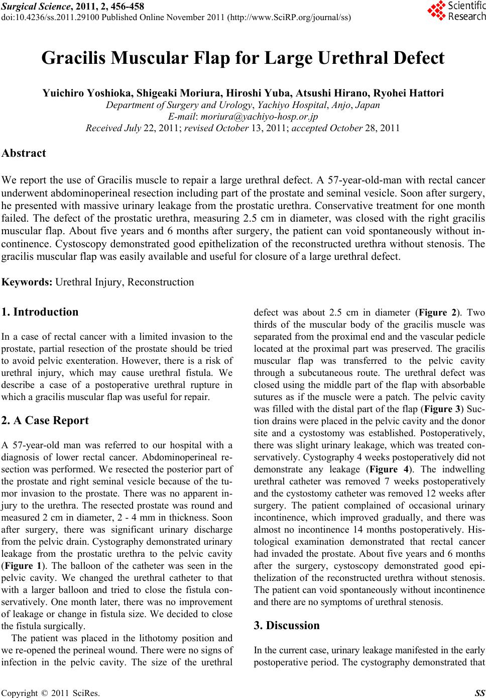
site and a cystostomy was established. Postoperatively,
there was slight urinary leakage, which was treated con-
servatively. Cysto graph y 4 w eeks po stoper atively d id no t
demonstrate any leakage (Figure 4). The indwelling
urethral catheter was removed 7 weeks postoperatively
and the cystostomy catheter was removed 12 weeks after
surgery. The patient complained of occasional urinary
incontinence, which improved gradually, and there was
almost no incontinence 14 months postoperatively. His-
tological examination demonstrated that rectal cancer
had invaded the prostate. About five years and 6 months
after the surgery, cystoscopy demonstrated good epi-
thelization of the reconstructed urethra without stenosis.
The patient can void spontaneously withou t incontinence
and there are no symptoms of urethral stenosis.
3. Discussion
In the current case, urinary leakage manifested in the early
postoperative period. The cystography demonstrated that