 Pharmacology & Pharmacy, 2011, 2, 289-298 doi:10.4236/pp.2011.24037 Published Online October 2011 (http://www.SciRP.org/journal/pp) Copyright © 2011 SciRes. PP 289 Design and Development of an in Vitro Assay for Evaluation of Solid Vaginal Dosage Forms Jyoti Gupta1, Jason Qihai Tao2, Sanjay Garg1, Raida Al-Kassas1* 1School of Pharmacy, Faculty of Medical and Health Sciences, The University of Auckland, Auckland, New Zealand; 2International Partnership for Microbicides, Silver Springs, USA. Email: *r.al-kassas@auckland.ac.nz Received February 27th, 2011; revised April 11th, 2011; accepted July 20th, 2011. ABSTRACT Vaginal dosage forms are seen as a viable option for empowering women to protect themselves from the risk of HIV transmission. Because of limited research in the field, there is a lack of suitable dissolution methods established for determination of drug release from vaginal formulations inside the vaginal tract. The main aim of this study was to de- velop a simple, reliable and reproducible in vitro release method for evaluation of solid vaginal dosage forms (VDFs) which was hoped to exhibit a close in vitro-in vivo correlation. Dapivirine, a drug being developed as a microbicide and a well estab lished marketed a nti fungal d rug, Clotrimazole were used as model drugs. Two doses (0.5 mg and 1.25 mg) of Dapivirine were prepared as novel rapidly disintegrating, bioadhesive tablets. Clotrimazole 100 mg, prepared in house as conventional release tablets and commercially available Canesten (Clotrimazole tablet 100 mg) were used. The in vitro drug release testing of these tablets was carried out using a designed system which consisted of modified USP dissolution Appa ratus II in conjunction with Enhancer cell (as sample holder) in 150 ml capacity flasks instead of the standard 900 ml flasks. The suitability of the system was investigated for variable parameters such as formulation types, drug concentration, stirring speeds, media volume and comparison of in house product with marketed product. The method was successfully optimized at a volume of 100 ml and a low speed of 25 rpm at pH 4 and was found sensi- tive enough to distinguish between formulation s and evaluate products of different streng ths. A linear drug release pro - file (R2 = 0.99) was obtained in case of Dapivirine, indicating that drug release is controlled by diffusion. The devel- oped dissolu tion system has a potential to exhibit a good in vitro-in vivo correlation in addition to carrying out routine dissolution tests for solid VDFs. Keywords: Vaginal Solid Dosage Forms, Enhancer Cell, Dapivirine, Clotrimazole 1. Introduction Administration of drugs in the vagina is believed to be as old as pharmacotherapy, first written documents dating from 19th century BC [1]. Vaginal drug delivery presents several advantages including, ease of administration, possibility of self-administration, hepatic first pass-effect bypass, low systemic drug exposure and increased per- meability for some drugs when compared to the oral or other routes [1]. Vaginal dosage forms available around the world include creams, gels, tablets, capsules, pessaries, foams, ointments, films, tampons, rings and douches. While majority of vaginal drugs so far have been in the form of gels, there is a growing interest in alternative dosage forms such as tablets, rings, and films. Conven- tional vaginal dosage forms are associated with limita- tions of poor retention, leakage and messiness causing inconvenience to users, leading to poor subject/patient compliance and loss of therapeutic efficacy [2]. In recent years vaginal bioadhesive tablets have been developed as a new type of controlled release form for the treatment of both topical and systemic diseases. The greatest advan- tages of such bioadhesive tablets are the release of drug at a controlled rate and the possibility of maintaining them in the vagina for extended periods of time [3]. Dissolution testing is routinely used in Quality Control (QC) and Research and Development (R & D). To date none of the official compendia [4-6] have included a standard method for evaluation of release pattern from vaginal preparations. Modified USP apparatus [7-10] and various other methods [11-13] have been employed by researchers to study the release of drugs from vaginal formulations. In these studies a standard dissolution ap- paratus with large volume of media has been employed  Design and Development of an in Vitro Assay for Evaluation of Solid Vaginal Dosage Forms 290 which does not simulate the actual physiological condi- tions of the vagina, characterized by small volume of fluid. Volume plays a significant role in the rate of drug released and absorbed. In addition, other parameters such as speed of agitation and pH should be considered in order to assure close simulation of in vivo situation. There are several systems used in the industry to study the release of drugs from various pharmaceutical dosage forms including vaginal formulations. These include USP apparatus 4 (Flow through cell), Franz cells and Enhan- cer cells [14,15]. Flow through cell could be used for solid and semi solid vaginal formulations; however the equipment is quite expensive and not available in many laboratories. On the other hand, Franz cell has been pro- posed to be one of the USP standard apparatus for topi- cal/transdermal formulations. However, it is mostly suit- able for semi-solid formulations and requires highly trained technician to operate it and a membrane to be a supportive physical barrier. The release of the drug from the formulation, in most instances, depends on the type of membrane to be used and its diffusion through it. En- hancer cell has also been used to evaluate release profiles of semi-solid formulations. The system consists of a paddle over enhancer cell with a small dissolution vessel. To date, most of the membranes used in this method were synthetic membranes or filters. This system utilizes the existing USP apparatus and is thus easy to set up, portable and reproducible. In the present study, we have employed Enhancer cell system in conjunction with USP dissolution apparatus II and 150 ml capacity flasks to replace the 900 ml flasks used in the standard apparatus. The key difference of our design is “mesh membrane” which replaced the synthetic membranes most commonly used in past. The mesh membrane of adequate pore size holds the formulation in place and provide physical barrier which prevents the formulation from being agitated too much while allowing the drug to migrate or release freely from the formulation matrix to the media and allowing some contact/exchange of the media and the sample in the cavity. This system adopts physiological parameters of the vagina (volume of fluid, speed of agitation and pH), in an attempt to obtain a standard quality control procedure for evaluation of vaginal tablets, to minimize variables in results and to achieve a close approximation to in vivo situation. The aims of our design and the study were to develop a simple, reliable and reproducible in vitro release method for evaluation of solid vaginal dosage forms (VDFs) which mimicked in vivo vaginal application of a tablet dosage and was hoped to exhibit an adequate in vitro-in vivo correlation for future developments. The drugs selected for the study were Dapivirine and Clotrimazole. Dapivirine, also known as TMC120, is a substituted diarylpyrimidine derivative and a non nucleo- side reverse transcriptase inhibitor (NNRTI) being de- veloped as a vaginal microbicide for prevention of HIV transmission [16]. Vaginal tablets of Dapivirine were prepared to disintegrate rapidly in the presence of small volume of fluid and form a bioadhesive gel from which the drug is slowly released over a period of time (there is no evidence yet for this bioadhesive tablet to be a con- trolled release formulation). Clotrimazole is a chlorinated imidazole derivative wide- ly prescribed for the treatment of vulvovaginal candidiasis VVC. Vaginal tablets of Clotrimazole were prepared in house as conventional release tablets. 2. Materials and Methods 2.1. Materials Dapivirine was obtained as a gift from International Part- nership for Microbicides (IPM). Clotrimazole was also received as a gift from Zhejiang Chemicals Import and Export Corporation, China. Canesten tablets (Clotrima- zole tablets 100 mg) were procured from a local phar- macy in New Zealand. The directly compressible lactose was procured from Fonterra (New Zealand), microcrys- talline cellulose (MCC) from Asahi Kasei Corp (Japan), magnesium stearate from Acros Organic (USA), sodium starch glycollate from Spectrum, USA. Ammonium ace- tate was purchased from BDH Laboratory (England), sodium phosphate dibasic from Riedel-de Haen (Ger- many), sodium phosphate monobasic, acetonitrile and methanol from Merck (Germany). Sodium lauryl sul- phate was purchased from Serva Electrophoresis and tween 80 from Sigma Aldrich (Germany). 2.2. Tablet Preparation Dapivirine was prepared in two different doses (0.05% and 0.125%) as novel rapidly disintegrating, long acting bioadhesive tablets. Dapivirine being a low dose drug was mixed geometrically with polymer, diluents, su- perdisintegrant and acid buffering agents to ensure a homogeneous mixing of the drug with the excipients. The mixing was carried out in a pilot scale cube mixer attached to a multipurpose unit operator (Erweka, Ger- many). The detailed composition and manufacturing procedure for Dapivirine cannot be disclosed due to In- tellectual Property reasons. Clotrimazole 100 mg conventional vaginal tablets were prepared as follows: Clotrimazole, passed through 60 mesh, was mixed with MCC (passed through 30 mesh) in the cube mixer for 30 minutes. Directly compressible grade lactose was passed through 30 mesh and added to the mix of Clotrimazole and MCC and mixed for one hour. Magnesium stearate and sodium starch glycollate Copyright © 2011 SciRes. PP 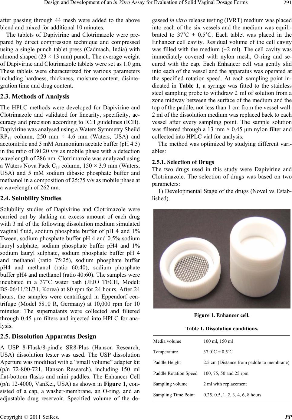 Design and Development of an in Vitro Assay for Evaluation of Solid Vaginal Dosage Forms291 after passing through 44 mesh were added to the above blend and mixed for additional 10 minutes. The tablets of Dapivirine and Clotrimazole were pre- pared by direct compression technique and compressed using a single punch tablet press (Cadmach, India) with almond shaped (23 × 13 mm) punch. The average weight of Dapivirine and Clotrimazole tablets were set as 1.0 gm. These tablets were characterized for various parameters including hardness, thickness, moisture content, disinte- gration time and drug content. 2.3. Methods of Analysis The HPLC methods were developed for Dapivirine and Clotrimazole and validated for linearity, specificity, ac- curacy and precision according to ICH guidelines (ICH). Dapivirine was analysed using a Waters Symmetry Sheild RP18 column, 250 mm × 4.6 mm (Waters, USA) and acetonitrile and 5 mM Ammonium acetate buffer (pH 4.5) in the ratio of 80:20 v/v as mobile phase with a detection wavelength of 286 nm. Clotrimazole was analyzed using a Waters Nova Pack C18 column, 150 × 3.9 mm (Waters, USA) and 5 mM sodium dibasic phosphate buffer and methanol in a composition of 25:75 v/v as mobile phase at a wavelength of 262 nm. 2.4. Solubility Studies Solubility studies of Dapivirine and Clotrimazole were carried out by shaking an excess amount of each drug with 3 ml of the following dissolution medium simulated vaginal fluid, sodium phosphate buffer of pH 4 and 1% Tween, sodium phosphate buffer pH 4 and 0.5% sodium lauryl sulphate, sodium phosphate buffer pH4 and 1% sodium lauryl sulphate, sodium phosphate buffer pH 4 and methanol (ratio 75:25), sodium phosphate buffer pH4 and methanol (ratio 60:40), sodium phosphate buffer pH4 and methanol (ratio 40:60). The samples were incubated in a 37˚C water bath (JEIO TECH, Model: BS-06/11/21/31, Korea) at 80 rpm for 24 hours. After 24 hours, the samples were centrifuged in Eppendorf cen- trifuge (Model 5810 R, Germany) at 10,000 rpm for 10 minutes. The supernatants were collected and filtered through 0.45 µm filters and injected into HPLC for ana- lysis. 2.5. Dissolution Apparatus Design A USP 8-Flask/8-pindle SR8-Plus (Hanson Research, USA) dissolution tester was used. The USP dissolution Aperture was modified with a “small volume” adapter kit (p/n 72-800-721, Hanson Research), including 150 ml flat-bottom flasks and mini paddles. The Enhancer Cell (p/n 12-4000, VanKel, USA) as shown in Figure 1, con- sisted of a cap, a washer-membrane, an O-ring, and an adjustable drug reservoir. Specified volume of the de- gassed in vitro release testing (IVRT) medium was placed into each of the six vessels and the medium was equili- brated to 37˚C ± 0.5˚C. Each tablet was placed in the Enhancer cell cavity. Residual volume of the cell cavity was filled with the medium (~2 ml). The cell cavity was immediately covered with nylon mesh, O-ring and se- cured with the cap. Each Enhancer cell was gently slid into each of the vessel and the apparatus was operated at the specified rotation speed. At each sampling point in- dicated in Table 1, a syringe was fitted to the stainless steel sampling probe to withdraw 2 ml of solution from a zone midway between the surface of the medium and the top of the paddle, not less than 1 cm from the vessel wall. 2 ml of the dissolution medium was replaced back to each vessel after every sampling point. The sample solution was filtered through a 13 mm × 0.45 μm nylon filter and collected into HPLC vial for analysis. The method was optimized by studying different vari- ables: 2.5.1. Selecti o n of Dru gs The two drugs used in this study were Dapivirine and Clotrimazole. The selection of drugs was based on two parameters: 1) Developmental Stage of the drugs (Novel vs Estab- lished). Figure 1. Enh a ncer cell. Table 1. Dissolution conditions. Media volume 100 ml, 150 ml Temperature 37.0˚C ± 0.5˚C Paddle Height 2.5 cm (Distance from paddle to membrane) Paddle Rotation Speed 100, 75, 50 and 25 rpm Sampling volume 2 ml with replacement Sampling Time Point 0.25, 0.5, 1, 2, 3, 4, 6, 8 hours Copyright © 2011 SciRes. PP 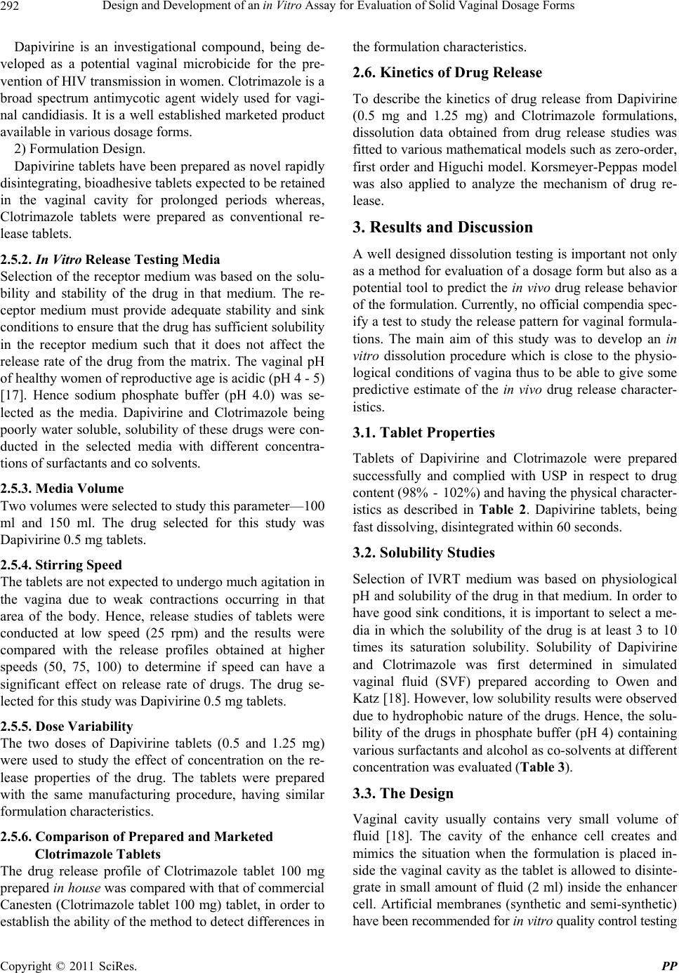 Design and Development of an in Vitro Assay for Evaluation of Solid Vaginal Dosage Forms 292 Dapivirine is an investigational compound, being de- veloped as a potential vaginal microbicide for the pre- vention of HIV transmission in women. Clotrimazole is a broad spectrum antimycotic agent widely used for vagi- nal candidiasis. It is a well established marketed product available in various dosage forms. 2) Formulation Design. Dapivirine tablets have been prepared as novel rapidly disintegrating, bioadhesive tablets expected to be retained in the vaginal cavity for prolonged periods whereas, Clotrimazole tablets were prepared as conventional re- lease tablets. 2.5.2. In Vitro Rel ease Testing Media Selection of the receptor medium was based on the solu- bility and stability of the drug in that medium. The re- ceptor medium must provide adequate stability and sink conditions to ensure that the drug has sufficient solubility in the receptor medium such that it does not affect the release rate of the drug from the matrix. The vaginal pH of healthy women of reproductive age is acidic (pH 4 - 5) [17]. Hence sodium phosphate buffer (pH 4.0) was se- lected as the media. Dapivirine and Clotrimazole being poorly water soluble, solubility of these drugs were con- ducted in the selected media with different concentra- tions of surfactants and co solvents. 2.5.3. Medi a Vol ume Two volumes were selected to study this parameter—100 ml and 150 ml. The drug selected for this study was Dapivirine 0.5 mg tablets. 2.5.4. Stirri ng Speed The tablets are not expected to undergo much agitation in the vagina due to weak contractions occurring in that area of the body. Hence, release studies of tablets were conducted at low speed (25 rpm) and the results were compared with the release profiles obtained at higher speeds (50, 75, 100) to determine if speed can have a significant effect on release rate of drugs. The drug se- lected for this study was Dapivirine 0.5 mg tablets. 2.5.5. Dose Variability The two doses of Dapivirine tablets (0.5 and 1.25 mg) were used to study the effect of concentration on the re- lease properties of the drug. The tablets were prepared with the same manufacturing procedure, having similar formulation characteristics. 2.5.6. Comp arison of Prepared and M arketed Clotrimazole Tablets The drug release profile of Clotrimazole tablet 100 mg prepared in house was compared with that of commercial Canesten (Clotrimazole tablet 100 mg) tablet, in order to establish the ability of the method to detect differences in the formulation characteristics. 2.6. Kinetics of Drug Release To describe the kinetics of drug release from Dapivirine (0.5 mg and 1.25 mg) and Clotrimazole formulations, dissolution data obtained from drug release studies was fitted to various mathematical models such as zero-order, first order and Higuchi model. Korsmeyer-Peppas model was also applied to analyze the mechanism of drug re- lease. 3. Results and Discussion A well designed dissolution testing is important not only as a method for evaluation of a dosage form but also as a potential tool to predict the in vivo drug release behavior of the formulation. Currently, no official compendia spec- ify a test to study the release pattern for vaginal formula- tions. The main aim of this study was to develop an in vitro dissolution procedure which is close to the physio- logical conditions of vagina thus to be able to give some predictive estimate of the in vivo drug release character- istics. 3.1. Tablet Properties Tablets of Dapivirine and Clotrimazole were prepared successfully and complied with USP in respect to drug content (98% - 102%) and having the physical character- istics as described in Table 2. Dapivirine tablets, being fast dissolving, disintegrated within 60 seconds. 3.2. Solubility Studies Selection of IVRT medium was based on physiological pH and solubility of the drug in that medium. In order to have good sink conditions, it is important to select a me- dia in which the solubility of the drug is at least 3 to 10 times its saturation solubility. Solubility of Dapivirine and Clotrimazole was first determined in simulated vaginal fluid (SVF) prepared according to Owen and Katz [18]. However, low solubility results were observed due to hydrophobic nature of the drugs. Hence, the solu- bility of the drugs in phosphate buffer (pH 4) containing various surfactants and alcohol as co-solvents at different concentration was evaluated (Table 3). 3.3. The Design Vaginal cavity usually contains very small volume of fluid [18]. The cavity of the enhance cell creates and mimics the situation when the formulation is placed in- side the vaginal cavity as the tablet is allowed to disinte- grate in small amount of fluid (2 ml) inside the enhancer cell. Artificial membranes (synthetic and semi-synthetic) have been recommended for i vitro quality control testing n Copyright © 2011 SciRes. PP  Design and Development of an in Vitro Assay for Evaluation of Solid Vaginal Dosage Forms Copyright © 2011 SciRes. PP 293 Table 2. Physical characteristics of Dapivirine 0.5 mg and 1.25 mg and Clotrimazole 100 mg tablets. Parameters Dapivirine 0.5 mg Dapivirine 1.25 mg Clotrimazole 100 mg Average weight (g) 1.0175 ± 0.003 1.010 ± 0.002 1.009 ± 0.12 Hardness (horizontal) (Kp) 11.028 ± 0.12 11.25 ± 0.42 14.015 ± 0.19 Hardness (vertical) (Kp) 7.93 ± 0.33 8.07 ± 0.50 11.39 ± 0.28 Thickness (mm) 5.36 ± 0.04 5.39 ± 0.01 4.95 ± 0.07 Friability (% w/w) 0.65 0.57 0.72 Disintegration time (min) 0.52 ± 0.05 0.58 ± 0.03 6.19 ± 0.10 Moisture content (% w/w) 2.54 ± 0.04 2.51 ± 0.02 1.25 ± 0.02 Table 3. Solubility data of Dapivirine and Clotrimazole in SVF and phosphate buffe r c ontaining surfactants and co-solvents. Media Concentration of Dapivirine (µg/ml) Concentration of Clotrimazole (µg/ml) Simulated Vaginal Fluid 0.53 21.30 Sodium Phosphate Buffer (pH 4.0) + 0.5% Tween 80 21.19 22.06 Sodium Phosphate Buffer (pH 4.0)+ 1.0% Tween 80 40.21 112.30 Sodium Phosphate Buffer (pH 4.0) + 0.5% SLS 36.82 494.27 Sodium Phosphate Buffer (pH 4.0) + 1.0% SLS 123.51 1338.60 Sodium Phosphate Buffer (pH 4.0):Methanol (75:25) 0.85 16.82 Sodium Phosphate Buffer (pH 4.0):Methanol (60:40) 7.18 102.08 Sodium Phosphate Buffer (pH 4.0):Methanol (40:60) 112.70 2323.11 due to the variability involved with biological mem- branes. The membranes should be permeable to the drug and its pore size and thickness should not affect the drug release. The membrane selected should provide an inert holding surface for the test formulation, and not act as a barrier as vaginal microbicidal formulations are not de- signed for systemic absorption but expected to act topi- cally. The Nylon Mesh used during this entire experi- ment served only as a holder first for the placed tablet and then for the gel-matrix obtained after disintegration of the tablet in the sample compartment in contact with the receptor solution. The mesh size was small enough to hold disintegrated tablet inside the sample cavity but still permitted the receptor solution to easily circulate from the 150ml dissolution vessel into the sample cavity of the Enhancer cell, thus facilitating homogenous distribution of the drug released from the formulation. 3.4. In Vitro Drug Release Studies 3.4.1. Selectio n of Media Dapivirine and Clotrimazole are poorly water soluble drugs. This necessitated the use of media with surfactant or organic solvent. Based on the solubility data of the drugs (Table 3), the two media selected to study the re- lease profile were sodium phosphate buffer containing 1) 1% SLS and 2) 60% methanol. The release study was conducted at a paddle rotation speed of 100 rpm and me- dia volume was 100 ml. Figures 2 and 3 show compara- tive release profiles of Dapivirine and Clotrimazole, re- spectively in two media. Dapivirine release profile from surfactant medium showed a sharp release in 15 minutes, forming a plateau after 6 hours. However, profile in aqueous organic medium demonstrated a slow and linear rise in the drug release during the 8 hours study. This can be explained by higher solubility of Dapivirine in surfac- tant media (123.5 µg/ml) as compared to aqueous or- ganic media (112.7 µg/ml). Due to high probability of air bubbles with the use of surfactant media, aqueous or- ganic media which shows a profile closer to the expected in vivo drug release behavior was selected as the media of choice. Previous reports [19,20] on Dapivirine have used aqueous organic media for drug release studies. On the other hand, Clotrimazole release in aqueous organic media was higher as compared to that in surfactant media, which is attributed to the drug’s higher solubility in queous organic media (2323 µg/ml). a 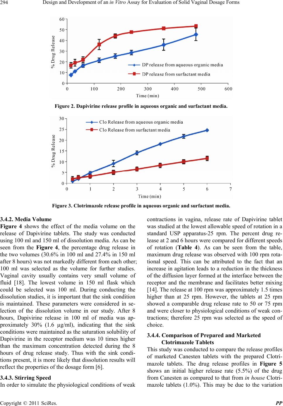 Design and Development of an in Vitro Assay for Evaluation of Solid Vaginal Dosage Forms 294 Figure 2. Dapivirine release profile in aqueous organic and surfactant media. Figure 3. Clotrimazole release profile in aqueous organic and surfactant media. 3.4.2. Medi a Vol ume Figure 4 shows the effect of the media volume on the release of Dapivirine tablets. The study was conducted using 100 ml and 150 ml of dissolution media. As can be seen from the Figure 4, the percentage drug release in the two volumes (30.6% in 100 ml and 27.4% in 150 ml after 8 hours) was not markedly different from each other; 100 ml was selected as the volume for further studies. Vaginal cavity usually contains very small volume of fluid [18]. The lowest volume in 150 ml flask which could be selected was 100 ml. During conducting the dissolution studies, it is important that the sink condition is maintained. These parameters were considered in se- lection of the dissolution volume in our study. After 8 hours, Dapivirine release in 100 ml of media was ap- proximately 30% (1.6 µg/ml), indicating that the sink conditions were maintained as the saturation solubility of Dapivirine in the receptor medium was 10 times higher than the maximum concentration detected during the 8 hours of drug release study. Thus with the sink condi- tions present, it is more likely that dissolution results will reflect the properties of the dosage form [6]. 3.4.3. Stirri ng Speed In order to simulate the physiological conditions of weak contractions in vagina, release rate of Dapivirine tablet was studied at the lowest allowable speed of rotation in a standard USP apparatus-25 rpm. The percent drug re- lease at 2 and 6 hours were compared for different speeds of rotation (Table 4). As can be seen from the table, maximum drug release was observed with 100 rpm rota- tional speed. This can be attributed to the fact that an increase in agitation leads to a reduction in the thickness of the diffusion layer formed at the interface between the receptor and the membrane and facilitates better mixing [14]. The release at 100 rpm was approximately 1.5 times higher than at 25 rpm. However, the tablets at 25 rpm showed a comparable drug release rate to 50 or 75 rpm and were closer to physiological conditions of weak con- tractions; therefore 25 rpm was selected as the speed of choice. 3.4.4. Comp arison of Prepared and M arketed Clotrimazole Tablets This study was conducted to compare the release profiles of marketed Canesten tablets with the prepared Clotri- mazole tablets. The drug release profiles in Figure 5 shows an initial higher release rate (5.5%) of the drug from Canesten as compared to that from in house Clotri- mazole tablets (1.0%). This e due to the variation may b Copyright © 2011 SciRes. PP  Design and Development of an in Vitro Assay for Evaluation of Solid Vaginal Dosage Forms295 Figure 4. Comparison of Dapivirine release in different media volumes. Figure 5. Release profile comparison of different formulations of Clotrimazole. Table 4. Comparison of Dapivirine release after 2 and 6 hours at different speeds of rotation. Paddle Speed (rpm) Release in 2 hours (%) Release in 6 hours (%) 25 14.7 23.6 50 20.2 29.9 75 18.6 27.8 100 21.6 36.0 in the disintegration time of the two products. The disin- tegration time of the two formulations when measured using standard BP Disintegration apparatus, was found to be 6 - 7 minutes for prepared Clotrimazole tablets and 45 seconds for Canesten. However, after a period of time the release profiles of the marketed Canesten and the pre- pared clotrimazole tablets became comparable. Thus, it is evident that the method is sensitive enough to differenti- ate variability in the method of manufacturing. 3.4.5. Dose Variability This parameter was based on release studies conducted on two different doses of Dapivirine (0.05% and 0.125%), in order to assess the sensitivity of the developed disso- lution method (Figure 6). The dissolution conditions were as reported in Table 1. From Figure 6, it can be seen that the shape of the two release profiles is similar showing a higher release from the higher dose, indicating the capability of the method to differentiate the variabil- ity in dose. 3.5. Comparison of Release Profile from Enhancer Cell and Standard USP Dissolution Method The drug release study was conducted using the standard dissolution method described in official compendia [4-6] for solid oral dosage forms. The release profile from standard method was compared with that from Enhancer cell method. Dapivirine 0.5 mg tablet was employed in this study and 500 ml of 0.1 N HCL was used as media. As can be seen from Figure 7, Enhancer cell method shows a slow release as only 20% of drug is released within 2 hours. On the other hand, the release rate of drug studied in standard dissolution apparatus is high as 80% of drug is released within 2 hours. With the standard USP dissolution method, employing Copyright © 2011 SciRes. PP  Design and Development of an in Vitro Assay for Evaluation of Solid Vaginal Dosage Forms 296 Figure 6. Comparison of Dapivirine release from two doses (0.5 mg and 1.25 mg). Figure 7. Comparative release profile of Dapivirine from standard and enhancer cell method at 50 rpm. 500 ml of media and 50 rpm rotational speed, the rapidly disintegrating tablets of Dapivirine dispersed quickly in the large media volume giving a solubility profile of the drug rather than predicting its release characteristics. These tablets were designed to disperse quickly and form a gel in small amount of fluid in the vagina. Enhancer cell has the advantage of containing the tablet in a pocket wherein it is allowed to disperse in small volume (2 ml) of media and form a bioadhesive gel from which the drug is released slowly. Comparative release studies of Da- pivirine from Enhancer cell and standard method at 50 rpm clearly show that the shape of the two profiles is same. The only difference is the rate of release of the drug which corresponds to the difference in the volume. 3.6. Kinetics and Mechanism of Drug Release The release data of Dapivirine 0.5 mg and 1.25 mg were fitted best to Higuchi’s model (R2 = 0.989 and 0.992 re- spectively) as indicated in Figures 8 and 9. This suggests that the drug release is controlled by diffusion. When the release data was fitted to the model of Korsmeyer-Peppas (Table 5), the release exponent n value was found to be close to 0.5 for both the doses thus indicating Fickian diffusion. The drug release data of Clotrimazole tablets was best fitted to zero-order model with an R2 value of 0.9957. This was confirmed when the data was applied to Korsmeyer Peppas model. The value of release exponent n was found to be close to 1.0 (Table 5) indicating zero- order release. 4. Conclusions A dissolution system was designed and optimized for evaluation of vaginal tablets. The system enabled con- tainment of the drug matrix in the cell to avoid complete distribution and dissolution of the drug in the medium. Drug release studies were successfully conducted on Dapivirine and Clotrimazole tablets. The suitability of the system was investigated for various parameters like different formulation design, dose variability, different stirring speeds, media volume and comparison of pre- pared and marketed product. The Enhancer cell method was found to be sensitive enough to distinguish between formulations and demonstrated the ability to detect products of different strengts. In conclusion, a simple, h Copyright © 2011 SciRes. PP  Design and Development of an in Vitro Assay for Evaluation of Solid Vaginal Dosage Forms297 Figure 8. Dapivirine 0.5 mg release per cm2 versus square root of time. Figure 9. Dapivirine 1.25 mg release per cm2 versus square root of time. Table 5. Model fitting using Korsmeyer-Peppas equation. Product R2 n k Dapivirine 0.5 mg 0.9272 0.6907 0.4090 Dapivirine 1.25 mg 0.9161 0.4742 1.6420 Clotrimazole 100 mg 0.9624 0.9122 0.0850 reliable, and reproducible dissolution method has been developed for solid vaginal dosage forms. This method can be used to support formulation development for early stage drug product development and potentially for qual- ity control and in vivo performance analysis of the final product. REFERENCES [1] J. Das Neves, “Vaginal Drug Delivery,” SciTopics, 2008. [2] J. Robinson and W. Bologna, “Vaginal and Reproductive System Treatments Using a Bioadhesive Polymer,” Journal of Controlled Release, Vol. 28, No. 1-3, 1994, pp. 87-94. doi:10.1016/0168-3659(94)90156-2 [3] A. Gursoy and A. Bayhan, “Testing of Drug Release from Bioadhesive Vaginal Tablets,” Drug Development and In- dustrial Pharmacy, Vol. 17, No. 18, 2007, pp. 2457-2475. doi:10.3109/03639049109048087 [4] European Pharmacopoeia 60: EDQM, 2007. [5] British Pharmacopoeia, Stationary Office, London, 2009. [6] United States Pharmacopoeia and National Formulary, 2009. [7] A. El-Kamel, M. Sokar and V. Naggar, “Bioadhesive Con- trolled Release Metronidazole Vaginal Tablets,” Acta Phar- maceutica, Vol. 52, 2002, pp. 171-179. [8] S. Garg, L. Jambu and K. Vermani, “Development of No- vel Sustained Release Bioadhesive Vaginal Tablets of Povi- done Iodine,” Drug Development and Industrial Pharmacy , Vol. 33, No. 12, 2007, pp. 1340-1349. doi:10.1080/03639040701385782 [9] F. Ahmad, M. Alam, Z. Khan, M. Khar and M. Ali, “De- velopment and in Vitro Evaluation of an Acid Buffering Bioadhesive Vaginal Gel for Mixed Infections,” Acta Pharmaceut ica, Vol. 58, No. 4, 2008, pp. 407-19. doi:10.2478/v10007-008-0023-2 [10] L. Wang and X. Tang, “A Novel Ketoconazole Bioadhe- sive Tablet for Vaginal Delivery: Design, in Vitro and in Vivo Evaluation,” International Journal of Pharmaceutics, Vol. 350, No. 1-2, 2008, pp. 181-187. [11] C. Lee and Y. Chien, “Development and Evaluation of Copyright © 2011 SciRes. PP  Design and Development of an in Vitro Assay for Evaluation of Solid Vaginal Dosage Forms 298 Mucoadhesive Drug Delivery System for Dualcontrolled Delivery of Nonoxynol-9,” Journal of Controlled Release, Vol. 39, No. 1, 1996, pp. 93-103. doi:10.1016/0168-3659(95)00142-5 [12] C. Valenta, C. Kast and I. Harich, “Development and in Vitro Evaluation of a Mucoadhesive Vaginal Delivery System for Progesterone,” Journal of Controlled Release, Vol. 77, No. 3, 2001, pp. 323-332. doi:10.1016/S0168-3659(01)00520-X [13] V. Kale, R. Trivedi and P. Muley, “Proposed Design of a Dissolution Apparatus for Vaginal Formulations Contain- ing Probiotics,” Dissolution Technologies, Vol. 15, No. 2, 2008. [14] P. Rege, V. Vilivalam and C. Collins, “Development in Release Testing of Topical Dosage Forms: Use of En- hancer Cell with Automated Sampling,” Journal of Phar- maceutical and Biomedical Analysis, Vol. 17, No. 8, 1998, pp. 1225-1233. doi:10.1016/S0731-7085(97)00184-2 [15] W. Liebenberg, E. Engelbrecht and A. Wessels, “A Com- parative Study of the Release of Active Ingredients from Semisolid Cosmeceuticals Measured with Franz, Enhancer or Flow through Cell Diffusion Apparatus,” Journal of Food and Drug Analysis, Vol. 12, No. 1, 2004, pp. 19-28. [16] J. Nuttall, D. Thake and M. Lewis, “Concentrations of Dapivirine in the Rhesus Macaque and Rabbit following Once Daily Intravaginal Administration of a Gel Formula- tion of [14C] Dapivirine for 7 Days,” Antimicrobial Agent s and Chemotherapy, Vol. 52, No. 3, 2008, pp. 909-914. doi:10.1128/AAC.00330-07 [17] K. Vermani and S. Garg, “The Scope and Potential of Vaginal Drug Delivery,” Pharmaceutical Science & Tech- nology Today, Vol. 3, No. 10, 2000, pp. 359-364. doi:10.1016/S1461-5347(00)00296-0 [18] D. Owen and D. Katz, “A Vaginal Fluid Stimulant,” Con- traception, Vol. 59, No. 2, 1999, pp. 91-95. [19] A. Woolfson, R. Malcom and R. Morrow, “Intravaginal Ring Delivery of the Reverse Transcriptase Inhibitor TMC 120 as an HIV Microbicide,” International Journal of Pharmaceutics, Vol. 325, No. 1-2, 2006, pp. 82-89. doi:10.1016/j.ijpharm.2006.06.026 [20] K. Gupta, S. Pearce and A. Poursaid, “Polyurethane In- travaginal Ring for Controlled Delivery of Dapivirine, a Nonnucleoside Reverse Transcriptase Inhibitor of HIV-1,” Journal of Pharmaceutical Sciences, Vol. 97, No. 10, 2008, pp. 4228-4239. doi:10.1002/jps.21331 Copyright © 2011 SciRes. PP
|