Journal of Cancer Therapy
Vol.4 No.6A1(2013), Article ID:34534,8 pages DOI:10.4236/jct.2013.46A1008
The Technique of Enhancing the Transdermal Penetration for the Gold Nanoparticles and Perspectives of Application
![]()
1Department of Human Anatomy, Medical Faculty, Ul’yanovsk State University, Ul’yanovsk, Russia; 2Department of Optics and Biophotonics, Faculty of Physics, Saratov State University, Saratov, Russia.
Email: *prof.khayrullin@gmail.com
Copyright © 2013 Radik M. Khayrullin et al. This is an open access article distributed under the Creative Commons Attribution License, which permits unrestricted use, distribution, and reproduction in any medium, provided the original work is properly cited.
Received May 20th, 2013; revised June 21st, 2013; accepted June 29th, 2013
Keywords: Nanoparticles; Skin Penetration; Dimethylsulfoxide; Tiofansulfoxide
ABSTRACT
Background: In recent years, worldwide attention of researchers focused on the practical implementation of nanoscale materials in biomedical technology. It’s proved that intravenous injected gold nanoparticles are accumulated in tumor tissues. However, when gold nanoparticles injected intravenously negative effects in the internal organs of experimental animals are observed. 160 nm diameter particles affect the wall of blood vessels, resulting in vacuolar degeneration of endothelial cells. Particles with a diameter of 50 nm lead to more expressed changes in the internal organs. Injection of the particles diameter of 15 nm causes moderate degeneration of parenchymal cells of internal organs and circulatory disorders. Materials and Methods: In current research, for the first time using the methods of experimental pathology the permeability of intact and damaged skin for nanoscale gold particles in combination with organosulfur compounds— dimethylsulfoxide and tiofansulfoxide were studied. We used 140 male outbred white rats with an average weight 150 - 200 grams. All the animals were divided into one control and three experimental groups. Results: Laser microperforation skin with ultrasound treatment can provide good skin permeability, but in contrast to use of agents with organosulfur compounds inflammatory reaction, the destruction of superficial and deep skin tissue structural elements are observed. The comparative efficacy of dimethylsulfoxide and tiofansulfoxide for transdermal permeability of gold nanoparticles was studied. It’s proved that in topical application solution of nanoparticles with organosulfur compounds negative effects of the accumulation of nanoparticles in the internal organs, disorders of organ and tissue microcirculation, development in the degenerative changes are not observed. We found that the depth of penetration of damaged skin (contact dermatitis) for the gold nanoparticles in combination with organosulfur compounds, and ultrasound exposure is substantially higher than the penetration of intact skin.
1. Introduction
A special place among nanoscale materials used for diagnostic and therapeutic purposes, take the gold nanoparticles. A high efficiency of gold nanoparticles in the therapy of malignant tumors of the skin and its appendages using local hyperthermia in the experiment was found. According to histological examination, after intravenous injection of gold nanoparticles dystrophic changes are detected not only in the liver and spleen, but also in other organs i.e. liver, spleen, lungs and kidneys [1]. Practically transdermal penetration of the intact and damaged skin for gold nanoscale materials is not explored. As a rule, the perspective of nanoparticle-mediated delivery of drugs into the epidermis and dermis without affecting their integrity has a little success. It is known that usual penetration of nanoparticles and microparticles into the skin is negligible [2]. Most exogenous nanoparticles, whether viruses, bacteria, dust, allergens or substances do not penetrate the human skin, if the skin barrier is not impaired [3,4]. Several anti-cancer and anti-proliferation drugs have been delivered with dendrimers and nanoparticles, including 5-aminolevulinic acid (ALA), 5-fluorouracil (5FU), paclitaxel, podophyllotoxin, and Realgar [5-13]. Photodynamic therapy is based on the delivery of photosensitive drugs to skin lesions followed by targeted light exposure. ALA and methylaminolevulinate (MAL) are commonly used as they both induce the production of the photosensitiser protoporphyrin IX in skin cells. Photodynamic therapy is now being used for an increasing number of skin conditions, including actinic keratosis and nonmelanoma skin cancers. Topical delivery has not been optimized. Intact skin is a natural barrier for nanoparticles [14-16]. Among all known compounds that enhance the transdermal diffusion, including low-molecular, the most currently effective are those organosulfur compounds such as dimethylsulfoxide and tiofansulfoxide. They provide practically instant percutaneous absorption. Considering most safety and accuracy of topical injection of nanoparticles directly into tissue of epidermal tumor in situ, with the possible exception of a side effect of accumulation in internal organs, the study of skin permeation for gold nanoparticles in conjunction with compounds, which enhancing their transdermal diffusion is actual. The aim of this work was to study the permeability of the intact and damaged skin of the gold nanoparticles and develop a way of enhancing their transdermal diffusion in complex with organosulfur compounds.
2. Materials and Methods
All the animals were divided into one control and three experimental groups (Figure 1). The control group was not applied by any effects. In first experimental group of animals (n = 18) aqueous solutions of gold nanoparticles were administered intravenously for determining pathological changes in the internal organs during parenterally injection. In second experimental group of animals (n = 40) effects without organosulfur compounds were applied (laser micro-perforation, phonophoresis, application to the skin gel with nanoparticles), in the third experimental group of animals (n = 76) were used drugs with organosulfur compounds and phonophoresis for exposure on intact skin and on contact chemical dermatitis. Resistant contact dermatitis was caused by a single topical application of 5% solution of 2,4-dinitrochloro-benzene. Studies carried out on a third day after application. In research for topical application in conjunction with gold nanoparticles organosulfur compound: tiofansulfoxide and dimethylsulfoxide were used. The animals, divided into 3 groups of 6 rats were entered solutions under the ciphers K-shells (nanoshells), NR (nanorods) with gold nanoparticles concentration 50 ug/ml in volumes of 1 ml/kg or 2 ml/kg of animal weight. The control group received physiological saline in similar volumes. Thus formed two control subgroups of 3 animals each. First of them was inject-
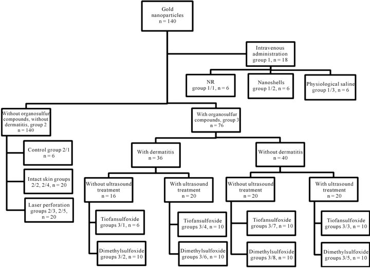
Figure 1. General scheme of the experiment.
ed 1 ml of physiological saline per kilogram of weight, the second −2 ml. Animals of the 2nd and 3rd experimental groups were conducted by optical coherence tomography (OCT). Scanning of each area was carried out twice: the first time—before effects, and the second time —after effects or elimination of the gel. Animals of the third experimental group were divided into 8 experimental subgroups. In animals of subgroups number 3/1, 3/2, 3/4, 3/6 was caused a resistant contact dermatitis, by a single topical application of 5% solution of 2,4-dinitrochlorobenzene. Duration of gel application on intact skin in all cases was 2 hours. Gel application on pre-perforated laser or affected by dermatitis skin duration was 15 minutes. As one of the methods for increasing the skin permeability during a superficial its previous microperforation was used. Source of radiation was erbium laser Palomar Lux 2940 (Palomar Medical Products Ltd., USA) with the following settings of radiation: the wavelength of 2940 nm, pulse energy of 0.5 - 3 J. Pulses had threestream structure. To enhance the tissue penetration of nanoparticles the phonophoresis was used (Dynatronics, Germany). Areas of rat skin after application of the drug were exposed to ultrasound effects at a frequency of 1 MHz, power 1.1 W and a duration of 2 minutes. In the study, the technique of optical coherence tomography was used to determine the areas of accumulation of gold nanoparticles in the skin. Studies carried out on the device model 022 OCP930SR (Thorlabs Inc, USA) at a wavelength 930 nm. Scanning of each area was carried out twice: the first time—before affects, and the second time —after gel elimination of the areas. Histological preparations of the skin and organs of animals were studied by standard techniques: with the coloring of histological sections of silver nitrate, the classical histological technique micropreparations skin and organs of the standard color. The histological preparations of skin and organs in the second group were additionally investigated by atomic force absorption spectrophotometry, and the third group —by the morphometric measurements. Morphometric data obtained were subjected to statistical analysis by calculating the arithmetic mean (M), the error of the arithmetic mean (± m), the accuracy of the difference of average (p) using the Student t-test at the 5% significance level.
3. Results
3.1. Characteristic of Pathological Changes in Internal Organs after Intravenous Injection Solutions of Gold Nanoparticles
After intravenous injection solution of gold nanoparticles experimentel animals, were observed for 14 days. During the experiment fatal outcome was not fixed. At the opening of the analyzed animals, taken out of the experiment on the 15th day of observation after injection of solutions of gold nanoparticles macroscopic changes in the liver and lungs were found. In the group of rats treated with NR, in 3 of 6 animals, liver inhomogeneous in color, with the underlined pattern, grain, with increased blood filling. In the experimental group of rats after injection of a solution of K-shells macroscopic changes in the liver and spleen were found. The liver is full-blooded; on the surface of the capsule multiple focuses of dull gray in color with a yellowish tint are marked. In the kidneys of the experimental group of animals there is some thickening of the cortex. The kidneys of the experimental animals do not vary in appearance from the kidneys of the control animals. In the lungs (of 1/6 rats) there are multiple macrofocal hemorrhage. In the control group of rats visible pathological changes were not observed. In the brain, of the animals exposed to injection of the solution NR in a volume of 2 ml/kg body weight on the 15th day hyperemia of blood vessels and cytolysis individual neurocytes are obsereved. Paracellular edema is determined in three out of six animals. In the liver of these animals there are decompensation of hepatic beams, the individual areas of hemorrhage and lysis of the nuclei of hepatocytes. In renal tissue hypochromic staining of the proximal convoluted tubules and glomeruli kollabirovanie. Histological examination on 15th day after injection of gold nanoparticles shows the ruptures of alveoli walls in the rat lungs. There are areas with thick walls interalveolar septums, where blood vessels and capillaries are enlarged and filled with densely erythrocytes. In a significant number of rats after intravenous injection necrosis of separate heaptocyte, and cells with nucleus pyknosis are found. The phenomenon of karyopyknosis, karyorhexis and hyperemia of blood vessels are detected. In the kidneys of some rats exposed to a solution of K-shells the individual convoluted tubules with lyse nuclei and vacuolated cytoplasm of epithelial cells are registered. In the spleen of rats treated with a solution of K-shells, there is a vasodilatation in the follicles and hyperemia of the red pulp.
3.2. Permeability of Intact Skin for Drugs of Gold Nanoparticles Using Organosulfur Compounds Dimethylsulfoxide and Tiofansulfoxide
According to the results of OCT imaging gold nanoparticles facilitate enhanced contrast tissue skin layers by using their in a drug composition containing dimethylsulfoxide. Such a contrast, according to the results, was absent on OCT-tomograms skin areas of experimental animals, which was applied drugs containing tiofansulfoxide. Histological findings confirmed the deeper penetration of gold nanoparticles using dimethylsulfoxide. The preparation containing dimethylsulfoxide, causes the penetration of nanoparticles to the reticular layer of dermis (Figure 2). By topical application the preparation of gold nanoparticles containing tiofansulfoxide, we have not found them to penetrate the subepidermal skin layers (Figure 3). Most of the nanoparticles were observed on the surface of the epidermis, as the remnants of the applied drug. In addition, a significant amount of them was observed on the surface of the cuticle and the inner epithelial sheath of hair, in the craters of hair follicles.
3.3. Pathological Assessment of the Skin and Internal Organs Changes after Laser Microperforation with the Application of Preparations of Gold Nanoparticles without Organosulfur Compounds
Accumulation of gold nanoparticles in the liver and spleen did not occur for any of the types of effects. In this regard, the images obtained by staining sections of liver and spleen in the control group did not differ from the images of the same organs sections in the experimental groups. Generally, the results of the study show that the physical effects on the skin as a factor for enhancing permeability of the nanoparticles of various sizes, can lead to destruction of both the surface (single layers of the epidermis) or deeper structural elements of the skin (collagen fibers papillary layer of dermis). This demonstrates the promising methods of application to the skin formulations with nanoparticles in solutions of organosulfur compounds that can enhance the total permeability of the skin without damaging effects.
3.4. Comparative Assessment the Permeability of Drugs of Gold Nanoparticles with Organosulfur Compounds under Ultrasound Treatment with Application to Intact Skin and Contact Chemical Dermatitis
Our studies have shown that the permeability of the skin of experimental animals with chemically induced contact dermatitis to gold nanoparticles in combination with organosulfur compounds with ultrasound treatment can reach the level of subcutaneous fat and subcutaneous muscle, which was not observed with the similar effects on intact skin. The permeability of the intact skin after application the preparation of nanoparticles with tiofansulfoxide under the ultrasound treatment provides a negligible and superficial penetration into the surface layers of the skin (Figure 4). At the same time, using the preparation of nanoparticles with dimethylsulfoxide and its application under the ultrasound treatment to the skin with chemical contact dermatitis revealed their deep penetration up to the subcutaneous fat and subcutaneous musculature (Figure 5). On Figure 4 the distribution of gold nanoparticles in skin layers is shown: 1-accumulations of gold nanoparticles in the reticular layer of dermis, and
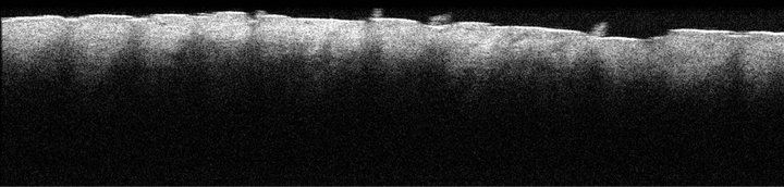
Figure 2. OKT-tomogram of skin of experimental group 3/8, with a local application to intact skin of gold nanoparticles in combination with dimethylsulfoxide.
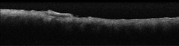
Figure 3. OKT-tomogram of skin of experimental group 3/7 with a local application to intact skin of gold nanoparticles in combination with tiofansulfoxide.
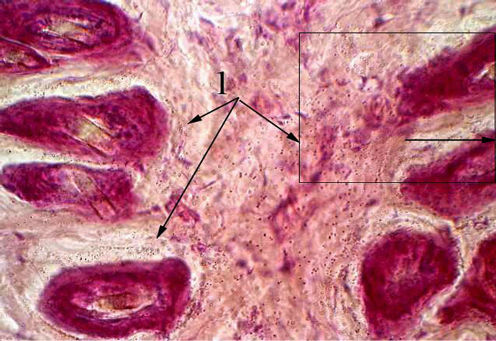
Figure 4. Skin of the experimental group 3/4 with a contact dermatitis and application of gold nanoparticles complexed with tiofansulfoxide and ultrasonic treatment. Stained with silver nitrate on Hacker G. W. and hematoxylin-eosin, microphotographs, × 600.
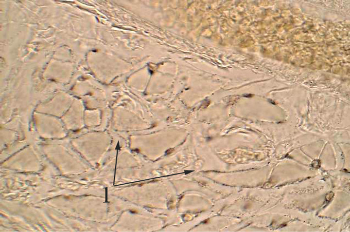
Figure 5. The subcutaneous muscles of the experimental group 3/6 with a contact dermatitis and application of gold nanoparticles complexed with dimethylsulfoxide. Stained with silver nitrate on Hacker G. W. Microphotographs, × 600.
in hair follicles. On Figure 5, best permeability of gold nanoparticles (1) is shown, which are defined essentially homogeneously in the dermis, concentrating in an area of the sebaceous glands. Also in significant quantities are defined in the subcutaneous muscles.
Thus, the overall assessment of the results the study of comparative efficiency enhance trasndermal permeability by organosulfur compounds gold nanoparticles under ultrasound treatment shows that the affected by chemical contact dermatitis skin is permeable better than intact. This is contradicting the data Baroli et al. [5], which demonstrated the worst permeability of the damaged skin as compared to intact. Perhaps the factor contributing these differences in our study is the additional ultrasound treatment. The nature of organosulfur compounds is also essential to enhancing transdermal penetration for nanoparticles. Mikromorphometry the depth of penetration of gold nanoparticles presented in Table 1 proves that dimethylsulfoxide has higher conducting properties than tiofansulfoxide both via intact and via the affected by dermatitis skin.
3.5. Comparison of Gold Nanoparticles Permeability after Application on the Rats Skin Areas with Artificially Induced Contact Dermatitis in Combination with Dimethylsulfoxide and Tiofansulfoxide and Ultrasound Treatment
During the experiment, it was found that topical application of gold nanoparticles complexed with dimethylsulfoxide on the skin with induced contact dermatitis with ultrasound treatment provides better permeability of the skin for the of gold nanoparticles than using gold nanoparticles in combination with at tiofansulfoxide with similar effects. Figure 6 shows the best permeability of gold nanoparticles, which are defined almost evenly in the dermis, concentrating in the area of the sebaceous glands. Also in significant quantities are defined in the subcutaneous muscles. Using gold nanoparticles in combination with dimethylsulfoxide provides deeper permeation of skin in the area of artificially induced contact dermatitis for the gold nanoparticles with ultrasound treatment significantly higher and reaches the subcutaneous fat and subcutaneous muscles, unlike the use of gold nanoparticles in complex with tiofansulfoxide which at similar effects provides little permeability. On Figure 7, gold nanoparticles are detected in the dermis, in the area of sebaceous glands and hair follicles.
4. Discussion
Among therapeutic agents, there are drugs including gold particles or its compounds [17]. It is known that gold can interact with proteases hydrolyzing collagen, as well as with elastase and other active components of connective tissue. Gold has anti-inflammatory, immunosuppressive, antitumoral, antifermental and some other therapeutic effects. Gold preparations used in the treatment of patients with rheumatoid and psoriatic arthritis, Felty’s syndrome, lupus erythematosus. One of the possible ways to improve the transdermal transport of nanocarriers can be
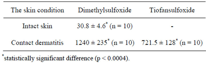
Table 1. The depth of skin penetration of drugs of gold nanoparticles with different organosulfur compounds under the ultrasound treatment (M ± m).
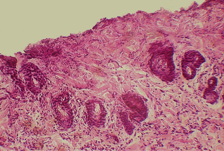
Figure 6. Skin of the experimental group 3/6 with a contact dermatitis and application of gold nanoparticles complexed with dimethylsulfoxide with ultrasound treatment. Hematoxylin-eosin, microphotograph, × 160.
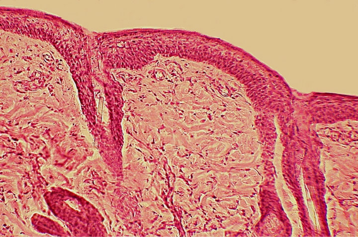
Figure 7. Skin of the experimental group 3/4 with a contact dermatitis and application of gold nanoparticles complexed with tiofansulfoxide with ultrasound treatment. Hematoxylin-eosin, microphotograph, × 160.
the use of organosulfur compounds with properties drastically improve the effect of the skin permeability for various chemical agents and drugs. Among such compounds should primarily attributed dimethylsulfoxide. The preparation according to modern concepts related to nanotechnology agent because its molecules are selectively carried out various drugs into the damaged tissues and cells. To date, the domestic and foreign scientific literature has more than 2000 messages on the study of the biological and clinical effects of that drug. There are reports on the use of dimethylsulfoxide and dissolved therein anticancer drugs topically in the form of applications, smearing the treatment of patients with melanoma, basal cell carcinoma, Bowen’s disease with a good clinical effect [18].
Another drug that is well proven for transdermal transport of drugs in veterinary practice is tiopfansuloxide. Tiofansuloxide as organosulfur compound has a high affinity to tissue composition of skin proteins because of its high sulfur content. Recent studies also prove promising use of the drug for transdermal transport of different sizes particles. According to the analysis of modern databases, both in foreign and domestic practice, there is not a precedent of the use of organosulfur compounds for targeted transdermal transport of nanocarriers. This suggests that this line of research to find the most promising ways to improve the transdermal nanocarriers transport of biological and pharmaceutical products. Laser microperforation provides good permeability of gold nanoparticles into the deeper layers of the skin, but causes severe destruction of tissue structures.
In the current study topical application of gold nanoparticles in combination with organosulfur compounds on the skin with artificially induced contact dermatitis and ultrasound treatment a higher permeability was shown than after application on intact skin. In the result of the experimental studies the higher permeability of gold nanoparticles in combination with DMSO with application on the skin with artificially induced contact dermatitis and ultrasound treatment was determined. The permeability of the gold nanoparticles in this method reaches the subcutaneous fat and subcutaneous muscles. Gold nanoparticles application on the skin with artificially induced contact dermatitis in combination with tiofansulfoxide and ultrasound treatment is achieved the skin permeability only to a mesh layer. As a result of our study was demonstrated that a significant modulatory effect on transdermal permeability of gold nanoshells size of 120 nm and 50 nm, the size of the nanorods and their intradermal transport possessed a number of physical and chemical effects. Regardless of the body (intact or damaged skin) a significant increase in the transdermal diffusion of nanoparticles without damaging effects can be achieved by using organosulfur compounds, and in particular dimethylsulfoxide under ultrasound treatment. The obtained results allow us to consider this line of research the most promising to find ways to improve the transdermal transport as functionalized as well as non-functionalized of gold nanoparticles for therapeutic purposes and nanocarries of pharmaceuticals and biologically active compounds.
5. Conclusion
In medicine and veterinary skin application for a long time used as one of the ways of the local delivery of drugs, and with the advent of well-studied ways transdermal diffusion of substances, the skin became widely used as an organ for their systemic delivery into the organism [19-21]. As a rule, even the prospect of nanoparticle-mediated delivery of drugs into the epidermis and dermis without damaging the integrity of the organ has little success. An analysis of special scientific literature on this issue shows that the statement that the nanoparticles greater than 20 nm in diameter, which do not penetrate into viable skin tissue is controversial. Compromising the integrity of the epidermal barrier in the intact or the diseased skin is ambiguous contribute permeability of the particles.
This study allowed us to estimate the permeability of the skin to the nanoparticles and their distribution in the organs of laboratory animals after topically application in combination with a variety of organosulfur compounds. It is proved that topically application of nanoparticles solutions with organosulfur compounds does not cause negative effects of the accumulation of nanoparticles in the internal organs, disorders of organ and tissue microcirculation and the development degenerative changes of these. General methodology and developed during of experiments techniques can be used to develop nanoparticle formulations of drugs for cosmetics, skin cancer treatment in human and veterinary medicine, the most common form of the epidermal malignancies.
REFERENCES
- R. M. Khayrullin, G. S. Terentyuk, M. V. Savenkova, et al., “Morphological Study of the Permeability the Skin and Placenta of the Rat for the Gold Nanoparticles,” International Journal of Experimental and Clinical Anatomy, Vol. 6, 2012, p. 84.
- L. Norlen, M. S. Roberts and K. A. Walters, “The Physical Structure of the Skin Barrier, Dermal Absorption and Toxicity Assessment,” CRC Press Inc., New York, 2008, pp. 37-68.
- K. Bhaskar, J. Anbu, V. Ravichandiran, et al., “Lipid Nanoparticles for Transdermal Delivery of Flurbiprofen: Formulation, in Vitro, ex Vivo and in Vivo Studies,” Lipids in Health and Disease, Vol. 8, No. 6, 2009, p. 6. doi:10.1186/1476-511X-8-6
- J. Luengo, B. Weiss, M. Schneider, et al., “Influence of Nanoencapsulation on Human Skin Transport of Flufenamic Acid,” Skin Pharmacology and Physiology, Vol. 19, No. 4, 2006, pp. 190-197. doi:10.1159/000093114
- B. Baroli, “Penetration of Nanoparticles and Nanomaterials in the Skin: Fiction or Reality?” The Journal of Pharmaceutical Sciences, Vol. 99, No. 1, 2010, pp. 21-50. doi:10.1002/jps.21817
- S. Battah, S. Balaratnam, A. Casas, et al., “Macromolecular Delivery of 5-Aminolaevulinic Acid for Photodynamic Therapy Using Dendrimer Conjugates,” Molecular Cancer Therapeutics, Vol. 6, No. 3, 2007, pp. 876-885. doi:10.1158/1535-7163.MCT-06-0359
- S. Battah, S. O’Neill, C. Edwards, et al., “Enhanced Porphyrin Accumulation Using Dendritic Derivatives of 5- Aminolaevulinic Acid for Photodynamic Therapy: An in Vitro Study,” The International Journal of Biochemistry & Cell Biology, Vol. 38, No. 8, 2006, pp. 1382-1392. doi:10.1016/j.biocel.2006.02.001
- S. Erdogan, “Liposomal Nanocarriers for Tumor Imaging,” Journal of Biomedical Nanotechnology, Vol. 5, No. 2, 2009, pp. 141-150. doi:10.1166/jbn.2009.1016
- N. Nishiyama, Y. Morimoto, W. D. Jang and K. Kataoka, “Design and Development of Dendrimer PhotosensitizerIncorporated Polymeric Micelles for Enhanced Photodynamic Therapy,” Advanced Drug Delivery Reviews, Vol. 61, No. 4, 2009, pp. 327-338. doi:10.1016/j.addr.2009.01.004
- G. Oberdorster, A. Maynard, K. Donaldson, et al., “Principles for Characterizing the Potential Human Health Effects from Exposure to Nanomaterials: Elements of a Screening Strategy,” Particle and Fibre Toxicology, Vol. 2, No. 8, 2005, p. 8. doi:10.1186/1743-8977-2-8
- T. W. Prow, W. A. Rose, N. Wang, et al., “BiosensorControlled Gene Therapy/Drug Delivery with Nanoparticles for Nanomedicine,” Advanced Biomedical and Clinical Diagnostic Systems III, Vol. 5629, No. 1, 2005, pp. 199-208. doi:10.1117/12.589422
- M. S. Roberts, “The Latest Science (Including Safety) on Nanotechnology and Skin Penetration,” FDA Public Hearing on the Science of Nanomaterials, Washington DC, 2006.
- V. V. Venuganti and O. P. Perumal, “Poly(Amidoamine) Dendrimers as Skin Penetration Enhancers: Influence of Charge, Generation, and Concentration,” Journal of Pharmaceutical Sciences, Vol. 98, No. 7, 2009, pp. 2345-2356. doi:10.1002/jps.21603
- M. S. Roberts, Y. Dancik, T. W. Prow, et al., “Non-Invasive Imaging of Skin Physiology and Percutaneous Penetration Using Fluorescence Spectral and Lifetime Imaging with Multiphoton and Confocal Microscopy,” European Journal of Pharmaceutics and Biopharmaceutics, Vol. 77, No. 3, 2011, pp. 469-488. doi:10.1016/j.ejpb.2010.12.023
- J. P. Ryman-Rasmussen, J. E. Riviere, N. A. Monteiro-Riviere, “Surface Coatings Determine Cytotoxicity and Irritation Potential of Quantum Dot Nanoparticles in Epidermal Keratinocytes,” Journal of Investigative Dermatology, Vol. 127, No. 1, 2007, pp. 143-153. doi:10.1038/sj.jid.5700508
- R. T. Tregear, “The Permeability of Mammalian Skin to Ions,” Journal of Investigative Dermatology, Vol. 46, No. 1, 1966, pp. 16-23.
- U. Munster, C. Nakamura, A. Haberland, et al., “RU 58841-Myristate—Prodrug Development for Topical Treatment of Acne and Androgenetic Alopecia,” Pharmazie, Vol. 60, No. 1, 2005, pp. 8-12.
- B. S. Kim, M. Won, K. M. Lee and C. S. Kim, “In Vitro Permeation Studies of Nanoemulsions Containing Ketoprofen as a Model Drug,” Drug Delivery, Vol. 15, No. 7, 2008, pp. 465-469. doi:10.1080/10717540802328599
- A. Vogt, B. Combadiere, S. Hadam, et al., “40 nm, But Not 750 or 1500 nm, Nanoparticles Enter Epidermal CD1a+ Cells after Transcutaneous Application on Human Skin,” Journal of Investigative Dermatology, Vol. 126, No. 6, 2006, pp. 1316-1322. doi:10.1038/sj.jid.5700226
- X. Wu, G. J. Price and R. H. Guy, “Disposition of Nanoparticles and an Associated Lipophilic Permeant Following Topical Application to the Skin,” Molecular Pharmaceutics, Vol. 6, No. 5, 2009, pp. 1441-1448. doi:10.1021/mp9001188
- Y. Zhao, M. B. Brown and S. A. Jones, “The Effects of Particle Properties on Nanoparticle Drug Retention and Release in Dynamic Minoxidil Foams,” International Journal of Pharmaceutics, Vol. 383, No. 1-2, 2010, pp. 277-284. doi:10.1016/j.ijpharm.2009.09.029
NOTES
*Corresponding author.