Open Journal of Radiology
Vol.4 No.2(2014), Article
ID:46906,6
pages
DOI:10.4236/ojrad.2014.42026
Short Report: A Sponge Phantom Provides a Homogeneous k-Space Pattern at MRI
Jan Menke
Radiology Center, University Hospital, Goettingen, Germany
Email: Menke-J@T-Online.de
Copyright © 2014 by author and Scientific Research Publishing Inc.
This work is licensed under the Creative Commons Attribution International License (CC BY).
http://creativecommons.org/licenses/by/4.0/


Received 24 April 2014; revised 20 May 2014; accepted 27 May 2014
ABSTRACT
Background: The spectral coverage of magnetic resonance (MR) sequences can be well assessed in k-space. However, many objects do not provide high signal intensities in the peripheral k-space. Purpose: To experimentally find a phantom that provides a homogeneous spectral pattern also at the high spatial frequencies of the k-space periphery. Material and Methods: Different phantoms were imaged on a 1.5 Tesla magnet, and the resulting MR images were viewed in k-space after fast Fourier transform. Results: Firstly, phantoms with a homogeneous physical structure were studied with a T2-weighted MR sequence, but they provided an inhomogeneous k-space pattern with dominant central low-frequency components. Secondly, phantoms with an inhomogeneous physical structure were studied. In this group, a water-soaked sponge showed a relatively homogeneous k-space pattern also at high spatial frequencies, owing to the fine porous structure. This sponge phantom can also be soaked with Gadolinium chelates for T1-weighted MR imaging. Conclusion: A simple sponge phantom provides a homogeneous k-space pattern, owing to its fine porous structure. This could be utilized in MR sequence development and for viewing the spectral coverage of MR sequences in k-space.
Keywords:Magnetic Resonance Imaging, Phantoms, Fourier Analysis, Spectrum Analysis, Computer-Assisted Image Processing

1. Introduction
All magnetic resonance (MR) images are primarily acquired in k-space that is the spatial frequency domain [1] -[3] . The central k-space represents low-frequency components and therefore determines image contrast and large-sized structures. The peripheral k-space represents high-frequency components and therefore determines fine image details such as vessel borders. In many imaged objects the central k-space has much higher magnitudes than the peripheral k-space. For MR sequence development and other purposes it could be useful to have an object with a homogeneous k-space pattern with magnitudes evenly distributed across the k-space also at its periphery. The purpose of this experimental study was to find such a phantom. It has been the preliminary study of research about viewing MR images in k-space, which is well possible [4] .
2. Material and Methods
2.1. Overview
MR imaging was performed on a 1.5 Tesla MR system (Symphony, Siemens Medical Solutions, Erlangen, Germany) using the standard head-coil. K-space visualization was performed with fast Fourier transform (FFT) [1] [2] [5] . Different phantoms and materials were tested to see which one provides the most homogeneous k-space pattern. Such homogeneous k-space pattern was assessed, if the magnitudes at the k-space center were comparable to the magnitudes at the k-space periphery.
2.2. Data Conversion and Processing
This study utilized MR magnitude images as provided by the MR system. Internally they are based on raw frequency-domain measurement data that are post-processed to optimize the image quality. The MR images were converted from DICOM format to ANALYZE format using the freeware medical image viewer MRIcro (MRIcro 1.40, http://www.mricro.com). This program was also used for viewing the original MR images as well as their k-space counterparts. These k-space images were generated by fast Fourier transform of the MR magnitude images back to k-space, utilizing a proprietary C++ program (Visual C++ 6.0, Microsoft Corporation) that incorporates the free software FFTW (FFTW 3.1.2., http://www.fftw.org) [5] . They were named the “back-transformed k-space images” and were stored in the ANALYZE format with complex numbers and 32 bit accuracy both for the real and the imaginary part [5] . In this article, complex k-space is shown with magnitude images on a logarithmic scale.
2.3. Testing of Different Phantoms
The study aim was to find a phantom with a homogeneous spectral pattern in k-space. Experiments were performed with different phantoms to study the effect of object structure on spectral k-space properties. Phantom testing was generally performed with a T2-weighted two-dimensional turbo spin echo sequence (TR 4100 ms, TE 98 ms, flip angle 180˚, matrix size 512 × 384, pixel size 0.45 × 0.45 mm) without and with elliptic filtering. MR imaging without elliptic filtering was done to visualize the complete k-space. Additional MR imaging with elliptic filtering imprinted a contrast in the back-transformed k-space images that originated not from the measured object but from the MR sequence. Phantoms with homogeneous and inhomogeneous structure were evaluated (Table 1). The study started with imaging free-in-air to have a measure for basic image noise. The phantom experiments proceeded with imaging a small water-filled sphere, a small water-filled cube, a large spherical phantom used for quality assurance (QA), and a QA-resolution phantom with rods. In the study design it was planed to include further phantoms by evolution of the method, which inherently is an interactive process between experimental results and consecutively refined methods. Among those phantoms were a cup with fresh corn, a sponge soaked with water, and a pack of such wet sponges. To fit into the field of view, the pack of sponges was imaged with an enlarged pixel size of 0.9 × 0.9 mm. The k-space homogeneity of the different tested phantoms was visually assessed in comparison to each other at an image gallery.
3. Results
3.1. Phantoms with Homogeneous Physical Structure
Imaging free-in-air exhibits low-magnitude Rician noise in the MR image with a corresponding noise spectrum in the back-transformed k-space magnitude image (Figure 1(a)). The small water sphere shows concentric rings in k-space similar to a droplet in a water basin (Figure 1(b), two-dimensional sombrero function). In contrast, the small water cube exhibits a very different harmonic with a cross-like spectral pattern comparable to two orthogonal sinc functions (Figure 1(c)). Comparison of Figure 1(b) and Figure 1(c) indicates that an object’s

Table 1. Phantoms.
*QA, quality assurance phantoms, provided by the manufacturer of the MR system.
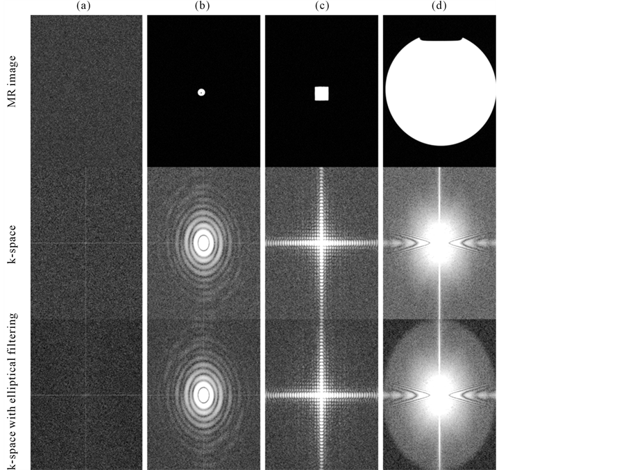
Figure 1. Phantoms with homogeneous physical structure. (a) air; (b) small water sphere; (c) small water cube; (d) large spherical phantom for quality assurance. These phantoms with homogeneous structures were imaged using a T2-weighted turbo spin echo sequence without and with elliptic filtering. The MR images are shown in the upper row, corresponding backtransformed k-space images without elliptic filtering in the central row, and back-transformed k-space images with elliptic filtering in the lower row. Only the largest phantom ((d) lower row) exhibits the elliptic filtering that was applied. For further details, see Results.
shape has a strong influence on the spectral power distribution in k-space. In the lower row of Figure 1(a) to Figure 1(c) the elliptic filtering is not visible. Only the largest phantom provided sufficient high spatial frequencies to display the elliptic filtering of the chosen MR sequence (Figure 1(d)). However, the bright central peak in the k-space magnitude image indicates a dominance of low spatial frequencies in this homogeneously structured large spherical phantom.
3.2. Phantoms with Inhomogeneous Physical Structure
The QA-resolution phantom also shows the sequence’s elliptic filtering in k-space (Figure 2(a)). However, there are harmonic stripes in the k-space image, originating from the parallel alignment of rods in the MR image (Figure 2(a)). In comparison, a bowl filled with fresh corn has a more randomly arranged structure in the MR image and shows no harmonic spectral components in k-space (Figure 2(b)). However, there are still not many high-frequency structures in this MR image, indicated by an under-representation of peripheral spectral magnitude in the corresponding k-space image (Figure 2(b)). In contrast, a wet sponge does have such high-frequency components, exhibits a relatively homogeneous spectral pattern in k-space, and shows the sequence’s elliptic filtering well (Figure 2(c)). Figure 2(d) shows that, when a larger field of view is required, a pack of wet sponges may be used.
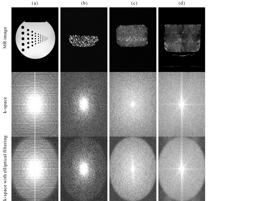
Figure 2. Phantoms with inhomogeneous physical structure. (a) resolution phantom with rods for quality assurance; (b) bowl with fresh corn; (c) wet sponge; (d) pack of six wet sponges (MR images in the upper row; corresponding back-transformed k-space images without elliptic filtering in the central row, and back-transformed k-space image with elliptic filtering in the lower row). These phantoms with inhomogeneous structures were imaged using a T2-weighted turbo spin echo sequence without and with elliptic filtering. The sponge phantoms (c) and (d) exhibit the most homogeneous spectral pattern and show the elliptic filtering to best effect. Further details are given in Results.
4. Discussion
4.1. Summary of Findings
In summary, phantoms with a homogeneous physical structure provided an inhomogeneous k-space pattern with dominance of central low-frequency components. In contrast, phantoms with an inhomogeneous physical structure and fine details provided a more homogeneous k-space pattern with higher magnitude of peripheral highfrequency components.
4.2. Phantoms with Homogeneous Physical Structure
One might expect that an object with a homogeneous physical structure should also provide a homogeneous k-space pattern. However, a homogeneously structured object contains only a small fraction of small details that contribute to the high-frequency parts of the k-space, and therefore the low-frequency component is dominant, as observed in the large spherical QA-phantom (Figure 1(d)). Imaging free-in-air provides a seemingly homogeneous k-space pattern, but this is just noise and no MR signal (Figure 1(a)). Although interesting, the MR experiments with a circular and a squared point source also showed no homogeneous k-space pattern (Figure 1(b) and Figure 1(c)). These both later experiments showed that the k-space pattern much depends on the object’s shape. At first glance the ring-like k-space pattern of the sphere (Figure 1(b)) seems to be completely different from the k-space pattern of the water-cube that has characteristics of two orthogonal sinc functions (Figure 1(c)). However, if the cube would be fast-rotating then it would look like a sphere. The same applies to the k-space, where the fast-rotating sinc functions would look like the concentric rings of a sombrero function.
4.3. Phantoms with Inhomogeneous Physical Structure
In the image gallery of Figure 2 the homogeneity of k-space pattern increased from the left (QA-resolution phantom) to the right (pack of wet sponges). Similarly, the amount of fine object details increased from the left to the right. The sponge is dominated by fine details due to its porosity. These fine details contribute to the k-space periphery. At the same time the whole sponge also is a large-sized object and therefore has central lowfrequency components in k-space. The sponge phantom exhibits no disturbing harmonics (as observed in the QA-resolution phantom) but shows a homogeneous k-space pattern. This can be explained by the fact that the sponge’s pores are not systematically arranged in a grid-like structure, but they are somewhat randomly arranged at close inspection.
4.4. Sponge Phantom Providing a Homogeneous k-Space Pattern
In this study, the sponge phantom evolved from experiments and exhibits a relatively homogeneous k-space pattern. The sponge is a porous medium with a somewhat random physical structure, which is the reason why it provides a relatively homogeneous mixture of low and high spatial frequencies at MR imaging. It is likely that other porous media may be similarly suitable.
4.5. Applications of the Method
The sponge phantom has already been used for viewing the spectral pattern of MR images in k-space, which is well possible [4] . This allows for a visual understanding of k-space not only in two-dimensional MR imaging, but also in the more complex three-dimensional MR imaging [4] . The sponge phantom may be also be used in the development and testing of new MR sequences in two-dimensional as well as three-dimensional imaging, and for sequence parameter optimization. For T2-weighted imaging the sponge can be simply soaked with water. For contrast-enhanced T1-weighted imaging the sponge can be soaked with a Gadolinium chelates solution [4] . Other fluids can also be used for soaking, if a corresponding homogeneous k-space pattern is desired.
4.6. Study Limitations
Many more phantoms could be imaged, and possibly other phantoms will provide an even more homogeneous k-space pattern than the presented sponge phantom. However, the sponge phantom has the advantage that it can be soaked with different fluids, including Gadolinium chelates for T1-weighted imaging. Furthermore, the sponge phantom is cheap and easily available from the shop.
5. Conclusion
A sponge phantom can provide a homogeneous k-space pattern at MR imaging, owing to its fine porous structure. This may be utilized for sequence development, sequence parameter optimization, and to enhance a visual understanding of k-space, particularly in three-dimensional MR imaging.
References
- Dhawan, A.P. (2003) Medical Image Analysis. IEEE Press, Wiley-Interscience, Hoboken, New Jersey, USA, 27-29, 37-39, 62-91, 111-174.
- Mezrich, R. (1995) A Perspective on k-Space. Radiology, 195, 297-315. http://dx.doi.org/10.1148/radiology.195.2.7724743
- Paschal, C.B. and Morris, H.D. (2004) K-Space in the Clinic. Journal of Magnetic Resonance Imaging, 19, 145-159. http://dx.doi.org/10.1002/jmri.10451
- Menke, J., Helms, G. and Larsen, J. (2010) Viewing the Effective k-Space Coverage of MR Images: Phantom Experiments with Fast Fourier Transform. Magnetic Resonance Imaging, 28, 87-94. http://dx.doi.org/10.1016/j.mri.2009.05.027
- Frigo, M. and Johnson, S.G. (2005) The Design and Implementation of FFTW3. Proceedings of IEEE, 93, 216-231. http://dx.doi.org/10.1109/JPROC.2004.840301