Open Journal of Pediatrics, 2011, 1, 34-36
doi:10.4236/ojped.2011.13009 Published Online September 2011 (http://www.SciRP.org/journal/ojped/ OJPed
).
Published Online September 2011 in SciR es. http://www.scirp.org/journal/OJPed
Anterior and posterior urethral valves: a rare association
Sahnoun Lassaad1*, Lamiri Rachida1, Ben Saad Manel1, Salem Randa2, Mekki Mongi1,
Maazoun Kais1, Krichene Imed1, Jamila Chahed1, Nouri Abdellatif1
1Department of Pediatric Surgery, Fattouma Bourguiba Hospital, Monastir, Tunisia;
2Department of Radiology, Fattouma Bourguiba Hospital, Monastir , Tunisia.
Email: *lassaad.sahnoun@rns.tn
Received 7 May 2011; revised 15 June 2011; accepted 22 July 2011.
ABSTRACT
Association of anterior and posterior urethral valve is
a rare congenital anomaly causing lower urinary
tract obstruction in children. This paper highlights
our experience with a two-month-old boy with double
urethral valves. Antenatal ultrasonography failed to
detect any abnormality. At the age of 1 month, he
presented with urinary dribbling, straining at mic-
turition, and febrile urinary tract infection. Clinically,
the urethra was dilated at micturition. Diagnosis was
confirmed by retrograde urethrogram and urethro-
scopy. Endoscopic ablation of both valves was done
by electrocautery hook.
Keywords: Anterior Urethral Valve; Posterior Urethral
Va lve; Renal Disorders; Double Infravesical Obstruction
1. INTRODUCTION
Posterior urethral valve (PUV) is the most common
cause of congenital obstructive uropathy in boys. How-
ever anterior urethral valve (AUV) is an uncommon
cause of a lower urinary tract obstruction. Association of
the two is extremely rare and only four cases have been
reported [1]. In this study, we describe our case.
2. CASE REPORT
A two-month-old boy with a history of febrile urinary
tract infection at the age of one month, was referred to
our department for evaluation of continuous dribbling of
urine and straining at micturition. Clinical examination
revealed a palpable bladder. The external genitalia were
normal. A swelling was palpable in the region of the
bulbar urethra. Urinalysis, urine culture, and renal func-
tion tests were normal. The catheterization was not suc-
cessful because the catheter did not pass through the
urethra. An anterior urethral valve or urethral diverticu-
lum was recognized.
Renal ultrasonography showed a thick walled bladder
without hydronephrosis (Figure 1).
A retrograde cysto-urethrogram revealed the double
infravesical obstruction and the dilated urethra sugges-
tive of concomitant anterior and posterior urethral valves.
The AUV was located at the bulbar urethra (Figure 2).
There was no vesicoureteral reflux.
With the patient under general anesthesia, an endo-
scopic ablation of both valves was done by electrocau-
tery hook. The bladder appeared trabeculated. An urinary
catheter was left in place.
Postoperatively, the patient was able to pass urine
with good stream and had normal stable renal function.
He was discharged 12 days after surgery.
Three months later, the patient presented with urinary
infection and dribbling. A renal ultrasou nd scan failed to
show any abnormality. On cystoscopy, there were obs-
Figure 1. Thic kened bl adder wal l.
S. Lassaad et al. / Open Journal of Pediatrics, 2011, 1, 34-36 35
Figure 2. Voiding cystography showing double obstruction and
urethral dilatation: (a) anterior urethral valve; there is a bul-
bous dilatation of the anterior urethra ending in a smooth bulge,
which marks the site of the AUV; (b) posterior urethral valve:
an elongated and dilated posterior urethra with abrupt nar-
rowing.
tructting anterior and posterior residual urethral valves.
An electrocautery hook was done to both the residual
valves.
The postoperative course was uneventful. The patient
is now 2 years old and continues to void with good
stream.
3. DISCUSSION
The association of anterior urethral valve (AUV) with
posterior urethral valve (PUV) is rare; only 4 cases have
been reported [1-3].
PUV is seven times more common than AUV; how-
ever, the obstructive effects of AUV can be equally dam-
aging.
The association of AUV and PUV could not be ex-
plained on an embryological basis as the source of AUV
and PUV development is different.
The etiology of AUV is still controversial, but faulty
union of the glandular and penile urethral segments, in-
complete formation of the corpus spongiosum, congeni-
tal cystic dilation of the periurethral glands, and abortive
attempt at urethral duplication have been proposed as
possible mechanisms [1,2,4].
AUV can be located anywhere distal to the membra-
nous urethra and, in terms of distribution; these valves
are most common in the bulbar urethra (40%) [1,2,5].
The clinical manifestation of AUV and PUV is highly
variable and depends on patient age and degree of ob-
struction. It may range from minimal obstruction to se-
vere obstruction with bilateral severe hydroureteroneph-
rosis, end-stage renal disease, and even bladder rupture
[1,2,6,7]. Physical examination fin di n gs ca n be no rmal.
Prenatal ultraso nography may sug gest the diagnosis of
AUV and PUV through visualization of urethral dilata-
tion and hydronephrosis, oligohydramnios and bladder
distension highly suggestive of infravesical obstruction.
(a) In our case, the antenatal and the postnatal ultrasono-
graphy were normal probably because the obstruction was
not complete.
(b)
Cystourethrography is the mainstay of the diagnosis.
It can reveal a thickened trabeculated bladder, a dilated
or elongated posterior urethra, a dilation of the anterior
urethra, and a hypertrophied bladder neck. Diverticula
and vesicoureteral reflux may also be present .
The spectrum of severity of AUV ranges from mild
urethral dilatation to bilateral hydronephrosis with renal
insufficiency (Figure 3) [8,9].
Figure 3. The four types of AUV.
C
opyright © 2011 SciRes. OJPed
S. Lassaad et al. / Open Journal of Pediatrics, 2011, 1, 34-36
Copyright © 2011 SciRes.
36
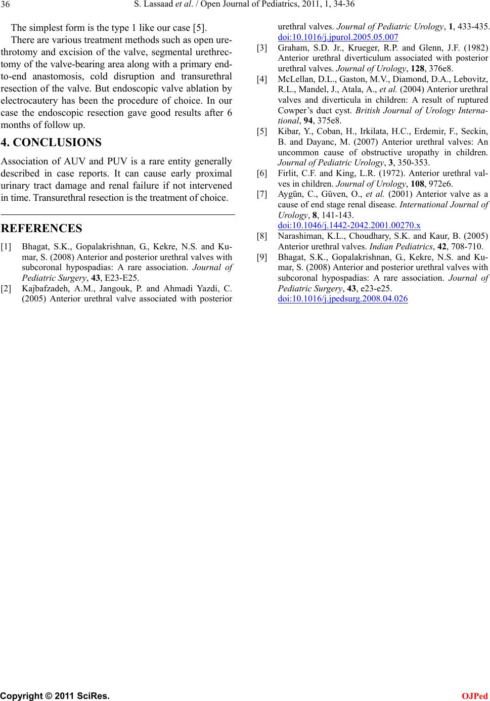
The simplest form is the type 1 like our case [5].
There are various treatment methods such as open ure-
throtomy and excision of the valve, segmental urethrec-
tomy of the valve-bearing area along with a primary end-
to-end anastomosis, cold disruption and transurethral
resection of the valve. But endoscopic valve ablation by
electrocautery has been the procedure of choice. In our
case the endoscopic resection gave good results after 6
months of follow up.
OJPed
4. CONCLUSIONS
Association of AUV and PUV is a rare entity generally
described in case reports. It can cause early proximal
urinary tract damage and renal failure if not intervened
in time. Transurethral resection is the treatment of choice.
REFERENCES
[1] Bhagat, S.K., Gopalakrishnan, G., Kekre, N.S. and Ku-
mar, S. (2008) Anterior and posterior urethral valves with
subcoronal hypospadias: A rare association. Journal of
Pediatric Surgery, 43, E23-E25.
[2] Kajbafzadeh, A.M., Jangouk, P. and Ahmadi Yazdi, C.
(2005) Anterior urethral valve associated with posterior
urethral valves. Journal of Pediatric Urology, 1, 433-435.
doi:10.1016/j.jpurol.2005.05.007
[3] Graham, S.D. Jr., Krueger, R.P. and Glenn, J.F. (1982)
Anterior urethral diverticulum associated with posterior
urethral valves. Journal of Urology, 128, 376e8.
[4] McLellan, D.L., Gaston, M.V., Diamond, D.A., Lebovitz,
R.L., Mandel, J. , Atal a, A., et al. (2004) Anterior urethral
valves and diverticula in children: A result of ruptured
Cowper’s duct cyst. British Journal of Urology Interna-
tional, 94, 375e8.
[5] Kibar, Y., Coban, H., Irkilata, H.C., Erdemir, F., Seckin,
B. and Dayanc, M. (2007) Anterior urethral valves: An
uncommon cause of obstructive uropathy in children.
Journal of Pediatric Urology, 3, 350-353.
[6] Firlit, C.F. and King, L.R. (1972). Anterior urethral val-
ves in children. Journal of Urology, 108, 972e6.
[7] Aygün, C., Güven, O., et al. (2001) Anterior valve as a
cause of end stage renal disease. International Journal of
Urology, 8, 141-143.
doi:10.1046/j.1442-2042.2001.00270.x
[8] Narashiman, K.L., Choudhary, S.K. and Kaur, B. (2005)
Anterior urethral valves. Indian Pediatrics, 42, 708-710.
[9] Bhagat, S.K., Gopalakrishnan, G., Kekre, N.S. and Ku-
mar, S. (2008) Anterior and posterior urethral valves with
subcoronal hypospadias: A rare association. Journal of
Pediatric Surgery, 43, e23-e25.
doi:10.1016/j.jpedsurg.2008.04.026