Open Journal of Pediatrics, 2011, 1, 21-26
doi:10.4236/ojped.2011.13006 Published Online September 2011 (http://www.SciRP.org/journal/ojped/ OJPed
).
Published Online September 2011 in SciR es. http://www.scirp.org/journal/OJPed
The child with chronic cough: when does double-channel pH
monitoring rule out gastroesophageal reflux?
Richard Kitz1*, Peter Ahrens2, Olaf Eickmeier1, Hansjosef Boehles1, Markus A. Rose1
1Children’s Hospital, Goethe University, Frankfurt, Germany;
2Prinzessin Margaret Children’s Hospital, Darmstadt, Germany.
Email: *R.Kitz@ckhf.de
Received 13 May 2011; revised 3 July 2011; accepted 19 July 2011.
ABSTRACT
Background: gastroesophageal reflux (GER) plays a
major role in the pathogenesis of pediatric chronic
airway disease. Esophageal pH-monitoring (epHM) is
the diagnostic gold standard for acid GER. To date,
there are no cut-off values for chronically coughing
children ruling out relevant GER. Methods: 24-hour,
double-channel epHM was performed in 549 children
(3 months to 16 years old) with chronic pulmonary
disease. We stratified according to age as follows:
1.5 years, > 1.5 - 4 years, > 4 - 8 years, > 8 < 12 years
and > 12 - 16 years. Following parameters were cal-
culated for both channels: total number of reflux epi-
sodes, number of reflux episodes > 5 minutes, dura-
tion of the longest reflux episode, and reflux index.
Results: according to the above given age classifica-
tion, the median number of reflux episodes in the
lower esophagus was 31, 27, 32, 34, and 42 and for
the upper esophagus 20, 13, 15, 14, and 11 respec-
tively. The median reflux index at the distal esopha-
gus was 2.55, 2.1, 2.3, 2.15, and 1.9; at the upper
esophagus it was 1.4, 1.0, 1.1, 0.9, and 0.6 respectively.
Conclusions: our data contribute useful support to
the evaluation of pediatric airway disease. We pro-
vide reference values for decisions in the exploration
of children with airway disorders and suspected
GER.
Keywords: Gastroesophageal Reflux; Pediatric Airway
Diseases; pH-Moni t o ri n g; C hronic Aspiration
1. INTRODUCTION
Numerous studies in chronic airway disease emphasize
the etiological role of gastroesophageal reflux (GER),
some of them also proving a significant epidemiological
correlation. The prevalence of respiratory symptoms
among patients with GER is high. Monitoring of eso-
phageal pH (epHM) is considered to be the gold stan-
dard in detecting classic GER. The North American So-
ciety for Pediatric Gastroenterology and Nutrition rec-
ommends performing epHM in patients with persistent
asthma, even in the absence of symptoms of GER, and
recommends medical treatment if epHM demonstrates
an increased frequency or duration of esophageal acid
exposure [1]. In asthmatic children with GER, antacid
treatment resulted in a significant reduction in asthma
medication [2].
Astonishingly, data upon normal values of acid expo-
sure of the esophagus in children with unspecific pul-
monary symptoms, measured by pH-monitoring, are
scarce. There are only three studies providing data on
multiple sites measuring in the esophagu s: Bagucka et al.
provided double-channel pH monitoring normal ranges
of gastroesophageal reflux parameters in the upper
esophagus in 200 infants aged 0.5 - 17 months, referred
with suspected GER disease [3]. Sondheimer [4] meas-
ured pH at three levels in the esophagus in only 11 chil-
dren without pathological GER and in 14 with patho-
logical GER. In an older study [5] on 27 healthy children
and adolescents (9.3 - 17.3 years old, mean 13.1) by
Gustafsson in 1988, values for the upper probe, 15 cm
above the cardia, were given as follows: RI 0.6%, num-
ber of reflux episodes in 19/24 h; the duration of longest
reflux episode being 3.3 minutes.
To date, there are no reference values available for the
upper esophagus in children with unspecific pulmonary
symptoms, but without typical gastrointestinal symptoms
of GER (e.g., vomiting). This is the first study to vali-
date the standard method in detecting GER in these chil-
dren from infancy to adolescence.
2. MATERIALS AND METHODS
Patients. At our institution, children undergo epHM in
search of evidence of GER when presenting chronic
cough throughout the year, recurrent wheezing, or re-
current bronchitis/pneumonia. Among those and over a
period of six years, 549 children (aged 3 months to 16
R. Kitz et al. / Open Journal of Pediatrics, 2011, 1, 21-26
22
years) had a normal epHM according to ESPGHAN-
protocol criteria (reflux index at the distal esophagus <
5%; see below) and were included into this study [6].
Exclusion criteria were other serious chronic diseases
(e.g., cystic fibrosis, perennial allergic disease, or food
allergy), neurological disorders with the risk of dys-
phagia, immunodeficiency, malformations of the tra-
cheobronchial tree, or chronic foreign body aspiration.
At the time of the study, none of the patients was on acid
suppression therapy or on theophylline antiasthmatic
treatment.
Examinations were performed on a walk-in basis. In-
formed consent was obtained from the patients or the
patients’ guardians ahead of the examination. Guardians
were asked to withhold their children’s normal diet and
allow customary sleeping positions throughout the 24-
hours period. Ethical approval was not obtained because
of the study’s retrospective design. For ethical reasons,
we renounced examining a heal t hy control group.
Continuous epHM was performed according to the
ESPGHAN standardized protocol. A monocrystalline
antimony double-channel pH probe (Medtronic Synec-
tics Medical, Sweden) was calibrated in buffers of pH
7.01 and pH 1.07 and then placed transnasaly into the
esophagus. The distance of the two pH sensors on the
probe was adjusted according to the body height of the
patient, using our previously published formula [7]. Af-
ter adjusting the probe for an optimal position within the
esophagu s, with th e use of a control chest x-ray, with the
upper probe position of the sensors between the clavicles.
The lower probe position was set in the distal esophagus.
Therefore we used three different distances between the
sensors: 5 cm in patients with a body height of 80 cm,
10 cm in patients with a body height of 120 cm, and 15
cm in patients with a body height of > 120 cm. This fa-
cilitates interindividual comparability for epHM. The
probes were then connected to a portable dig ital reco rder
(Digitrapper MK III, Synectics Medical AB, Sweden).
Patients and their guardians were then asked to keep a
diary for the next 24 hours. This diary included data on
the time and kind of consumed meals, beverages and
drugs as well as posture. After 24 hours, data were ana-
lyzed by the software “Esophogram®” (Synectics Medi-
cal AB, Sweden). Reflux episodes were defined as a
decrease of esophageal ph below 4 for longer than five
seconds followed by an increase of pH for minimum of
pH 4.5, thus avoiding oscillating phenomena. This en-
abled us to detect the number of reflux episodes at each
sensor in the past 24 hours, the longest reflux episode
and the number of long lasting reflux episodes (> 5 min-
utes). The summarized time of all reflux episodes di-
vided by the total recording time (in general over a pe-
riod of 24 hours) is the “reflux index” (RI).
Statistical analysis:
Statistical analysis was done with the software pack-
age SPSS for Windows® version 11.0 (SPSS Inc., Chi-
cago, Illinois/USA). Data of the study population were
tested for Gaussian distribution, however, Kolmogorov-
Smirnoff´s delta showed no normal distribution. Thus,
we decided to choose nonparametrical statistic testing.
Medians and interquartile ranges (IQR) between the
25% and 75% percentile, provide relevant descriptive
parameters. A Kruskal-Wallis analysis was performed to
compare epHM parameters of all age groups. In order to
facilitate a comparison with literature data, we addition-
ally provide mean values as well as standard deviations.
3. RESULTS
Among 549 patients with normal lower reflux index (RI)
values (< 5%), 126 were younger than 18 months, 148
were 18 months to 4 year s old, 195 were 4 to 8 year s old,
47 were 8 to 12 years old, and 33 were 12 to 16 years of
age.
Results for our four epHM parameters, studied in pa-
tients with distal RI < 5%, are listed in Table 1. Overall,
there was less reflux in the upper esophagus than in the
lower esophagus. Infants between 3 to 18 months of age
had the highest reflux activity of all examined age gr-
oups. As expected, physiological reflux diminishes as
maturation of the cardia proceeds. In all age groups, re-
flux episodes lasting longer than five minutes are scarce
and longer reflux episodes tended to occur in older chil-
dren, while the total number of reflux episodes was
higher in younger childr e n.
When comparing data of the age groups older th an 18
months, we only found a trend toward slight reflux ac-
tivity depending on age. No statistical significant differ-
ences for the epHM parameters could be found in the
Kruskal-Wallis analysis. Therefore, we decided to pool
children between the ages of 18 months to 16 years as
one group (Table 2). This was also true for children with
higher lower esophageal RIs (data not shown).
In infants younger than 18 months of age and normal
RIs in the lower esophagus, the median number of reflux
episodes in the upper esophagus was 21 (IQR 10 - 31)
over a 24-hour period. While the longest reflux episode
lasted 4 min. (IQR 2.0 - 8.2 minutes), the median num-
ber of episodes lasting more than 5 minutes was 0 (IQR
0 - 1) over a 24-hour period. The resulting reflux index
(RI) in the upper esophagus was 1.4 (IQR 0.5 - 2.5).
Children between the ages of 18 months to 16 years had
a total number of 14 (IQR 7 - 24) reflux episodes in the
upper esophagus. Their longest episode lasted 4 minutes
(IQR 2 - 8 min). The median number of reflux episodes
longer than 5 minutes was 0 (IQR 0 - 1). The resulting
reflux index RI was 1.0 (IQR 0.4 - 2) in this age group.
C
opyright © 2011 SciRes. OJPed
R. Kitz et al. / Open Journal of Pediatrics, 2011, 1, 21-26 23
Table 1. Upper and lower esophageal pH monitoring parameters in patients with normal di stal reflux index (RI) values (< 5%) strati-
fied for patient age.
Age (years) 3 - <18 months
n = 126
1.5 - <4 years
n = 148
4 - <8 years
n = 195
8 - <12 years
n = 47
12 - 16 years
n = 33
Mean 0.87 2.60 5.51 9.46 13.91
SD 0.34 0.74 1.10 1.10 1.14
25% quartile 0.59 1.86 4.55 8.59 13.04
Median 0.85 2.46 5.33 9.16 13.70
SEM 0.03 0.06 0.08 0.16 0.20
75% quartile 1.22 3.28 6.36 10.28 14.75
No. of reflux episodes
upper lower upper lower upper lower upper lower upper upper
Mean 21.80 31.96 17.20 29.75 16.93 35.50 19.53 47.48 15.30 44.82
SD 14.90 18.04 13.56 19.31 12.75 26.27 19.78 64.88 14.03 24.65
25% quartile 10.00 21.00 7.00 17.25 7.00 18.00 8.00 16.00 6.00 27.50
Median 20.50 31.00 13.00 27.00 15.00 32.00 14.00 34.00 11.00 42.00
SEM 1.33 1.61 1.11 1.59 0.91 1.88 2.89 9.36 2.44 4.29
75% quartile 31.00 40.25 26.75 39.00 23.00 47.00 26.00 53.75 23.50 55.50
p (dist./prox.) < 0.01 < 0.01 < 0.01 0.01 < 0.01
No. reflux-episodes 5 minutes
Mean 0.70 0.85 0.95 1.00 0.76 0.99 0.55 0.77 0.81 1.15
SD 1.20 1.07 1.49 1.18 1.07 1.02 1.33 0.90 1.65 1.32
25% quartile 0.00 0.00 0.00 0.00 0.00 0.00 0.00 0.00 0.00 0.00
Median 0.00 0.00 0.00 1.00 0.00 1.00 0.00 0.50 0.00 1.00
SEM 0.10 0.09 0.12 0.10 0.08 0.07 0.19 0.13 0.29 0.23
75% quartile 1.00 1.00 1.00 1.75 1.00 2.00 1.00 1.00 1.00 2.00
p (dist./prox.) < 0.01 < 0.01 < 0.01 0.01 < 0.01
Longest reflux episode
Mean 6.20 7.18 6.88 7.43 8.10 8.18 6.42 7.37 12.39 8.76
SD 6.50 5.99 7.80 6.35 11.21 7.10 9.67 6.78 23.41 8.42
25% quartile 2.00 3.00 2.00 3.00 2.00 3.00 1.00 3.00 1.00 2.50
Median 4.00 5.00 5.00 6.00 5.00 6.00 3.00 4.50 3.00 6.00
SEM 0.58 0.54 0.64 0.52 0.80 0.51 1.41 0.98 4.08 1.47
75% quartile 8.20 11.00 8.00 10.00 9.00 11.00 7.00 10.50 13.50 11.00
p (dist./prox.) < 0.01 < 0.01 < 0.01 0.01 < 0.01
Reflux-Index (%)
Mean 1.70 2.43 1.64 2.20 1.51 2.31 1.37 2.19 1.96 2.44
SD 1.60 1.43 1.97 1.37 1.59 1.33 1.93 1.41 3.46 1.50
25% quartile 0.50 1.20 0.40 1.00 0.50 1.20 0.30 1.12 0.20 1.15
Median 1.40 2.55 1.00 2.10 1.10 2.30 0.90 2.15 0.60 1.90
SEM 0.14 0.13 0.16 0.11 0.11 0.09 0.28 0.20 0.60 0.26
75% quartile 2.50 3.52 2.17 3.30 2.00 3.30 1.60 3.40 2.45 4.00
p (dist./prox.) < 0.01 < 0.01 < 0.01 0.01 < 0.01
SEM = standard error of means; SD = standard deviation.
C
opyright © 2011 SciRes. OJPed
R. Kitz et al. / Open Journal of Pediatrics, 2011, 1, 21-26
Copyright © 2011 SciRes.
24
OJPed
Table 2. Upper and lower esophageal pH monitoring parameters in patients with normal distal reflux index (RI) values (< 5%). Com-
parison of infants and children up to 16 years of age.
Age (years) Infants (3 - <18 months)
n = 126
Children (18 months - 16 years)
n = 423
Mean 0.87 5.62
SD 0.34 3.37
25% quartile 0.59 3.19
Median 0.85 4.89
SEM 0.03 0.16
75% quartile 1.22 7.18
No. of reflux episodes
upper lower upper lower
Mean 21.80 31.96 17.19 35.57
SD 14.90 18.04 14.05 31.53
25% quartile 10.00 21.00 7.00 18.00
Median 20.50 31.00 14.00 31.00
SEM 1.33 1.61 0.68 1.53
75% quartile 31.00 40.25 24.00 45.00
No. of reflux episodes lasting longer than 5 minutes
mean 0.70 0.85 0.81 0.98
SD 1.20 1.07 1.31 1.09
25% quartile 0.00 0.00 0.00 0.00
median 0.00 0.00 0.00 1.00
SEM 0.10 0.09 0.06 0.05
75% quartile 1.00 1.00 1.00 2.00
Longest reflux episode (minutes)
mean 6.20 7.18 7.82 7.87
SD 6.50 5.99 11.53 6.91
25% quartile 2.00 3.00 2.00 3.00
median 4.00 5.00 4.00 6.00
SEM 0.58 0.54 0.56 0.34
75% quartile 8.20 11.00 8.00 11.00
Reflux-Index
mean 1.70 2.43 1.57 2.27
SD 1.60 1.43 1.96 1.37
25% quartile 0.50 1.20 0.40 1.10
median 1.40 2.55 1.00 2.20
SEM 0.14 0.13 0.10 0.07
75% quartile 2.50 3.52 2.00 3.37
SEM = standard error of means; SD = standard deviation.
Statistical significant differences for epHM parame-
ters were found for the total number of reflux episodes
(p < 0.01) and the RI (p < 0.01), but not for the longest
episode and the number of episod es lasting longer than 5
minutes.
4. DISCUSSION
When GER is considered as a cause of chronic airway
disease, 2-channel epHM is the diagnostic tool of choice
to confirm diagnosis. In clinical practice, GER as the
cause of coughing may no t be readily appa rent, or cough
may be the sole presen ting symptom of GER. Each level
of the airways can be affected by GER [8]. Cough can
result from laryngeal irritation, from esophageal vagal
induced bronchoconstriction, or from pulmonary micro-
aspiration. Similar mechanisms have been postulated to
explain chronic GER-related respiratory disease. None-
theless, most of these children do not present gastroin-
testinal complaints [9]. It is therefore obvious that re-
cording of esophageal pH may detect acid- induced bron-
choconstriction (asthma) and measuring the proximal es-
ophageal acid exposure may help to better detect mi-
croaspiration.
In adults, Harding identified 18% of 1983 patients
with pathologic pH-recordings with self-reported asthma
according to ATS criteria [10]. The absence of clinical
reflux symptoms in these asthmatics with GER (known
as “silent reflux”) was frequent (18%). The German
ProGERD study in 6215 adult patients with GER found
prevalences for chronic extraoesophageal disorders in
chronic cough of 32.8%, in laryngeal disorders of 4.8%,
and in asthma of 13% [11]. An American case-control
study compared 1980 children aged 2 - 18 years with
GERD and 7920 controls without GER, showing a sev-
R. Kitz et al. / Open Journal of Pediatrics, 2011, 1, 21-26 25
eral-fold increased risk for sinusitis, asthma, pneumonia,
and bronchiectasis [12]. Others found a high incidence
of GER in asthmatic pediatric populations [13,14]. Val-
ues of esophageal pH (epH) are the highest in a sub-
group of infants with chronic respiratory disorders un-
dergoing 24-hour pH-monitoring [15]. Studies dealing
with the impact of epHM on respiratory diseases should
reflect these theories and normal values of acid exposure
are of great importance. This is even more important,
since overestimation of GER as a cause of asthma
symptoms may lead to nonjustified therapy of GER as
asthma treatment [16].
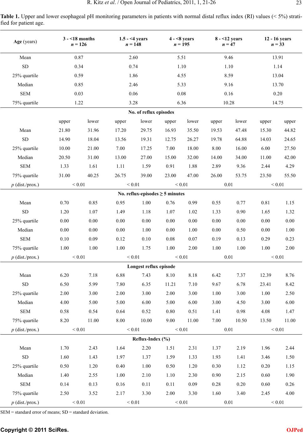
Reference values are mandatory to distinguish be-
tween physiological and pathologic reflux activity. De-
tecting the reflux in the upper esop hagus may be of spe-
cial interest in children with extraesophageal disorders.
While older studies propose the 95th percentile as the
cut-off value [17], more recent publications favor the
SEM as range of normal values [3]. Our study provides
both limits in order to give a solid base for data interpre-
tation. In order to avoid an over-treatment, we recom-
mend the conserv ative approach of taking valu es beyond
the 75th interquartile range as an indication for an anti-
acid treatment.
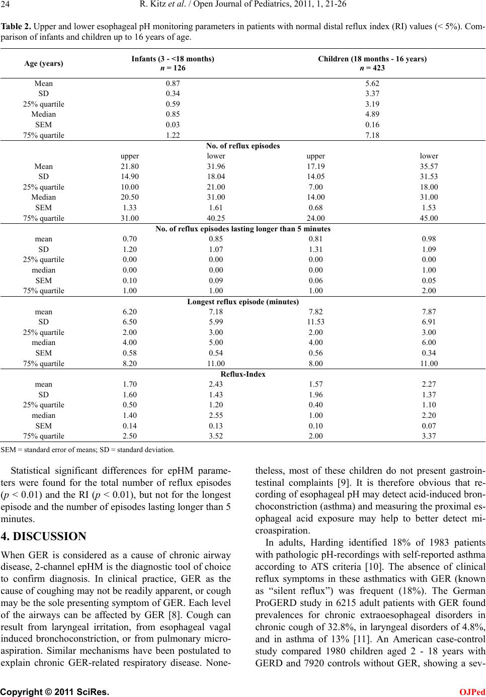
Bagucka et al. [3] published normal data for the upper
esophagus in children initially referred for exclusion of
suspected GER. According to ESPGHAN-recommenda-
tions, they stratified their study group depending on the
RI in the lower esophagus. While their values encom-
passed infants younger than 17 months of age, we now
extended the age range up to 16 year-old adolescents.
When comparing the data, we found consistencies in the
total number of reflux episodes and the absence of epi-
sodes lasting longer than five minutes. Discrepancies
were found for the RI (0.5% vs 1.2%) in the present
study. These findings could be explained by the different
mean ages of the groups < 1.5 years. Bagucka et al.
studied infants as young as 0.5 months old, whereas in
our study no child was younger than 3 months. Their
median age was 3.0 months, whereas our children had a
median age of 10.2 months. Taking into account the
more buffered stomach content of milk-fed children, less
amount of reflux can be detected by measuring only
pH-condition [18]. In other words: the longer the pH in
the stomach is below 4, the more reflux episodes can be
detected in the esophagus. More precisely, simultane-
ously performed intraluminal impedance measurement
of the esophagus can detect bolus movements, thus
broadening our diagnostic spectrum as to detect nonacid
reflux activity in the future [19,20].
Problems in the application of reference values may
occur due to the type of recording device and electrodes.
At present, mostly antimony electrodes are used. They
are less accurate than glass electrodes, however, they
provide multi-channel recording on different levels in
the esophagus and are easier to handle as well as to place
into the esophagus.
5. CONCLUSIONS
The present study prov ides reference values of proximal
esophageal pH-monitoring from infancy to adolescence.
The inter-quartile ranges appear as suitable threshold
levels when anti-acid treatment for the control of re-
flux-associated airway disease is considered.
REFERENCES
[1] Rudolph, C.D., Mazur, L.J., Liptak, G.S., Baker, R.D.,
Boyle, J.T., et al. (2001) Guidelines for evaluation and
treatment of gastroesophageal reflux in infants and chil-
dren: recommendations of the North American Society
for Pediatric Gastroenterology and Nutrition. Journal of
Pediatric Gastroenterology and Nutrition, 32, S1-S31.
doi:10.1097/00005176-200100002-00001
[2] Khoshoo, V., Le, T., Haydel, R.M., Landry, L. and Nelson,
C. (2003) Role of gastroesophageal reflux in older children
with persistent asthma. Chest, 123, 1008-1013.
doi:10.1378/chest.123.4.1008
[3] Bagucka, B., Badriul, H., Vandemaele, K., Troch, E. and
Vandenplas, Y. (2000) Normal ranges of continuous pH-
monitoring in the proximal esophagus. Journal of Pedi-
atric Gastroenterology and Nutrition, 31, 244-247.
doi:10.1097/00005176-200009000-00008
[4] Sondheimer, J.M. and Haase, G.M. (1988) Simultaneous
pH-recordings from multiple sites in children with and
without distal gastroesophageal reflux. Journal of Pedi-
atric Gastroenterology and Nutrition, 7, 46-51.
[5] Gustafsson, P. and Tibbling, L. (1988) 24-hour oeso-
phageal two-level pH-monitoring in healthy children and
adolescents. Scandinavian Journal of Gastroenterology,
23, 91-94.
[6] ESPGHAN—Working Group of the European Society of
Pediatric Gastroenterology, Hepatology and Nutrition
(1992) A standardized protocol for the methodology of
esophageal pH-monitoring and interpretation of data for
the diagnosis of gastroesophageal reflux. Journal of Pe-
diatric Gastroenterology and Nutrition, 14, 467-471.
[7] Ahrens, P., Haas, S. and Kitz, R. (2003) Standardization
and optimization of 2-channel pH-monitoring in children
with gastroesophageal reflux-associated pulmonary dis-
ease. Monatsschrift Kinderheilkunde, 151, 1298-1305.
doi:10.1007/s00112-003-0843-6
[8] Dobhan, R. and Castell, D.O. (1993) Normal and abnormal
proximal esopha geal acid exposure: Results of am bulatory
dual-probe pH-monitoring. The American Journal of Gas-
troenterology, 88, 25-29.
[9] Gorenstein, A., Levine, A., Boaz, M., Mandelberg, A.
and Serour, F. (2003) Severity of acid gastroesophageal
reflux assessed by pH metry: Is it associated with respi-
ratory disease? Pediatric Pulmonology, 36, 330-334.
doi:10.1002/ppul.10361
[10] Harding, S.M., Guzzo, M.R. and Richter, J.E. (1999)
24-h Esophageal pH-Testing in Asthmatics. Chest, 115,
C
opyright © 2011 SciRes. OJPed
R. Kitz et al. / Open Journal of Pediatrics, 2011, 1, 21-26
Copyright © 2011 SciRes.
26
OJPed
654-659. doi:10.1378/chest.115.3.654
[11] Jaspersen, D., Kulig, M., Labenz, J., Leodolter, A., Lind,
T., et al. (2003) Prevalence of extra-oesophageal mani-
festations in gastro-oesophageal reflux disease: An analy-
sis based on the ProGERD Study. Alimentary Pharma-
cology & Therapeutics, 17, 1515-1520.
doi:10.1046/j.1365-2036.2003.01606.x
[12] El-Serag, H.B., Gilger, M., Kuebeler, M. and Rabeneck,
L. (2001) Extraesophageal association of gastroesophag-
eal reflux disease in children without neurological de-
fects. G a stro en t ero log y, 121, 1294-1299.
doi:10.1053/gast.2001.29545
[13] Coletti, R.B., Christie, D.L. and Orenstein, S.R. (1995)
Indications for pediatric esophageal pH-monitoring. State-
ment of the NASPGN. Journal of Pediatric Gastroen-
terology and Nutrition, 21, 253-262.
doi:10.1097/00005176-199510000-00002
[14] Yüksel, H., Yilmaz, O., Kirmaz, C., Aydogldu, S. and
Kasirga, E. (2006) Frequency of gastroesophageal reflux
disease in nonatopic children with asthma-like airway
disease. Respiratory Medicine, 100, 393-398.
[15] Arana, A., Bagucka, B., Hauser, B., Hegar, B. and Urbain,
D., et al. (2001) pH-Monitoring in the distal and proxi-
mal esophagus in symptomatic infants. Journal of Pedi-
atric Gastroenterology and Nutrition, 32, 259-264.
doi:10.1097/00005176-200103000-00005
[16] Stordal, K., Johannesdottir, G.B., Bentsen, B.S., Knudsen,
P.K. and Carlsen, K.C., et al. (2005) Acid suppression
does not change respiratory symptoms in children with
asthma and gastroesophageal reflux disease. Archives of
Disease in Childhood, 90, 956-960.
doi:10.1136/adc.2004.068890
[17] Vandenplas, Y., Goyvaerts, H. and Helven, R. (1991) Gas-
troesophageal reflux, as measured by 24-hour pH-moni-
toring, in 509 healthy infants screened for risk of sudden
infant death syndrome. Pediatric s, 88, 834-840.
[18] Mitchell, D.J., McClure, B.G. and Tubman, T.R. (2001)
Simultanous monitoring of gastric and oesophageal pH
reveals limitations of conventional oesophageal pH-mo-
nitoring in milk-fed infants. Archives of Disease in Child-
hood, 84, 273-276. doi:10.1136/adc.84.3.273
[19] Wenzel, T.G., Moroder, C., Trachterna, M., Thomson, M.
and Silny, J., et al. (2002) Esophageal pH-monitoring and
impedance measurement: A comparison of two diagnos-
tic tests for gastroesophageal reflux. Journal of Pediatric
Gastroenterology and Nutrition, 34, 519-523.
doi:10.1097/00005176-200205000-00009
[20] Rosen, R., Lord, C. and Nurko, S. (2006) The sensitivity of
multichannel intraluminal impedance and the pH probe in
the evaluation of gastroesophageal reflux in children.
Clinical Gastr oenterology and Hepatology, 4, 167-172.
doi:10.1016/S1542-3565(05)00854-2