Surgical Science, 2011, 2, 356-359
doi:10.4236/ss.2011.26077 Published Online August 2011 (http://www.SciRP.org/journal/ss)
Copyright © 2011 SciRes. SS
Neoadjuvant Therapy for L ocally Advanced Renal
Cell Carcinoma with Sorafenib in a Reference
Center in Mexico
Axel Costilla-Montero1, Benjamín Guadarrama-Benítez1, Marco A. Aragón-Castro1,
Omar Morales-Ordaz1, Rubén Gutiérrez-Rosales1, Claudia Carrillo-Ponce2,
Marcela Janka-Zires2, Luis Gabriel Vázquez-Lavista1*
1Departament of Ur ology, Centro Médico ISSEMYM Toluca, Tol uca, México
2Departament of Pathology, Centro Médico ISSEMYM Toluca, Toluca , México
3Department of Internal Medicine Fundacion, Centro Médi co IS SEMYM Toluca, Toluca, México
*E-mail: lugavala@yahoo.com
Received May 5, 2011; revised June 24, 2011; accepted July 7, 2011
Abstract
Background: neo-adjuvant therapy is usually indicated in locally advanced tumors, the aim is to decrease
the tumoral burden and enhance overall survival. Renal cell carcinoma is a chemo and radio resistant neo-
plasm and this type of approach is not as effective as in other solid tumors. On the other hand immunother-
apy is indicated in metastatic disease, demonstrating a better overall survival. Sorafenib is an antiangiogenic
drug approved for locally advanced or metastatic RCC. We postulated that it can be used in a neoadjuvant
way to decrease the vascularization of selected tumors. Report of the Case: 57 years old male referred to
our service with a right renal mass and metastatic disease to lumbar spine and suprarenal gland. He was
treated with three months of sorafenib previous to the surgery. Results: the patient went into surgery three
months after initiating the antiangiogenic drug, during the surgery we found less neo-formance vessels; the
dissection was subjectively easier, due to peri-renal edema. The pathologic analysis of the specimen was re-
nal cell carcinoma. Interestingly, 40% of central ischemic (coagulative) necrosis was found. Conclusion:
there are no neoadjuvant drugs accepted for the treatment of renal cell carcinoma; using an antiagiogenic
drug to decrease the vascular burden characteristic of this type of tumors could be a viable option in selected
cases. We used a lower dose of the drug with an acceptable safety profile.
Keywords: Renal Cell Carcinoma, Antiagiogenic Drugs, Neoadyvant Therapy, Sorafenib
1. Introduction
Kidney cancer is the seventh leading malignant co ndition
among m en and the twelfth a mong women, a ccounting for
2.6% of all cancers in the United States [1] and is the tenth
cause in México [2]. The most frequent histology is renal
clear cell carcinoma (RCCC). When RCCC in an organ
confined stage, it is surgically treated with good overall
survival rate, whoe ver, 30% of the cases are detected in an
advanced stage, in the world and in México, with a high
mortality rate [3,4]. RCCC shows poor response to che-
motherapy due to multiple mechanisms [5]. Currently
FDA approved treatment strategies for metastatic stages
are immunotherapy with cytokines [interferon alpha and
interleukine-2] and the use of antibodies against specific
proteins (vascular endothelial growth factor, platelet de-
rived growth factor, and tumor growth factor alpha) such
as sunitinib [6], sorafenib [7] or bevacizumab [8].
Neo-adjuvant therapy is usually indicated in locally
advanced tumors, and the aim is to decrease the tumoral
burden and enhance overall survival . As m entioned a bove,
due to the chemo-resitance of RCCC, neoadjuvant studies
are scarce. On the other hand, the response to immuno-
therapy is limited and tox icicity is h igh. Th ere are stud ies
using immunotherapy as an adjuvant therapy along with
cito-reducti ve nefrectom y wi th a better overal l survival o f
the patients; [6-8] the aim of this study is to report our
experience in one case treated with sorafenib as a neoad-
juvant drug.
Scientific evidence suggests that the concept of
A. COSTILLA-MONTERO ET AL.357
neoadjuvant therapy for RCCC should be revisited [9],
especially in selected patients according to the NCCN
guidelines [10].
2. Case Report
Fifty seven year old male referred to our service with a
right renal mass and metastatic disease to lumbar spine
and suprarenal gland.
CT scan was performed with the evidence of a renal
mass. He was treated with three months of sorafenib pre-
vious to the surgery. He received 200 mgs PO every 12
hrs for a month, the subsequent month no drug was ad-
ministered, and the cycle was repeated for a third month.
Figure 1 shows two slides of the CT scan demonstrating
densities changes in the center of the mass before and
after the neoadjuvant treatment.
3. Results
The patient went into surgery three months after initiating
the antiangiogenic drug, during the surgery we found less
neo-formance vessels; the dissection was subjectively
easier, due to peri-renal e dema. The pathologic analysis of
the specimen was T4 renal cell carcinoma Fuhrman III in
95% of the kidney. I nteresti ngl y, 40% o f central ischem i c
(coagulative) necrosis was found. The suprarenal gland
was found with metastatic disease Figure 2.
4. Discussion
Controlled fase III studies have demonstrated superiority
in those patients treated with INF and targeted therapies.
Sorafenib is a multi kinase inhibitor including VEGFr,
PDGFr and RAF. Using it as a unique drug the results
are promising, showing a better overall and progression
free survival in metastatic disease [7]. One of the first
case reports is bi Di Silverio et al. where they describe A
71-year-old man with advanced left renal cell carcinoma
(lymph node involvement and vena cava thrombus) was
submitted to 6 months of neoadjuvant treatment with
sorafenib before open radical nephrectomy. After soraf-
enib treatment and before surgery a new CT scan con-
firmed the presence of a 9.0 cm in diameter solid mass in
the left kidney but a reduction in thrombus extension,
limited to the left renal v ein. At histological examination
after radical nephrectomy, over 90% of the renal mass
was substituted by necrotic tissue [11].
Our patient showed 50% necrosis in the pre surgery
CT scans and this was corroborated in the final patho-
logical specimen with 40% of central ischemic (coagula-
tive) necrosis. It is worth to mention that the neoplastic
necrosis is usually a liquefactive one, and it is character-
ized by digestion of the dead cells resulting in transfor-
mation of the tissues into a liquid viscous mass. In this
case the architecture of the dead tissue was preserved and
the affected tissue exhibited a firm texture so it was sec-
ondary to ischemia. Subjectively during the dissection we
found a peri renal edema that made easier the surgery. It
is also worth mentioning th at the therapeutic dose used in
this patient was lower than that used in a palliative scen-
ery, and only a few adverse effects were reported, no
important co morbidity was found during the follow up.
The biggest case control series with tyrosine kinase
inhibition is with 14 patients who underwent cytoreduc-
tive nephrectomy for advanced renal cell carcinoma with
prior treatment with sorafenib or sunitinib, and were
compared with 73 patients without neoadjuvant therapy,
Figure 1. Pre- and post-neo-adjuvant treatment with sorafenib CT scan of the renal mass.
Copyright © 2011 SciRes. SS
A. COSTILLA-MONTERO ET AL.
Copyright © 2011 SciRes. SS
358
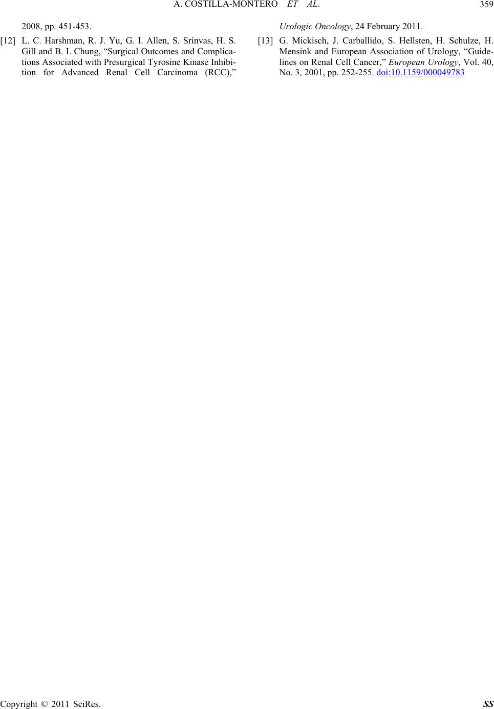
Figure 2. Pahtological specimen of right kidney with central
necrosis and metastatic disease to suprarrenal gland.
the parameters studied were: the incidence of periopera-
tive complications and outcomes after surgical procedures
between the two cohorts. The median preoperative renal
mass size was 11 cm (6.7 - 4.2 cm). Primary tumor
shrinkage was seen in 57%; median shrinkage was 18%
(8% - 25%). The median treatment period was 17 weeks,
and the median time from TKI discontinuation was 2
weeks. Compared with a control group and after adjust-
ing for confounding covariates, presurgical TKI use was
not associated with a significant increase in perioperative
complications (50% vs 40%, P = 0.25) or perioperative
bleeding (36% vs 34%, P = 0.97) but was associated
with increased incidence and grade of intraoperative ad-
hesions (86% vs 58%, P = 0.001; grade 3 vs 1, P =
0.002). They concluded that they found less hemorrhagic
and wound healing issues but a significant increase in
incidence and severity of intraoperative adhesions, which
can present a formidable technical challenge. The pre-
surgical TKI therapy can permit effective surgical cy-
toreduction with a safety and complication profile
equivalent to that of non-TKI-nephrectomy; however
safety data continue to evolve, and preoperative TKI use
requires further prospective investigation [12].
5. Conclusions
Sorafenib is a drug that inhibits specific proteins in neo-
plastic cells, it modulates transduction signal and has
shown to create intratumoral ischemic necrosis. The
NCCN [9] and the EUA [13] guidelines have suggested
that this drug can be used as a first line of treatment and
the neoadjuvant setting should be investigated thor-
oughly.
6. References
[1] A. Jemal, T. Murray and E. Ward, “Cancer Statistics,”
CA: A Cancer Journal for Clinicians, Vol. 55, No. 1,
2005, pp. 10-30. doi:10.3322/canjclin.55.1.10
[2] Instituto Nacionall de Oncologia, “Manual de Oncologia
Médica,” Procedimentos Medico Quirurgicos, 2ª Edición,
McGraw Hill, Boston, 2002.
[3] L. M. Hock, J. Lynch and K. C. Balaji, “Increasing Inci-
dences of all Stages of Kidney Cancer in the United
States: An Analysis of Surveillance, Epidemiology and
End Results Program Data,” Journal of Urology, Vol.
167, No. 1, 2002, pp. 57-60.
doi:10.1016/S0022-5347(05)65382-7
[4] S. Ramsay and M. Aithkinson, “Treatment of Renal Cell
Carcinoma: Are We beyond the Cytokine Era?” Nature
Clinical Practice Urology, Vol. 3, No. 9, 2006, pp.
478-484. doi:10.1038/ncpuro0581
[5] E. Soto-Vega, C. Arroyo, Y. Richaud-Patin, M. Gar-
cía-Carrasco, L. G. Vázquez-Lavista and L. Llorente “P-
Glycoprotein Activity in Renal Clear Cell Carcinoma,”
Urologic Oncology, Vol. 27, No. 4, 2009, pp. 363-366.
doi:10.1016/j.urolonc.2008.01.011
[6] R. J. Motzer, T. E. Hutson, P. Tomczak, et al., “Sunitinib
versus Interferon Alpha in Metastatci Renal Cell Carci-
noma,” The New England Journal of Medicine, Vol. 356,
No. 2, 2007, pp. 115-124. doi:10.1056/NEJMoa065044
[7] B. Escudier, “Sorafenib in Advanced Clear-Cell Re-
nal-Cell Carcinoma,” The New England Journal of Medi-
cine, Vol. 356, No. 2, 2007, pp. 125-134.
doi:10.1056/NEJMoa060655
[8] J. C. Young, L. Haworth, R. M. Sherry, et al., “A Ran-
domized Trial of Bevacizumav an Antivascular Endothe-
lial Growth Factor Antibody for Metastatic Renal Cell
Cancer,” The New England Journal of Medicine, Vol.
349, No. 5, 2003, pp. 427-434.
doi:10.1056/NEJMoa021491
[9] C. Wood, et al., “Neoadjuvant (Presurgical) Therapy for
Renal Cell Carcinoma: A New Treatment Paradigm for
Locally Advanced and Metastatic Disease,” Cancer, Vol.
115, No. S10, 2009, pp. 2355-2360.
doi:10.1002/cncr.24240
[10] National Comprehensive Cancer Network, “Clinical
Practice Guidelines in OncologyTM,” Kidney Cancer, V2,
2010. www.nccn.org
[11] F. Di siverio, A. Sciarra, U. Parente, A. Andrea, M. Von
Heland, V. Panebianco and R. Passariello, “Neoadjuvant
Therapy with Sorafenib in Advanced Renal Cell Carci-
noma with Vena Cava Extension Submitted to Radical
Nephrectomy,” Urologia Internationalis, Vol. 80, No. 4,
A. COSTILLA-MONTERO ET AL.359
2008, pp. 451-453.
[12] L. C. Harshman, R. J. Yu, G. I. Allen, S. Srinvas, H. S.
Gill and B. I. Chung, “Surgical Outcomes and Complica-
tions Associated with Presurgical Tyrosine Kinase Inhibi-
tion for Advanced Renal Cell Carcinoma (RCC),”
Urologic Oncology, 24 February 2011.
[13] G. Mickisch, J. Carballido, S. Hellsten, H. Schulze, H.
Mensink and European Association of Urology, “Guide-
lines on Renal Cell Cancer,” European Urology, Vol. 40,
No. 3, 2001, pp. 252-255. doi:10.1159/000049783
Copyright © 2011 SciRes. SS