Open Access Library Journal
Vol.03 No.01(2016), Article ID:68176,9 pages
10.4236/oalib.1102291
A Voyage to Beyond the Human Eye by Microscope, Leeuwenheokʼs Invention
Nasser Pouyan
Tehran, Iran

Copyright © 2016 by author and OALib.
This work is licensed under the Creative Commons Attribution International License (CC BY).
http://creativecommons.org/licenses/by/4.0/


Received 25 December 2015; accepted 10 January 2016; published 15 January 2016

ABSTRACT
Microscope, an instrument used for obtaining magnified image of small objects. The term of microscope was coined by Johannes Faber of Bamberg. The identity of its inventor has not been clearly established. Aristotle about 24 centuries before Leeuwenhoek described the working of microscope in some detail. The earliest records of optical lenses date from the late 13th century, when spectacles came into use. Roger Bacon, in his “Opus Magnus” of 1268 spoke of the use of lenses for magnifying objects. About 4 centuries later Leeuwenhoek built over 200 simple microscopes and became the father of protozoology and bacteriology. Leeuwenhoek was born in Delft, Holland. He is commonly known as the father of microbiology and considered the first microbiologist. He was raised in Delft, worked as a linen draper in his youth and founded his own shop (1654) and made a name for himself in municipal politics, and eventually developed an interest in lens making. Leeuwenhoek with his simple microscope for which he ground the lenses, achieved magnification of 270 times. Anton van Leeuwenhoek, during the last quarter of the seventeenth century with exquisitely polished homemade lenses, studied a great variety of natural materials such as pond water, vinegar, and blood. He observed protozoa (microscopic animals) in mixture of pepper and water, and bacteria in scrapings of human teeth. He described discovery of “animalcules”, as he called them, raised protozoa, bacteria, blood corpuscles, spermatozoa and the striated fibers found in bundles in voluntary muscles, and many other microscopic creatures and structures. He also had many findings in dentistry. Leeuwenhoek earned for himself a place of honor as a Fellow of the Royal Society in London. During his lifetime he sent 375 scientific papers to the Royal Society and 27 papers to the French Académie des Sciences. After the creation of the microscope it evolved slowly, hampered both by the lack of theoretical understanding and mechanical technology needed for making precision instruments. About 1800 the compound microscopes of the better makers began to resemble their modern counterparts. In 20th century the fundamental principles which were discovered led immediately to the development of oil-immer- sion objective and remain as the basis of microscope design.
Keywords:
Microscope, Leeuwenhoek
Subject Areas: Microbiology

1. Introduction
Microscope is an optical instrument with lenses, which makes very small objects appear larger. The simple microscope, or magnifier, is merely a lens held near the eye and usually it is limited to very low powers of magnification by various technical factors. It is notable that Antoni van Leeuwenhoekʼs pioneer observations of yeast cells and other microorganisms were made with simple microscope. Ordinarily the term microscope refers to the compound (several lenses) microscope, which is similar in principle to the astronomical telescope [1] (an optical instrument shaped like a tube, with lenses to make distant objects appear larger and nearer). The specimen is illuminated with light from below which shines through it, up towards the eye. The objective lens just above the specimen on the stage could be changed for different magnifications [2] .
The earliest records of optical lenses date from the late 13th century, when spectacles came into use, but when the simple lens began to be used as a microscope it is not clear.
Microscope from mid 17th century revealed a new world of tissues and cells. Marcello Malpighi (1628-1694), an Italian physician, microscopist and founder of histology studied micro anatomy in animals and humans, and described the embryology of a chick, graafian follicles, capillary circulation and vesicular structure of lungs. Johannes Peter Müller (1801-1858), a German pioneering physiologist (someone who studied how the body works), pioneered the microscopic study of diseased tissues of cells, which is called cellular pathology.
Mullerʼs pupil Theodor Schwann (1810-1882), a German physiologist, formulated the now-familiar idea of cell theory. Friedrich G.J. Henle (1809-1885), the greatest histologist of his time, wrote the first textbook of anatomy to describe cells and tissues as well as larger parts. He also wrote on the suprarenals, the pituitary, epithelial tissues, on ovarian function and erection. Rudolf Virchow (1821-1902), a German founder of cellular pathology by using microscope emphasised on the cell as the seat of pathology did much to discredit the theory that disease resulted from an imbalance of humors in the body, and regarded diseases as a cellular response to change. He also stated “all cells come from cell”.
About 1880 the advances of microscope were climaxed by Ernst Abbeʼs theoretical studies, which clarified the relation between angular aperture and resolving power. The fundamental principles he discovered led immediately to the development of the oil-immersion objective and remain as the basis of microscope design [3] (Figure 1).

Figure 1. Ernst Abbé an ophthalmologist and physicist and partner at the optical company of Carl Zeiss.
2. Microscope en Route of Development
Aristotle (384-322) Greek philosopher and father of biology whose achievement was scientific, described the workings of microscope in some detail.
Ancient Egyptians in North Africa and Romans in Europe also used different curved lenses although no reference to a compound microscope has been found. As Greek and Romans and Chinese applied their infinitive visdom to the issue but there is no known record regarding to use of artificial light or multiple lenses.
In second century, Ptolemy, also known as Claudius Ptolemaeus (c.100-170), Egyptian astronomer, mathematician, and geographer who spent much of his life in Alexandria described a stick appearing to bend in a pool of water. Ptolemy accurately recorded the angles to within half a degree. In 13th and 14th centuries, two Italian men claimed to have independently invented spectacles. According to their tomb stones:
One of them SalvinodʼArmato of the Armati of Florence (died 1284/1317) claimed to have kept the process secret.
The other, Allessandrodella Spina died in 1317, claimed to have revealed his process.
A local monk, Girodina da Rivalta, in 1306 gave a sermon in which he enthusiastically endorced spectacles as a terrific invention and in passing, indicated that they had been in use for about 20 years.
In 1289, finally another local from the Popozo family be moaned that “I am so delitated by age that without the glasses known as spectacle, I would no longer be able to read or to write”.
Roger Bacon (c.1214-1298) of Ilchester, an English monk of the Franciscan order, gave the first description of a primitive form of camera obscura in his treatise Perspectiva in 1267 [4] (the camera obscura was first suggested by the Arabian mathematician, Alhazen or Ibn al-Haitham, AD 965-1038 of Basra, Iraq) (Figure 2).
With the invention of printing art by Johann Gutenberg (c.1394-1468) and a complete Bible in Latin, “Gutenberg Bible or Mazarine Bible or 42-line [5] ”, printed in Mainz, Germany (c.1450-1455) by him, ideas and development could be spread easily and rapidly. For instance, Thomas Diggesʼ work on telescope in England in mid 16th century and Hans Lippersheyʼs work which including applying for a telescope patent, were transferred to others including Galileo.
Johannes Faber (1574-1629) of Bamberg, one of the original members of Accademia de Lincei coined the term “microscope”.
In 1606, Galileo (Galilei Galileo) (1564-1642), an Italian mathematician and scientist (regarded as the father of the science of moving bodies or dynamics), developed a compound microscope, consists of several convergent lenses with one having a short focal length. He was the first to apply the compound microscope to scientific studies. We should also tribute to sir Isaac Newton (1642-1727), the greatest figure in the history of the exact sciences who around the same time in the United Kingdom invented the reflecting telescope.

Figure 2. Roger Bacon who gave the first description of a primitive camera obscura.
Hans Lippershey or Lipperhey (1570-1619) with his son, Zacharias Jansen, Dutch instrument makers of Middleburg, generally credited with inventing the telescope. A probably apocryphal story is that Hans held a piece of spectacle lens in each hand, one in front of the other and by chance, he focused on a neibouring church steeple and noticed that thus devising the first telescope. This instrument was improved and used for astronomical observation by Galileo and others [3] . It is also believed that Zaccharias Jansenʼs Father Hans Lippershey, helped him build the first compound microscope in 1595. Zacharias wrote to a Dutch diplomat William Boreel, about the invention. When the physician of the French king inquired about the invention in the 1650ʼs, Boreel recounted the design of the microscope.
Cornelius Jacobson Drebbel (1570-1633) of Holland, is said to have invented the microscope about 1620. (But the identity of the inventor of microscope has not been clearly established.) Some evidences however, indicate that both Drebbel and Galileo may have been anticipated by Hans Lippershey and Zacharias Jansen, about 1590.
A Jesuit priest, Athanasius Kircher (1601-1680) of Geysen used a primitive microscope of 32 magnification to view blood cells in 1658. (The invention of first magic lantern is also ascribed to him.)
Robert Hooke (1635-1703), English scientist and pioneer in microscopy made improvements to the compound microscope. He published his micrographia (1665), an outstanding text describing his compound microscopic observations. The extract below from Hookʼs Micrographia demonstrates his perspective on how the microscope is utilised to enhance on how the microscope is utilized to enhance the senses… “In the collection of most of which I made use of microscopes and some other glasses and instruments that improve the senses… only to promote the use of mechanical helps for the senses, both in the surveying the already visible world, and for the discovery or many others hitherlo unknown.” (Figure 3).
Anton van Leeuwenhoek (1632-1723), a self-taught microscopist discovered and accurately described the protozoa in 1674 and bacteria in 1676.
Phillip Bonnai, in 1698 published an account of two compound microscopes.
In 1677, a French professor, le Père Cherubin was the first to view small objects under the microscope conjointly by both eyes.
In 1685, the first complete early microscopes was given by Johann Zahn (1641-1707) [6] .
In 1740, Benjamin Martin (1704-1782), British lexicographer and spectacle maker improved the microscope and sold pocket version of it.
Henry Baker of London (1698-1774) who was a known microscopical instruments maker recapitulated much of Leeuwenhoekʼs work in his book “The Microscope Made Easy” (1743). He was also one of the founders of the Society of Arts (1754).
In 1824, a diamond microscope was made by Andrew Pritcard (1804-1882) an English naturalist and natural history dealer. He also introduced “test objects” to compare the quality of various microscopes and made significant improvements to microscopy and studied microscopic organisms.
Jackson Lister (1786-1869), the father of Joseph Lister (1827-1912) British surgeon and father of antiseptic surgery, calculated the curvature of the magnifying lenses to reduce chromatic aberration, a problem where coloured fringes appear around objects. His model of 1826 was the first to have such lenses [7] .

Figure 3. Robert Hoek, brilliant British philosopher who developed the early compound microscope.
In 1839, “The Microscopical Society” was established in London. An Italian astronomer, Giovan?i Batista Amici (1786-1863) from Firenze of Florence constructed a reflecting microscope, and improved its achromatic object, in 1812.
In 1865, a spectrum microscope capable of detecting one millionth of a grain of blood was exhibited by an English chemist, microscopist and geologist, Henry Clifton Sorby (1826-1908). His major contribution was the development of techniques for studying iron and steel with microscope which paved the way for the mass production of steel.
In 1878, a German physicist Ernst Abbé (1840-1905) who was a partner of Carl Zeis (1816-1888) German optician and industrialist, modernized the microscopes by adding the achromatic objective and the oil immersion device. He added the substage condenser in 1886, and improved the technique of phase contrast microscopy, in 192. Abe and Zeis built factories throughout Europe, the firm gained fame for the excellence of its many types of optical instruments.
In 1935, the modern phase contrast technique was introduced by Dutch physicist, Frits Zernike [8] .
Fritz Zernike (1888-1966) received the 1953 Nobel Prize in physics for his development of the phase contrast microscope. Among its many uses, this microscope permits the viewing of living cells without the staining.
Richard Zsigmondy (1865-1929), Austrian chemist whose fundamental researches on colloid chemistry gave a solid basis for development of this branch of science, in collaboration with H.F.W. Siedentopf developed the ultramicroscope in 1903. He received the Noble Prize in chemistry in 1926 [9] .
3. Anton Van Leeuwenhoek, One of the Two Giants of Microscopy
One of the two giants of seventeenth-century microscopy (the other gain of microscopy was Marcello Malpighi (1628-1694) Italian physician and founder of histology), was Anton van Leeuwenhoek (1632-1723), a self- taught Dutch linen draper who is commonly known and considered to be the first microbiologist. He was an amateur grinder of lenses and a maker of microscopes, through which he discovered and accurately described the protozoa in 1674 and the bacteria in 1676. For his contributions to the sciences of bacteriology and protozoology, Leeuwenhoek has been called the father of microbiology. He was elected a fellow in the Royal Society in London (1680), and reports on 375 of his discoveries appeared in the “Philosophical Transactions” of the society.
Leeuwenhoek , the Dutch ingenious microscopist, by holding up his tiny one-lens simple microscope close to his eye, he observed blood cells, sperm, muscle fibers and single-celled organisms which he called “animalcules”. Ordinarily a simple microscope is limited to very low power of magnification by various technical factors, but it is notable that Leeuwenhoek’s pioneer observations of yeast cell and other microorganisms were made with simple microscopes [1] .
He examined biological materials included studies in anatomy, histology, physiology, embryology, botany, chemistry and physics (Figure 4).
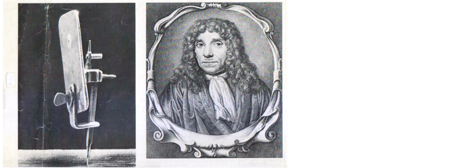
Figure 4. Anton van Leeuwenhoek and his microscope, through which he made many scientific studies that led to de discovery of spermatozoa. (Sebastian, Anton. Dates in Medicine. The Parthenon Publishing Group, New York-London, 2000, p. 27.)
Leeuwenhoek’s revealing investigations of corpuscles and capillary circulation are classic. His comparative study of animal spermatozoa and the life history of the ant, and his descriptions of the different structure of the stem in monocotyledonous and dicotyledonous plants are other examples of Leeuwenhoek’s versatility as a scientist and of his interest in microscopic life. Some of his observations on metamorphosing and developing animals helped to refute the then accepted and widespread belief that some living things evolved from nonliving materials [10] .
Leeuwenhoek’s great invention reached university medicine in the 1840s, and microscope was central in diagnosing the true causes of death from tissue taken at autopsy. It was also highly useful for studying blood samples taken from the patients to determine if they were anemic and to judge from the size and shape of the red blood cells the kind of anemia. One could also look urine specimens under the microscope for evidence of pus to see if, say, an infection of the urinary tract were causing that deep pelvic pain. Or one could try to find the bacteria of pneumonia, tuberculosis, or bronchitis in sample of sputum. Also a microscope “looked good” in one’s office. Daniel Cathell snickered in 1882 of the microscope and similar equipment, “If, at your office and elsewhere, you make up of instruments of precision… they will not only assist you in diagnosis, etc., but will also aid you greatly in curing people by heightening their confidence in you and enlisting their co-operation [11] .”
Among his findings of importance to dentistry were the tubules in the dentin and the microorganisms, including bacteria, that he found in the “materia alba” adhering to the teeth. When the president of the Royal Society in London sent him several worms that, he was told, had been taken from a carious tooth, Leeuwenhoek effectively disproved that they were tooth worms by proving microscopically that they were identical to the maggots that infest over ripe cheese. He postulated that the maggots had entered the carious lesion when the owner of the tooth ate the cheese, for, as he said, he had extracted maggots from the damaged teeth of his own wife after she had partaken of infested cheese [12] .
3.1. The Highlights
The study of organisms and the process which produces fermentation started with the invention of the microscope and the first description with drawings of yeast cells was submitted to the Royal Society in London (1680) by Leeuwenhoek.
He ground his own lenses and constructed over 200 microscopes in the 17th century.
Anton examined lake water which in summer became cloudy, whereas in winter it was clear. He noticed hundreds of tiny creature, swimming in a single drop through his microscope.
Leeuwenhoek made special microscopes so that he could see the flow of blood in rabitʼs ear, batʼs wing, and tadpoleʼs tail.
Leeuwenhoek was the first to observe bacteria, spermatozoa and protozoa.
The first protozoan parasite to be observed, “Giardia lamblia”, was identified by Leeuwenhoek, who noted it in his stools, in 1681.
He described the use of the crocus or saffron to stain muscle fibers, to the Royal Society, London in 1714.
In crystallography (the study of the structure, forms and properties of crystals), he showed the morphological variation of crystals in different salts, in 1695.
In 1698, Leeuwenhoek was invited in the boat of Peter I known as Peter the Great (1672-1725), Tsar of Russia (1682-1725). On the occasion Leeuwenhoek presented him an “eel-viewer”, so Tsar could study the blood circulation, whenever he wanted.
Physicians met scientists (or natural philosopher as they were then called) at such venues as the Royal Society and exchanged ideas and techniques. Physicians felt there was all to gain from making their doctrine more “scientific”, Leeuwenhoek’s microscope was a new aid and taken up by Robert Hooke (1635-1703) the brilliant British experimental philosopher who developed the early compound microscope.
He was visited by Gottfried Wilhelm Leibniz (1646-1716); German philosopher; William III (William of Orange) (1650-1702), King of England, Scotland, and Ireland (1689-1702) and his wife Anne (1665-1714), Queen of England and Scotland (1702-1707); the Amsterdam burgomaster (the mayor), Johan Huydecoper; and all gazed at the Leeuwenhoek’s tiny microscope.
He made more than 500 optical lenses and at least 25 microscopes of different types, of which only 9 of them survived. Those that have survived are capable of magnification up to 275 times. The microscopes were made of silver or copper frames, holding hand-made lenses.
Leeuwenhoek maintained throughout his life that there are aspects of microscope construction “which I only keep for myself”, in particular his most critical secret of how he created lenses. For many years no-one was able to reconstruct Leeuwenhoek’s design techniques. However, in 1957 C.L. Stong used thin glass thread fusing instead of polishing, and successfully created some working samples of a Leeuwenhoek design microscope. Such a method was also discovered independently by A. Mosolov and A. Belkin at the Russian Novosibirsk State Medical Institute.
He used samples and measurements to estimate numbers of microorganisms in units of water.
3.2. Life
Anton or Antonie Philips van Leeuwenhoek or Leuwenhoek was born in Delft1, on October 24, 1632. His father, a basket maker, died when Anton was only five years old. His mother, Margaretha married Jacbon Janz Molijan, painter after Philips’ death. Leeuwenhoek had four older sisters. Anton attended school near Leiden, city of the Western Netherlands, about 9 mi. northeast of the Hague, for a short time before being sent to live with his uncle, Benthuizen, an attorney and town clerk. At the age of 16, he became an apprentice at a linen draper’s shop in Amsterdam.
He married Barbara de Mey in July 1654, with whom Anton would have one surving daughter, Maria (four other children died in infancy). In 1654 he returned Delft where he lived and studied for the rest of his life. He opened a draper’s shop which he ran throughout the 1650 s.
Barbara died in 1666, and Anton married Cornelia Swalmius, with whom he had no surviving children. His status in Delft grew throughout the following years. Although he would remain an obscure figure outside of the city. He received a lucrative municipal title as chamberlain for the Delft sheriffs’ assembly chamber in 1660, a position which he would hold for almost 40 years. In 1669 he was named a surveyor by the Court of Holland; later he would become a municipal “wine-gauger” in charge of the city’s wine imports.
After developing his method for making powerful lenses, he introduced the invention to his friend Regnier de Graaf (1641-1673) the eminent Dutch physician2. When the Royal Society in London published an Italian lens maker’s work in the “Philosophical Transactions”, Regnier de Graaf wrote to its secretary, Henry Oldenburg (1615-1667)3 with endorsement of Leeuwenhoek’s microscopes. The Royal Society, in 1673 published a letter from Leeuwenhoek, including his microscopic observations on mold, bees, and lice. Until his death he wrote countless letters in his own colloquial flavor of Dutch to the Royal Society in London, detailing his findings in a wide variety of field, centered around his work microbiology.
He was elected to the Royal Society in London, in 1680 by nomination of William Croone (1633-1684), the British eminent physician [13] . Leeuwenhoek was “taken aback” which he considered a high honor, although he did not attend a Royal Society meeting.
3.3. Death
Leeuwenhoek suffered from a rare illness, an uncontrolled movement of the midriff, which is now named Van Leeuwenhoek’s disease. He died on August 26, 1723, at the age of ninety, and was buried four days later in the Oude Kerk in Delft, Holland. No one had seen his fine microscopes and nobody was able to make lenses as good as his, for about one century.
3.4. Works
Anton van Leeuwenhoek, although is best known for his work on microscopes and contributions towards the founding of microbiology, he did not wrote any books. His discovery came to light only through correspondence with Royal Society in London; the monthly Journal “Philosophical Transactions” published his scientific letters (Figure 5).
4. Conclusion and Impact
The microscope has given a solid basis for development to branches of sciences. For instance:
1) It helped to bury the old humoral theory (any of the four liquids including “blood, phlegm, choler, and melancholy” in the body that were once thought to determine a person’s physical and mental qualities), a concept that dominated medicine since the time of ancient Greek physician and father of medicine Hippocrates (460-370 BC).
2) Until 1665 the cell [Latin: cella, a small room], a term used by Robert Hoke (1635-1703) in his work Micrographia (1665) was unknown, and it was not until over a century later that it was first described as the basic structural until of all living matter. Because most cells were invisible to the unaided eye, their discovery was not possible until the important invention of the microscope (Figure 6).
Before the invention of microscope, physicians and surgeons did not understand how the body became infected or how diseases spread. The creation of microscope and the discovery of one-celled animals invisible to naked eye in 1675 called attention to the existence of microscopic organisms [14] , and raised the question of their origin. Many scientists and most laymen believed that certain organisms could be generated spontaneously from nonliving materials that frogs could arise from raindrops and maggots from the carcasses of dead horses. But the theory of “spontaneous generation” was finally resolved by Louis Pasteur (1822-1895), a French chemist and the father of bacteriology, who in 1864 by microscope, demonstrated that microorganisms arise from living “germs” rather than from nonliving matters. Pasteur from his studies concluded that there exist a great variety of microorganisms, each capable of reproducing its own kind. According to this theory, the different fermentations and diseases are caused by different types of microorganisms.

Figure 5. Leeuwenhoekʼs microscopic observations.

Figure 6. A microbiological microscope.
Indeed Leeuwenhoek’s invention of microscope opened a new world for scientific study of microorganisms, such as bacteria, viruses, yeasts, molds, protozoa, and primitive algae, and the application of the knowledge derived from this study to the fields of medicine, agriculture, and industry. Of all forms of life, microorganisms are the smallest and simplest, and in many cases their bodies consist of single cells. The largest are barely visible to naked eye, and the smallest viruses can be visualized only with electron microscope [15] .
All in all, a linen draper, who was not a university-trained scientist developed microscope lenses so efficient that they were unsurpassed until the 19th century, and advanced and revolutionized knowledge immeasurably [16] .
Cite this paper
Nasser Pouyan, (2016) A Voyage to Beyond the Human Eye by Microscope, Leeuwenheok's Invention. Open Access Library Journal,03,1-9. doi: 10.4236/oalib.1102291
References
- 1. Humphry, E (1975) Encyclopedia International, Vol. 12. Grolier, New York, 53.
- 2. Parker, S. (1996) Eyewitness Science Medicine. Dorling Kindersley, London-New York-Stuttgart, 30.
- 3. Humphry, E. (1975) Encyclopedia International, Vol. 12. Grolier, New York, 54.
- 4. Sebastian, A. (2001) A Dictionary of the History of Science. The Parthenon Publishing Group, New York-London, 66.
- 5. Humphry, E. (1975) Encyclopedia International, Vol. 8. Grolier, New York, 238.
- 6. Sebastian, A. (2001) A Dictionary of the History of Science. New York-London, 227.
- 7. Parker, S. (1996) Eyewitness Science Medicine. Dorling Kindersley, London-New York-Stuttgart, 30.
- 8. Sebastian, A. (2001) A Dictionary of the History of Medicine. New York-London, 228.
- 9. Humphry, E. (1975) Encyclopedia International, Vol. 19. Grolier, New York, 585, 598.
- 10. Humphry, E. (1975) Encyclopedia International, Vol. 10. Grolier, New York, 451.
- 11. Porter, R. (1998) The Cambridge Illustrated History of Medicine. Cambridge University Press, Cambridge, 140.
- 12. Ring, M.E. (1985) Dentistry, an Illustrated History. Abradale Press, New York, 145.
- 13. Lee, H.S.J. (2000) The Medical Millennium. The Parthenon Publishing Group, New York, London, 60.
- 14. Chocrane, J. (1996) An Illustrated History of Medicine. Tiger Book International, London, 60.
- 15. Humphry, E. (1975) Encyclopedia International, Vol. 12. Grolier, New York, 47-48.
- 16. Lyons, A.S. (1987) Medicine, an Illustrated History. Abradale Press, New York, 145.
Bibliography and Websites
1) Loudon, Irvin. Western Medicine, An Illustrated History. Oxford, Oxford University Press, 1977, pp. 9, 102 and 108.
2) Cartledge, Paul. The Cambridge Illustrated History of Ancient Greece, Cambridge: Cambridge University Press, 1998, pp. 312-314, 325 and 347.
3) Riverain, Jean. Dictionaire Des Médicins Ćélèbres. Librairie Larousse, pp. 94, 95, 112-114.
4) http://www.google.com.ua/search.
5) https://en.wikipedia.org/wiki/Microscope.
6) http://en.wikipedia.org/wiki/Robert-Hooke.
7) http://en.wikipedia.org/wiki/Roger-Bacon.
9) www.history-of-the-microscope.org/history-of-the-microscope-who-invented-the-microscope.php.
NOTES

1Delft, a historic and industrial center in the west-central Netherlands, between Rotterdam and Gravenhage (The Hague). Jan Vermeer (known also as Jan van der Meer van Delft) (1632-1675), Dutch painter was born in Delft and worked here.
2Graaf was one of the first to experiment on the pancreas, and wrote on pancreatic juice. He also described the egg-containing follicle (Graaf follicle), and coined the term “ovary”.
3Henry Oldenbrg from Bremen published the monthly “Philosophical Transactions” at his own expense.