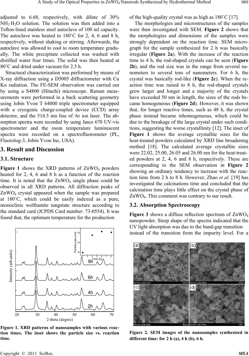
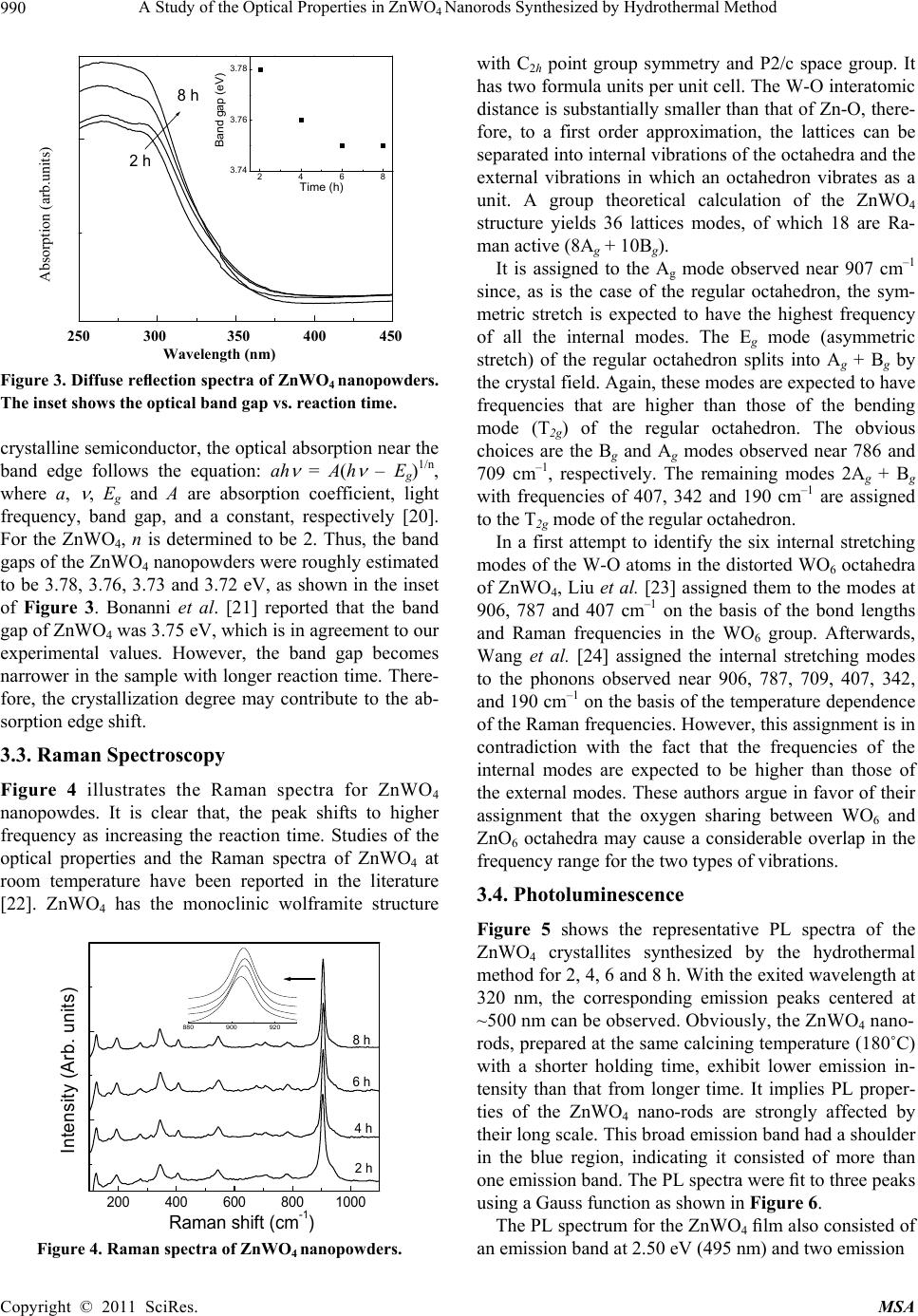
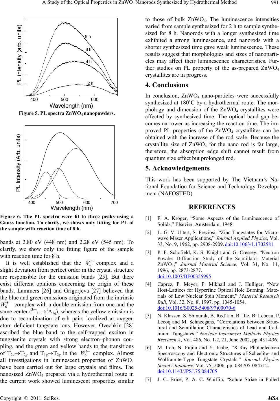
A Study of the Optical Properties in ZnWO4 Nanorods Synthesized by Hydrothermal Method
Copyright © 2011 SciRes. MSA
992
Crystals of Zinc Tungstate,” British Journal Applied
Physics, Vol. 18, No. 5, 1967, pp. 581-586.
doi:10.1088/0508-3443/18/5/304
[8] A. R. Phani, M. Passacantando, L. Lozzi and S. Santucci,
“Structural Characterization of Bulk ZnWO4 Prepared by
Solid State Method,” Journal Material Science, Vol. 35,
No. 19, 2000, pp. 4879-4883.
doi:10.1023/A:1004809804206
[9] L. Honeycutt and A. Kuzmin, J. Purans “Communication
and Design Course; Local atomic and electronic structure
of tungsten ions in AWO4 Crystals of Scheelite and
Wolframite Types,” Radiat Measurement, Vol. 33, No. 5,
2001, pp. 583-586.
[10] A. Henglein, “Estimated Distributions of Electronic
Redox Levels in aq/eaq
−, Haq+/Haq and Some Other
Systems,” General Introductory Chemistry, Vol. 78, No.
10, 1974, pp. 1078-1084.
doi:10.1002/bbpc.19740781016
[11] M. Bonanni, L. Spanhel, M. Lerch, E. Fuglein and G.
Muller, “Conversion of Colloidal ZnO-WO3 Heteroag-
gregates into Strongly Blue Luminescing ZnWO4 Xe-
rogels and Films,” Chemistry Material, Vol. 10, No. 1,
1998, pp. 304-310. doi:10.1021/cm9704591
[12] F.-S. Wen, X. Zhao, H. Huo, J.-S. Chen, E. Shu-Lin and
J.-H. Zhang, “Hydrothermal Synthesis and Photolumi-
nescent Properties of ZnWO4 and Eu3+-Doped ZnWO4,”
Materials Letters, Vol. 55, No. 3, 2002, pp. 152-157.
doi:10.1016/S0167-577X(01)00638-3
[13] M. H. Huang, S. Mao, H. Feick, H. Yan, Y. Wu, H. Kind,
E. Weber, R. Russo and P. Yang, “Room-Temperature
Ultraviolet Nanowire Nanolasers,” Science, Vol. 292,
2001, pp. 1897-1899. doi:10.1126/science.1060367
[14] Y. Cui and C. M. Lieber, “Functional Nanoscale
Electronic Devices Assembled Using Silicon Nanowire
Building Blocks,” Science, Vol. 2, 2001, pp. 851-853.
doi:10.1126/science.291.5505.851
[15] K. Kuribayashi, M. Yoshimura, T. Ohta and T. Sata,
“Processes in the Reaction of Yttrium Oxide with. Tung-
sten Trioxide,” Bull Chemistry Sciences Japanese, Vol.
50, No. 11, 1977, pp. 2932-2934.
doi:10.1246/bcsj.50.2932
[16] R. C. Pullar, S. Farrah and N. M. Alford, “MgWO4,
ZnWO4, NiWO4 and CoWO4 Microwave Dielectric Ce-
ramics,” Journal of the European Ceramic Society, Vol.
27, No. 2-3, 2007, pp. 1059-1063
[17] H. Fu, J. Lin, L. Zhang and Y. Zhu, “Photocatalytic Ac-
tivities of a Novel ZnWO4 Catalyst Prepared by a Hydro-
thermal Process,” Applied Catalysis A: General, Vol. 306,
No. 7, 2006, pp. 58-67.
doi:10.1016/j.apcata.2006.03.040
[18] K. N. P. Kumar, K. Keizer and A. J. Burggraaf, “Textural
Evolution and Phase Transformation in Titania Membran-
es: Part 1 Unsupported Membranes,” Journal Material
Chemistry, Vol. 3, No. 11, 1993, pp. 1141-1149.
doi:10.1039/jm9930301141
[19] X. Zhao, W. Yao, Y. Wu, S. Zhang, H. Yang and Y. Zhu,
“Fabrication and Photoelectrochemical Properties of Po-
rous ZnWO4 Film,” Journal of Solid State Chemistry, Vol.
179, No. 8, 2006, pp. 2562-2570.
[20] M. A. Butler, “Photoelectrolysis and Physical Properties
of the Semiconducting Electrode WO2,” Applied Physics,
Vol. 48, No. 5, 1977, pp. 1914-1920.
[21] M. Bonanni, L. Spanhel, M. Lerch, E. Fuglein and G.
Muller, “Conversion of Colloidal ZnO-WO3 Heteroag-
gregates into Strongly Blue Luminescing ZnWO4 Xe-
rogels and Films,” Chemics Material, Vol. 10, No. 1,
1998, pp. 304-310. doi:10.1021/cm9704591
[22] A. Kalinko and A. Kuzmin, “Raman and Photolumines-
cence Spectroscopy of Zinc Tungstate Powders”, Journal
of Luminescence, Vol. 129, No. 10, 2009, pp. 1144-1147.
doi:10.1016/j.jlumin.2009.05.010
[23] Y. Liu, H. Wang, G. Chen, Y. D. Zhou, B. Y. Gu and B.
Q. Hu, “Analysis of Raman spectra of ZnWO4 single
crystals,” Journal Applied Physics, Vol. 64, No. 9, 1988,
pp. 4651-4653. doi:10.1063/1.341245
[24] H. Wang, F. D. Medina, Y. D. Zhou and Q. N. Zhang,
“Temperature Dependence of the Polarized Raman
Spectra of ZnWO4 Single Crystals,” Physics Re-
views B, Vol. 45, No. 18, 1992, pp. 10356-10362.
doi:10.1103/PhysRev B.45.103 56
[25] G. Blasse, M. J. J. Lammers and D.S. Robertson,
“Structure and Bonding, the Luminescence of Cad-
mium Tungstate (CdWO4),” Physics Status Solidi A,
Vol. 63, 1981, pp. 569-572.
[26] L. Grigorjeva, R. Deych, D. Millers and S. Chernov,
“Time-Resolved Luminescence and Absorption in Cd-
WO4,” Radiation Measurement, Vol. 29, No. 3-4, 1998,
pp. 267-271.
[27] A. E. Ovechkin, V. D. Ryzhikov, G. Tamulaitis and A.
Žukauskas, “Luminescence of ZnWO4 and CdWO4 Cry-
stals,” Physics Status Solidity A, Vol. 103, No. 1, 1987,
285-290. doi:10.1002/pssa.2211030133