Surgical Science, 2011, 2, 294-296
doi:10.4236/ss.2011.25063 Published Online July 2011 (http://www.SciRP.org/journal/ss)
Copyright © 2011 SciRes. SS
Anatomy in Patients with 22q11 Deletion and Pulmonary
Atresia with Ventricular Septal Defect and Major
Aortopulmonary Collaterals
Ashish O. Sureka1, Lynn F. Peng2, Olaf Reinhartz2, V. Mohan Reddy2, Frank L. Hanley2
1Pediatrix Cardiology of North Texas, Dallas, USA
2Lucille Packard Childrens Hospital, Palo Alto, USA
E-mail: ashish_sureka@pediatrix.com
Received April 30, 2011; revised June 20, 2011; accepted July 6, 2011
Abstracts
We performed a retrospective analysis of patients with and without 22q11 deletion undergoing surgery for
pulmonary atresia with ventricular septal defect and major aortopulmonary collaterals between January 2004
and August 2009 at our institutions. Information was collected on collateral origin, arch sidedness, presence
of central pulmonary arteries, and presence of an aberrant subclavian vessel. While patients with 22q11 dele-
tion were more likely to have collateral origin from brachiocephalic vessels, patients without 22q11 deletion
were more likely to have collateral origin from the descending aorta. There was no significant difference in
arch sidedness or the presence of central pulmonary arteries. Patients with 22q11 deletion were more likely
to have an aberrant subclavian artery (15/46 vs 5/54, p < 0.05), whether a left or right arch was present. Nine
of the fifteen 22q11 deletion patients had a collateral originating from an aberrant subclavian artery. In time,
genomic and embryologic research may help determine the exact mechanisms by which 22q11 deletion con-
tributes to the development of congenital heart disease such as pulmonary atresia with ventricular septal de-
fect and major aortopulmonary collaterals.
Keywords: Tetralogy of Fallot with Pulmonary Atresia, Pulmonary Atresia with Ventricular Septal Defect,
22q11 Deletion
1. Introduction
The disease spectrum known as 22q11 deletion syndrome
is associated with cardiac, immunologic and psychiatric
abnormalities. Although the exact mechanism of the de-
letion’s effect is unknown, abnormal neural crest cell mi-
gration seems to contribute [1]. The T-Box 1(TBX1) ge-
netic pathway appears to be involved in the formation of
the pharyngeal arches and the cardiac outflow tracts [2,3].
Mice heterozygous for the TBX1 deletion have a high
frequency of defects of the fourth aortic arch [1,4]. In
addition, haploinsufficiency of TBX1 appears to cause
poor development of the distal outflow tracts [5-9].
A particularly challenging lesion common in these pa-
tients is pulmonary atresia with ventricular septal defect
with major aortopulmonary collaterals (PA/VSD/ MAP-
CA’s), also known as tetralogy of Fallo t with pulmonary
atresia and major aortopulmonary collaterals. Among
patients with this lesion, several studies have attempted
to highlight the differences between the patients with and
without 22q11 deletion. In one study comparing 23 cases
to controls, patien ts with the deletion were more likely to
have a right aortic arch, aberrant subclavian artery, and
MAPCA’s than patients without the deletion. There was
no difference in the incidence of absent true confluent
central pulmonary arteries [10].
A prospective study describing 25 patients compared
to controls foun d tho se with PA/VSD and 22 q11 de letion
were more likely to have MAPCA’s and absen t conflu en t
central pulmonary arteries than those without the dele-
tion. There was no difference in the incidence of an ab-
errant subclavian artery. Among the 25 patients, ten had
MAPCA’s, all originating from the descending aorta
[11].
This study sought to examine the comparative anat-
omy in patients with pulmonary atresia with ventricular
septal defect with major aortopulmonary collaterals with
and without 22q11 deletion.
A. O. SUREKA ET AL.295
2. Materials and Methods
We reviewed the data for all patients with PA/VSD/ MA-
PCA’s who underwent surgery at our institutions be-
tween January 2004 and August 2009. All patients un-
derwent cardiac catheterization and echocardiogram be-
fore operation; in some cases, the catheterization was
performed at outside institutions. All angiograms were
reviewed by the investigators at our institution. All cathe-
terizations were performed systematically to establish the
following information:
a) collateral origin(s), whether from brachiocephalic
arterial vessels, ductus-like collatera l vessels, descend-
ing aorta, or coronary arteries (patients often have mul-
tiple collaterals from various origins)
b) presence of central pulmonary arteries
c) arch sidedness
d) presence of an aberrant subclavian artery
Patients without selective angiograms that adequately
defined collateral origin or arch sidedness were excluded
from the study. Collaterals originating from the under-
surfac e of the aor ta but not en circ led by the r ecurren t lar-
yngeal nerve were termed ‘ductus-like collateral vessels,’
while other collaterals originated from brachiocephalic
arteries, the descending aorta, or coronary arteries.
All patients underwent FISH testing for 22q11 dele-
tion. Many patients were referred; the method of 22q11
flourescent in situ hybridization (FISH) testing v aried by
referring institution.
Statistical analysis was performed by Fisher’s exact
test. Analysis was performed with SAS version 9.1 (SAS
Institutes Inc., Cary, NC). Significance was determined
at a p value of less than 0.05. All p values were two-
tailed.
3. Results
Table 1 outlines the anatomical differences of PA/VSD
/MAPCA’s patients with 22q11 deletion compared to the
patients without the deletion. Patients with 22q11 dele-
tion were significantly more likely to have an aberrant
subclavian artery, regardless of arch sidedness (p < 0.05).
Among the 15 patients with an aberrant subclavian artery,
6 had a left aortic arch with an aberrant right subclavian
artery, while 9 had a right aortic arch with an aberrant
left subclavian artery. Nine of the 15 patients with an ab-
errant subclavian artery (60%) had a collateral originat-
ing from that vessel.
Patients with 22q11 deletion were significantly more
likely to have a collateral vessel arise from a brachioce-
phalic artery (p < 0.01). Patients without 22q11 deletion
were more likely to have a collateral vessel arising from
the descending aorta (p < 0.05).
Table 1. Anatomical differences in PA/VSD/MAPCA’s pa-
tients with and without 22q11 deletion.
Characteristic 22q11 Positive 22q11 Negativep-value
Brachiocephalic
artery collateral
vessels 25 of 46 (54%) 13 of 54 (24%)0.035*
Ductus—like
collateral vessels 7 of 46 (15%) 2 of 54 (4%) 0.076
Descending aorta
collateral vessels 40 of 46 (87%) 53 of 54 (98%)0.046*
Coronary arter y
collateral vessels 2 of 46 (4%) 4 of 54 (7%) 0.68
Presence of central
pulmonary a rteries35 of 46 (76%) 45 of 54 (83%)0.45
Presence of right
aortic arch 20 of 46 (43%) 21 of 54 (39%)0.69
Presence of aberrant
subclavian artery 15 of 46 (33%) 5 of 54 (9%) 0.0052*
*= p value < 0.05.
4. Discussion
To our knowledge, this is the first report detailing the
origin of collateral vessels in PA/VSD/MAPCA’s with
and without 22q11 deletion. As in previous reports, 22q11
deletion was associated with an aberrant subclavian ar-
tery [12,13]. Notably, in our study the presence of the
22q11 deletion was not significantly p r edictiv e for a right
aortic arch among patients with PA/VSD/ MAPCA’s.
These findings could help guide diagnostic testing in
patients with PA/VSD/MAPCA’s, with and without
22q11 deletion. Testing for sources of collateral circula-
tion is generally not methodical—the aorta, coronary art-
eries, and brachiocephalic vessels are assessed by cathe-
terization. In the era of widespread availability of FISH
testing, we believe our findings may help direct assess-
ment for collaterals and abnormalities of vessel branch-
ing based on the patient’s 22q11 status. Patients with
22q11 deletion are more likely to have collaterals from
brachiocephalic vessels and an aberrant subclavian artery,
regardless of arch sidedness.
The results of this study may also have long-term im-
plications for determining the mechanism by which 22q11
deletion interferes with the normal development of the
cardiac outflow tracts. Though abnormal migration of
neural crest-derived tissues has been implicated, it is un-
clear why and how patients with the deletion would more
often develop collateral circulation from brachiocephalic
vessels. More precise molecular characterization could
be helpful.
This study had several limitations. Referral bias was a
significant concern. As our institution is a referral center
for PA/VSD/MAPCA’s, our patients may not have
Copyright © 2011 SciRes. SS
A. O. SUREKA ET AL.
Copyright © 2011 SciRes. SS
296
anatomy representative of the entire population of PA/
VSD/MAPCA’s patients, with and without 22q11 dele-
tion. Also, the fact that some of the patients had studies
(including the FISH test and catheterization) at various
institutions introduces bias.
Collaterals can regress over time, and these patients
entered our system at various ages. A cohort study of all
newborns may yield different results.
Studies with more PA/VSD/MAPCA’s patients would
be helpful to better characterize the anatomy in those
with and without 22q11 deletion. In time, genomic and
embryologic research may help determine the exact me-
chanisms by which 22q11 deletion contributes to the
development of congenital heart disease such as PA/VSD
/MAPCA’s.
5. References
[1] L. A. Jerome and V. E. Papaioannou, “DiGeorge Syn-
drome Phenotype in Mice Mutant for the T-Box Gene,
Tbx1,” Nature Genetics, Vol. 27, No. 3, 2001, pp. 286-291.
doi:10.1038/85845
[2] A. Baldini, “DiGeorge Syndrome: An Update,” Current
Opinion in Cardiology, Vol. 19, No. 3, 20 04, pp. 201-204.
doi:10.1097/00001573-200405000-00002
[3] A. Baldini, “Dissecting Contiguous Gene Defects: TBX1,”
Current Opinion in Genetics & Development, Vol. 15, No.
3, 2005, pp 279-284. doi:10.1016/j.gde.2005.03.001
[4] E. A. Lindsay, F. Vitelli, et al, “Tbx1 Haploinsufficieny
in the DiGeorge Syndrome Region Causes Aortic Arch
Defects in Mice,” Nature, Vol. 410, No. 6824, 2001, pp.
97-101. doi:10.1038/35065105
[5] T. Hu, H. Yamagishi, et al. “Tbx1 Regulates Fibroblast
Growth Factors in the Anterior Heart Field through a
Rein Forcing Autoregulatory Loop Involving Forkhead
Transcrition Factors,” Development, Vol. 131, No. 21,
2004, pp. 5491-502. doi:10.1242/dev.01399
[6] R. G. Kelly and V. E. Papaioannou, “Visualization of
Outflow Tract Development in the Absence of Tbx1 Us-
ing an FgF10 Enhancer Trap Transgene,” Developmental
Dynamics, Vol. 236, No. 3, 2007, pp. 821-828.
doi:10.1002/dvdy.21063
[7] F. Vitelli, M. Morishima, et al, “Tbx1 Mutation Causes
Multiple Cardiovascular Defects and Disrupts Neural
Crest and Cranial Nerve Migratory Pathways,” Human
Molecular Genetics, Vol. 11, No. 8, 2002, pp. 915-922.
doi:10.1093/hmg/11.8.915
[8] H. Xu, M. Morishima, et al, “Tbx1 has a Dual Role in the
Morphogenesis of the Cardiac Outflow Tract,” Develop-
ment, Vol. 131, No. 13, 2004, pp. 3217-3227.
doi:10.1242/dev.01174
[9] Z. Zhang, T. Huynh, et al, “Mesodermal Expression of
Tbx1 is Necessary and Sufficient for Pharyngeal Arch
and Cardiac Outflow Tract Development,” Development
Vol. 133, No. 18, 2006, pp. 3587-3595.
doi:10.1242/dev.02539
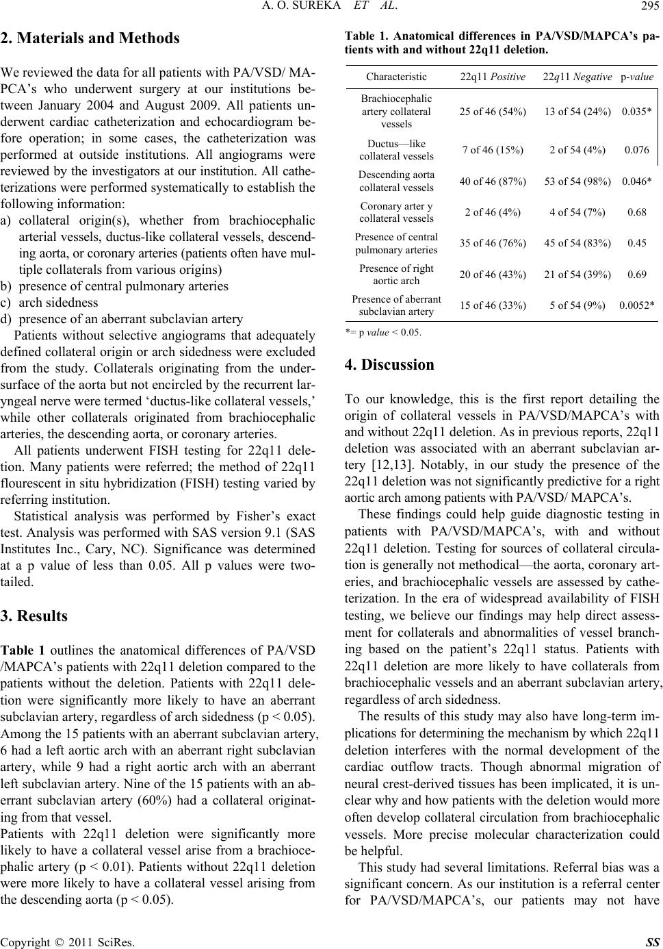
[10] K. Momma, C. Kondo, et al, “Tetralogy of Fallot with
Pulmonary Atresia Associated with Chromosome 22q11
Deletion,” Journal of the Americ an College of Cardiology,
Vol. 27, No. 1, 1996, pp. 198-202.
doi:10.1016/0735-1097( 95)0041 5-7
[11] M. Chessa, G. Butera, et al, “Relation of Genotype 22q11
Deletion to Phenotype of Pulmonary Vessels in Tetralogy
of Fallot and Pulmonary Atresia-Ventricular Septal De-
fect,” Heart, Vol. 79, No. 2, 1998, pp. 186-190.
[12] D. B. McElhinney, B. J. Clark, et al, “Association of
Chromosome 22q11 Deletion with Isolated Anomalies of
Aortic Arch Laterality and Branching,” Journal of the
American College of Cardiology, Vol. 37, No. 8, 2001, pp.
2114-2119. doi:10.1016/S0735-1097(01)01286-4
[13] R. Rauch, A. Rauch, et al, “Laterality of the Aortic Arch
and Anomalies of the Subclavian Artery-Reliable Indica-
tors for 22q11.2 Deletion Syndromes?” European Jour-
nal of Pediatrics, Vol. 163, No. 11, 2004, pp. 642-645.