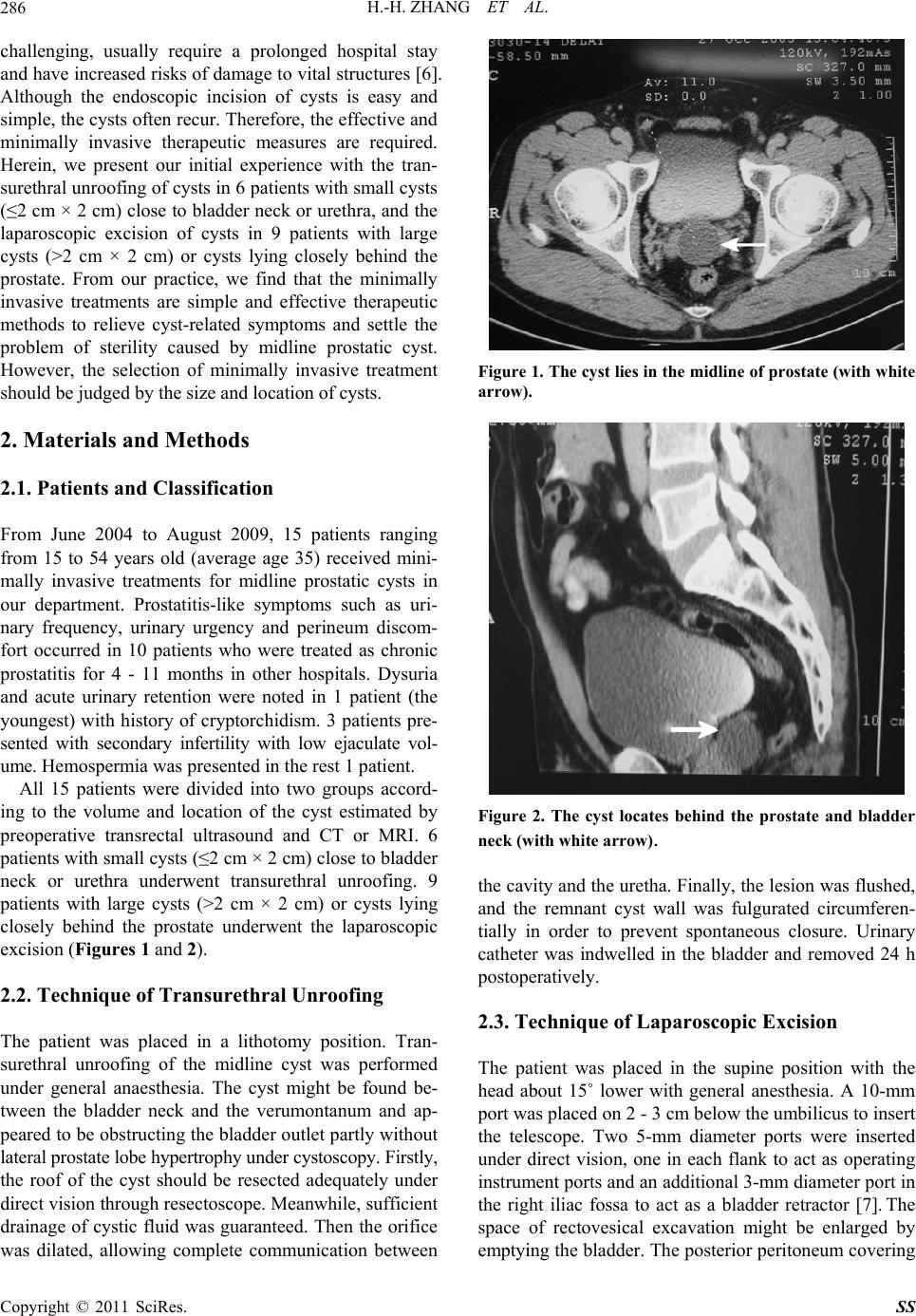
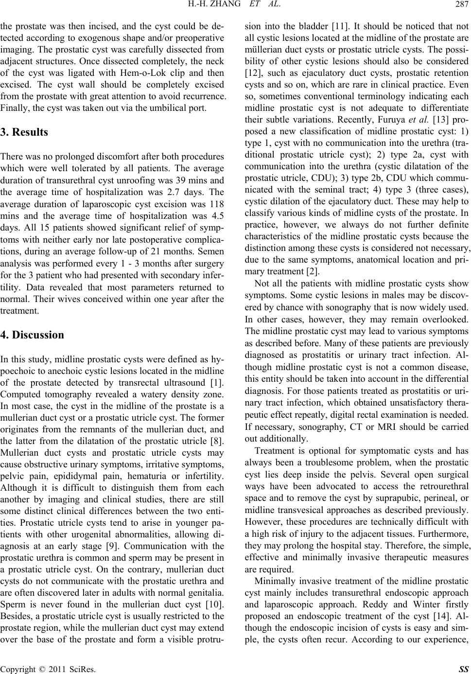
Surgical Science, 2011, 2, 285-289
doi:10.4236/ss.2011.25061 Published Online July 2011 (http://www.SciRP.org/journal/ss)
Copyright © 2011 SciRes. SS
Midline Prostatic Cysts Pres enting with Chro nic Prostatitis
or Secondary Infertility and Minimally Invasive Treatment:
Endoscopic or Laparoscopic Approach?
Hui-hui Zhang, Fan Qi, Jun Wang, Min-feng Chen, Zhuo Li, Xiong-bing Zu
Department of Urology, Xiangya Hospital, Central Sout h Uni versi t y, Changsha, China
E-mail: whzuxb
163.com
Received February 26, 2011; revised May 16, 2011; accepted Jun e 10, 2011
Abstract
Backgrounds: Surgical interventions especially minimally invasive treatments are recommended for symp-
tomatic midline prostatic cysts. The endoscopic unroofing of cysts close to urethra is easy and simple, but it
has little effect on the large cysts and cysts lying deeply, in contrast with the laparoscopic approach. There-
fore, the selection of minimally invasive therapeutic approaches is important. The aim of this study is to de-
scribe our experience in the diagnosis and selection of minimally invasive treatment for midline prostatic
cyst. Methods: 15 cases of midline prostatic cyst were studied.10 cases presented with prostatitis-like symp-
toms, 1 with dysuria and acute urinary retention, 3 with secondary infertility and the rest 1 with hemospermia.
6 patients presented with small cysts (2 cm × 2 cm) close to urethra and underwent transurethral unroofing.
The other 9 patients with large cysts (>2 cm × 2 cm) or cysts lying closely behind the prostate received the
laparoscopic excision. Results: The average duration of transurethral unroofing and laparoscopic excision
was 39 mins and 118 mins respectively, whereas the average time of hospitalization was 2.7 days and 4.5
days respectively. After a follow-up of 21 months, all cases were treated successfully without complications
and recurrence. Their prostatitis-like symptoms disappeared, and the three patients presented with secondary
infertility achieved conception within one year after the operation. Conclusions: A midline prostatic cyst can
present with chronic prostatitis-like symptoms and secondary infertility. It can be cured by minimally inva-
sive treatments, but these procedures should be carefully selected according to the size and location of the
cyst.
Keywords: Midline Prostatic Cyst, Chronic Prostatitis, Secondary Infertility, Minimally Invasive Treatment,
Transurethral Cyst Unroofing, Laparoscopy
1. Introduction
Midline prostatic cysts are infrequent and mostly detect-
ed incidentally during physical examination. Recently,
the widespread availability of transrectal ultrasound has
led to an increase in diagnosis of prostatic cysts. It has
been reported that midline prostatic cysts were observed
in 7.6% of healthy men and 5% of symptomatic outpa-
tients [1,2]. The majority of patients are symptom-free.
However, the enlarged prostatic cyst can compress the
adjacent tissues, such as posterior urethra or bladder neck.
Then, the patient may suffer from lower urinary tract
irritative or obstructive symptoms, epididymitis, perineal
and/or inguinal pain [2], which is often misdiagnosed as .
Likewise, the enlarged prostatic cyst can compress the
seminal vesicles and cause hematospermia, low ejaculate
volume or even infertility [3]. Worse still, malignant
degeneration can occur [4]. Thus, although midline
prostatic cyst is a rare disease, it should never be ne-
glected. When a patient suffers from recurrent lower
urinary tract infection, hematospermia or infertility, the
possible of a midline prostatic cyst should be regarded,
especially when they had received regular treatments.
Surgical intervention is recommended for sympto-
matic cysts. Various approaches have been described
including endoscopic incision or surgical excision by
suprapubic, perineal, or midline transvesical approaches
[5]. However, the open surgical excisions are technically
H.-H. ZHANG ET AL.
286
challenging, usually require a prolonged hospital stay
and have increased risks of damage to vital structures [6].
Although the endoscopic incision of cysts is easy and
simple, the cysts often recur. Therefore, the effective and
minimally invasive therapeutic measures are required.
Herein, we present our initial experience with the tran-
surethral unroofing of cysts in 6 patients with small cysts
(2 cm × 2 cm) close to bladder neck or urethra, and the
laparoscopic excision of cysts in 9 patients with large
cysts (>2 cm × 2 cm) or cysts lying closely behind the
prostate. From our practice, we find that the minimally
invasive treatments are simple and effective therapeutic
methods to relieve cyst-related symptoms and settle the
problem of sterility caused by midline prostatic cyst.
However, the selection of minimally invasive treatment
should be judged by the size and location of cysts.
2. Materials and Methods
2.1. Patients and Classification
From June 2004 to August 2009, 15 patients ranging
from 15 to 54 years old (average age 35) received mini-
mally invasive treatments for midline prostatic cysts in
our department. Prostatitis-like symptoms such as uri-
nary frequency, urinary urgency and perineum discom-
fort occurred in 10 patients who were treated as chronic
prostatitis for 4 - 11 months in other hospitals. Dysuria
and acute urinary retention were noted in 1 patient (the
youngest) with history of cryptorchidism. 3 patients pre-
sented with secondary infertility with low ejaculate vol-
ume. Hemospermia was presented in the rest 1 patient.
All 15 patients were divided into two groups accord-
ing to the volume and location of the cyst estimated by
preoperative transrectal ultrasound and CT or MRI. 6
patients with small cysts (2 cm × 2 cm) close to bladder
neck or urethra underwent transurethral unroofing. 9
patients with large cysts (>2 cm × 2 cm) or cysts lying
closely behind the prostate underwent the laparoscopic
excision (Figures 1 and 2).
2.2. Technique of Transurethral Unroofing
The patient was placed in a lithotomy position. Tran-
surethral unroofing of the midline cyst was performed
under general anaesthesia. The cyst might be found be-
tween the bladder neck and the verumontanum and ap-
peared to be obstructing the bladder outlet partly without
lateral prostate lobe hypertrophy under cystoscopy. Firstly,
the roof of the cyst should be resected adequately under
direct vision through resectoscope. Meanwhile, sufficient
drainage of cystic fluid was guaranteed. Then the orifice
was dilated, allowing complete communication between
Figure 1. The cyst l ies in the midline of prostate (with white
arrow).
Figure 2. The cyst locates behind the prostate and bladder
neck (with white arrow).
the cavity and the uretha. Finally, the lesion was flushed,
and the remnant cyst wall was fulgurated circumferen-
tially in order to prevent spontaneous closure. Urinary
catheter was indwelled in the bladder and removed 24 h
postoperatively.
2.3. Technique of Laparoscopic Excision
The patient was placed in the supine position with the
head about 15˚ lower with general anesthesia. A 10-mm
port was placed on 2 - 3 cm below the umbilicus to insert
the telescope. Two 5-mm diameter ports were inserted
under direct vision, one in each flank to act as operating
instrument ports and an additional 3-mm diameter port in
the right iliac fossa to act as a bladder retractor [7]. The
space of rectovesical excavation might be enlarged by
emptying the bladder. The posterior peritoneum covering
Copyright © 2011 SciRes. SS
H.-H. ZHANG ET AL.287
the prostate was then incised, and the cyst could be de-
tected according to exogenous shape and/or preoperative
imaging. The prostatic cyst was carefully dissected from
adjacent structures. Once dissected completely, the neck
of the cyst was ligated with Hem-o-Lok clip and then
excised. The cyst wall should be completely excised
from the prostate with great attention to avoid recurrence.
Finally, the cyst was taken out via the umbilical port.
3. Results
There was no prolonged discomfort after both procedures
which were well tolerated by all patients. The average
duration of transurethral cyst unroofing was 39 mins and
the average time of hospitalization was 2.7 days. The
average duration of laparoscopic cyst excision was 118
mins and the average time of hospitalization was 4.5
days. All 15 patients showed significant relief of symp-
toms with neither early nor late postoperative complica-
tions, during an average follow-up of 21 months. Semen
analysis was performed every 1 - 3 months after surgery
for the 3 patient who had presented with secondary infer-
tility. Data revealed that most parameters returned to
normal. Their wives conceived within one year after the
treatment.
4. Discussion
In this study, midline prostatic cysts were defined as hy-
poechoic to anechoic cystic lesions located in the midline
of the prostate detected by transrectal ultrasound [1].
Computed tomography revealed a watery density zone.
In most case, the cyst in the midline of the prostate is a
mullerian duct cyst or a prostatic utricle cyst. The former
originates from the remnants of the mullerian duct, and
the latter from the dilatation of the prostatic utricle [8].
Mullerian duct cysts and prostatic utricle cysts may
cause obstructive urinary symptoms, irritative symptoms,
pelvic pain, epididymal pain, hematuria or infertility.
Although it is difficult to distinguish them from each
another by imaging and clinical studies, there are still
some distinct clinical differences between the two enti-
ties. Prostatic utricle cysts tend to arise in younger pa-
tients with other urogenital abnormalities, allowing di-
agnosis at an early stage [9]. Communication with the
prostatic urethra is common and sperm may be present in
a prostatic utricle cyst. On the contrary, mullerian duct
cysts do not communicate with the prostatic urethra and
are often discovered later in adults with normal genitalia.
Sperm is never found in the mullerian duct cyst [10].
Besides, a prostatic utricle cyst is usually restricted to the
prostate region, while the mullerian duct cyst may extend
over the base of the prostate and form a visible protru-
sion into the bladder [11]. It should be noticed that not
all cystic lesions located at the midline of the prostate are
müllerian duct cysts or prostatic utricle cysts. The possi-
bility of other cystic lesions should also be considered
[12], such as ejaculatory duct cysts, prostatic retention
cysts and so on, which are rare in clinical practice. Even
so, sometimes conventional terminology indicating each
midline prostatic cyst is not adequate to differentiate
their subtle variations. Recently, Furuya et al. [13] pro-
posed a new classification of midline prostatic cyst: 1)
type 1, cyst with no communication into the urethra (tra-
ditional prostatic utricle cyst); 2) type 2a, cyst with
communication into the urethra (cystic dilatation of the
prostatic utricle, CDU); 3) type 2b, CDU which commu-
nicated with the seminal tract; 4) type 3 (three cases),
cystic dilation of the ejaculatory duct. These may help to
classify various kinds of midline cysts of the prostate. In
practice, however, we always do not further definite
characteristics of the midline prostatic cysts because the
distinction among these cysts is considered not necessary,
due to the same symptoms, anatomical location and pri-
mary treatment [2].
Not all the patients with midline prostatic cysts show
symptoms. Some cystic lesions in males may be discov-
ered by chance with sonography that is now widely used.
In other cases, however, they may remain overlooked.
The midline prostatic cyst may lead to various symptoms
as described before. Many of these patients are previously
diagnosed as prostatitis or urinary tract infection. Al-
though midline prostatic cyst is not a common disease,
this entity should be taken into account in the differential
diagnosis. For those patients treated as prostatitis or uri-
nary tract infection, which obtained unsatisfactory thera-
peutic effect repeatly, digital rectal examination is needed.
If necessary, sonography, CT or MRI should be carried
out additionally.
Treatment is optional for symptomatic cysts and has
always been a troublesome problem, when the prostatic
cyst lies deep inside the pelvis. Several open surgical
ways have been advocated to access the retrourethral
space and to remove the cyst by suprapubic, perineal, or
midline transvesical approaches as described previously.
However, these procedures are technically difficult with
a high risk of injury to the adjacent tissues. Furthermore,
they may prolong the hospital stay. Therefore, the simple,
effective and minimally invasive therapeutic measures
are required.
Minimally invasive treatment of the midline prostatic
cyst mainly includes transurethral endoscopic approach
and laparoscopic approach. Reddy and Winter firstly
proposed an endoscopic treatment of the cyst [14]. Al-
though the endoscopic incision of cysts is easy and sim-
ple, the cysts often recur. According to our experience,
Copyright © 2011 SciRes. SS
H.-H. ZHANG ET AL.
288
transurethral unroofing is well suitable for small prostatic
cysts (2 cm × 2 cm) close to bladder cavity or urethra.
This procedure is also technically simple, with lower risk
of complications and shorter hospital stay than open sur-
gical procedures. Moreover, it differs from the endo-
scopic incision because it causes a thorough drainage,
avoiding recurrence. Despite these, disadvantages still
exist, because this procedure may forms a wide connec-
tive passage from the cyst to the urethra, bringing the
risk of retrograde ejaculation into the open cyst cavity
and postvoid dribbling. Even worse, It may lead to the
infertility subsequently [15]. We noticed that Cornel, et
al. [16] had reported only 46% patients demonstrated an
improvement in seminal volume and in one patient im-
provement of sperm concentration was seen, after tran-
surethral unfoofing of midline prostatic cyst for subfer-
tile men. They listed two reasons to explain the poor ef-
ficiency. Firstly, vasography and vesicography were not
used in the diagnosis process, a function relationship
between the midline prostatic cyst and obstruction was
not established. Secondly, the cyst walls were not re-
sected. The edges may heal together once again thus al-
lowing the obstruction to return. From our point of view,
an improved patient selection might also influence the
result. Patients with large cysts or cysts lying deep often
receive unsatisfactory effect after transurethral unfoofing
procedure, because it’s difficult to accomplish thorough
unroofing. Thus, we suggest transurethral unfoofing sh-
ould only be done in small cysts close to bladder cavity
or urethra, ensuring that the cysts can be unroofed thor-
oughly as to be adequately open to urethra cavity. Mc-
Dougall, et al. [17] reported the first case of laparoscopic
treatment of prostatic cyst. Although this procedure is a
litter more difficult than transurethral unroofing and need
more time during the operation, it allows a thorough ex-
cision of prostatic cysts effectively obviating the disad-
vantages of transurethral procedure such as postoperative
recurrence, retrograde ejaculation and postvoid dribbling.
Laparoscopic approach could accomplish complete re-
moval of the cyst in a deep and narrow pelvic cavity with
minimal trauma to the normal structures, because it pre-
sents a good surgical view with an excellent exposure of
all surrounding structures, due to the magnification of
surgical field. So this procedure is well suitable for the
resection of large midline prostatic cysts and cysts lying
deep in the pelvis. The common advantages of laparo-
scopic approach also include minor incision, less post-
operative pain, and earlier return to full recovery [18].
In our present study, we designed a criterion that clas-
sified midline prostatic cysts into two groups according
to the size and location of cysts through the transrectal
ultrasound and CT or MRI. Each group received either
the transurethral unroofing or laparoscopic excision of
the cyst based on the criterion. Finally, 6 patients with
small cysts (2 cm × 2 cm) close to bladder cavity or
urethra underwent transurethral unroofing. 9 patients
with large cysts (>2 cm × 2 cm) or cysts lying closely
behind the prostate received the laparoscopic excision.
With the removal of cysts, the symptoms of the present
patients were significantly relieved. No complications
were recorded. It’s worth mentioning that the three pa-
tients presented with secondary infertility with low ejacu-
late volume had remarkable improvement in the ejaculate
volume. In addition, in all of them, conception was ach-
ieved within one year after the operation.
In conclusion, a midline prostatic cyst can cause chro-
nic prostatitis-like symptoms, and secondary infertility,
which is easily neglected. In patients with these symp-
toms, the prostatic midline cyst must be taken into ac-
count for the differential diagnosis. Treatment is neces-
sary for symptomatic patients. Our preliminary results in
15 patients with midline prostatic cysts after minimally
invasive treatment showed encouraging therapeutic ef-
fects. We suggest that minimally invasive treatments are
simple, safe and effective procedures for midline pros-
tatic cysts, but should be selected according to the size
and location. Only if we have a correct selection, a con-
venient operation and good therapeutic effect would be
guaranteed. Despite this, our experience is limited by
insufficient cases. In the future, more cases and longer
follow-up period are required to confirm our experience.
5. References
[1] M. Ishikawa, H.Okabe and T. Oya, “Midline Prostatic
Cyst in Healthy Men: Incidence and Transabdominal
Sonographic Findings,” American Journal of Roent-
genology, Vol. 181, No. 6, 2003, pp. 1669-1672.
[2] P. Dik, T. M. Lock and B. P. Schrier, “Transurethral
Marsupialization of a Medial Prostatic Cyst in Patients
with Prostatitis-Like Symptoms,” Journal of Urology,
Vol. 155, No. 4, 1996, pp. 1301-1304.
doi:10.1016/S0022-5347(01)66251-7
[3] J. S. Mayersak, “Urogenital Sinus-Ejaculatory Duct Cyst:
a Case Report with a Proposed Clinical Classification and
Review of the Literature,” Journal of Urology, Vol. 142,
No. 5, 1989, pp. 1330-1332.
[4] S. W. Warmann, M. Vogel and M. Wehrmann, “Giant
Mullerian Duct Cyst with Malignant Transformation in
15-Year-Old Boy,” Urology, Vol. 67, No. 2, 2006, pp.
424-426. doi:10.1016/j.urology.2005.09.009
[5] L. Coppens, P. Bonnet and R. Andrianne, “Adult Mulle-
rian Duct or Utricle Cyst:clinical Significance and Thera-
peutic Management of 65 Cases,” Journal of Urology,
Vol. 167, No. 4, 2002, pp. 1740-1744.
doi:10.1016/S0022-5347(05)65190-7
[6] C. K. Yeung, J. D. Sihoe and Y. H. Tam, “Laparoscopic
Excision of Prostatic Utricles in Children,” Brithish Jour-
Copyright © 2011 SciRes. SS
H.-H. ZHANG ET AL.
Copyright © 2011 SciRes. SS
289
nal of Urology International, Vol. 87, No. 6, 2001, pp.
505-508.
[7] I. E. Willetts, J. P. Roberts and A. E. MacKinnon,
“Laparoscopic Excision of a Prostatic Utricle in a Child,”
Pediatric Surgery International, Vol. 19, No. 7, 2003, pp.
557-558. doi:10.1007/s00383-003-0993-6
[8] S. Curran, O. Akin and A. M. Agildere, “Endorectal MRI
of Prostatic and Periprostatic Cystic Lesions and Their
Mimics,” American Journal of Roentgenology, Vol.188,
No. 5, 2007, pp. 1373-1379. doi:10.2214/AJR.06.0759
[9] T. D. Schuhrke and G. W. Kaplan, “Prostatic Utricle
Cysts (Mullerian Duct Cysts),” Journal of Urology, Vol.
119, No. 6, 1978, pp. 765-767.
[10] S. G. Chang, I. C. Hwang and J. H. Lee, “Infravesical
Obstruction due to Benign Intraurethral Prostatic Cyst,”
Journal of Korean Medical Science, Vol. 119, No. 6,
2003, pp. 125-126.
[11] M. Barzilai and Y. Ginesin, “A Mullerian Prostatic Cyst
Protruding into the Base of the Urinary Bladder,” Urolo-
gia Internationalis, Vol. 60, No. 3, 1998, pp. 194-196.
doi:10.1159/000030251
[12] R. Yasumoto, M. Kawano and T. Tsujino, “Is a Cystic
Lesion in the Midline of the Prostate a Mullerian Duct
Cyst?” European Urology, Vol. 31, No. 2, 1997, pp. 187-
189.
[13] R. Furuya, S. Furuya and H. Kato, “New Classification of
Midline Cysts of the Prostate in Adults via a Transrectal
Ultrasonography-Guided Opacification and Dye-Injection
Study,” Brithish Journal of Urology International, Vol.
102, No. 4, 2008, pp. 475-478.
[14] Y. N. Reddy and C. C. Winter, “Cyst of the Seminal
Vesicle: A Case Report and Review of the Literature,”
Journal of Urology, Vol. 108, No. 1, 1972, pp. 134-135.
[15] R. Anding, F. Steinbach and T. M. Bernhardt, “Urology
Treatment of Large Prostatic Cyst with Retropubic Inser-
tion of a Fat Tissue Flap,” Journal of Urology, Vol. 164,
No. 2, 2000, pp. 454-455.
doi:10.1016/S0022-5347(05)67390-9
[16] E. B. Cornel, G. R. Dohle and E. J. Meuleman, “Tran-
surethral Deroofing of Midline Prostatic Cyst for Subfer-
tile Men,” Human Reproduction, Vol. 14, No. 9, 1999, pp.
2297-2300. doi:10.1093/humrep/14.9.2297
[17] E. M. McDougall, R. V. Clayman and W. T. Bowles,
“Laparoscopic Excision of Müllerian Duct Remnant,”
Journal of Urology, Vol. 152, No. 2, 1972, pp. 482-484.
[18] J. H. Luo, W. Chen and J. J. Sun, “Laparoscopic Man-
agement of Müllerian Duct Remnants: 4 Cases Report
and Review of the Literature,” Journal of Andrology, Vol.
29, No. 6, 2008, pp. 638-642.
doi:10.2164/jandrol.108.005496