144 Inhibitory Effect of Fentanyl on Phenylephrine-Induced Contraction on Rabbit Aorta
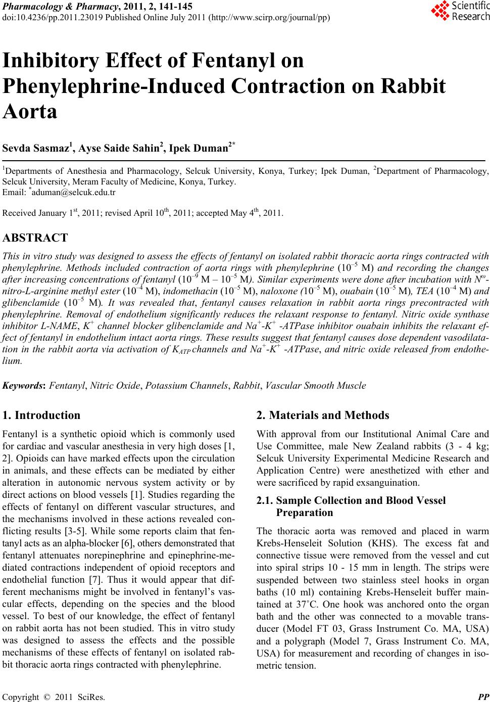
NAME, which is an inhibitor of nitric oxide synthase
enzyme. This finding suggests that NO released from
endothelium may play a partial role in fentanyl-caused
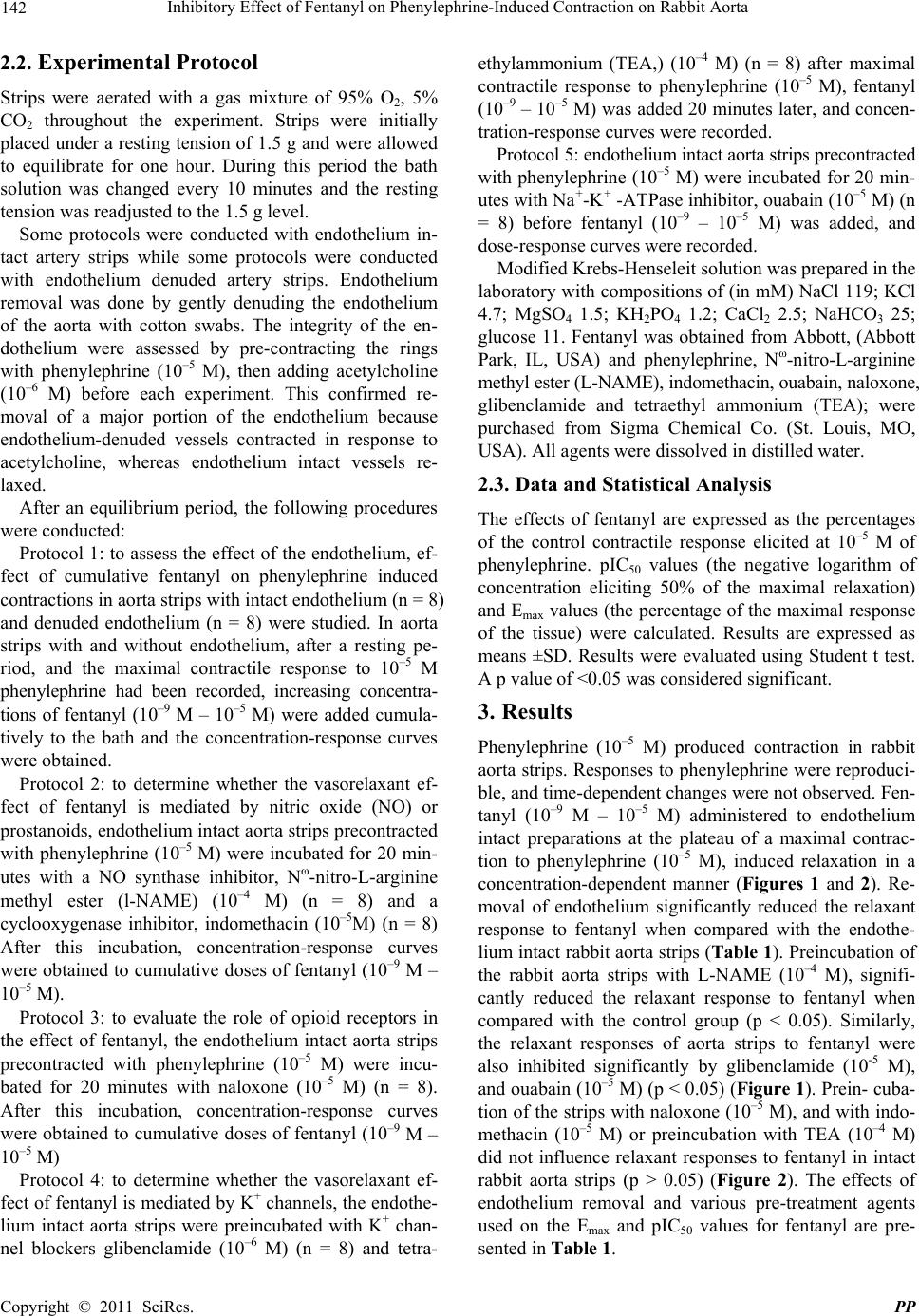
arterial dilation. On the other hand, the block of prostacy-
clin synthesis with indomethacin did not change these
responses. These results suggest that the relaxant effects
of fentanyl on rabbit aorta are partially mediated by en-
dothelium derived NO rather than prostacyclin. Previous
research on the effect of fentanyl on vascular reactivity
on different species and vessels revealed different results.
In studies conducted on human radial artery and porcine
coronary artery, it was shown that the relaxant effect of
fentanyl is independent of the endothelium 3,10. Simi-
larly, in another study conducted on human saphenous
veins the vasorelaxant effect of fentanyl was not reversed
by NO and prostacyclin synthase blockade 11.
Kaye et al. demonstrated that fentanyl has potent va-
sodepressor activity in the pulmonary vascular bed of the
cat and this response may be mediated opiate receptor
sensitive pathways 12. To determine if vascular re-
sponses were mediated by an opioid receptor, dose-re-
sponse studies for fentanyl were repeated in the presence
of naloxone, an opioid-receptor antagonist. Similar to
Sohn et al. 13 who studied the effects alfentanil, an
opioid with a similar structure to fentanyl on phenyle-
phrine-induced contractions in rat aorta, our results also
show that, in the presence of naloxone, fentanyl’s effect
was unchanged, indicating that vasodilatation was not
mediated by opioid receptors in rabbit aorta.
Potassium (K+) channels play an important role in the
regulation of vascular smooth muscle cell membrane
excitability and tonus 14. The activation of K+ chan-
nels in the vascular smooth muscles hyperpolarizes the
cell membranes and closes voltage dependent Ca2+ chan-
nels. These actions decrease intracellular Ca2+ and cause
vascular smooth muscle relaxation. On the contrary, the
inhibition of the channels produces membrane depolari-
zation and vascular smooth muscles contraction. In the
present study, glibenclamide, ATP-sensitive K+ channel
blocker, and TEA, Ca2+-activated K+ channel blocker,
were used to characterize the mechanism of fentanyl-
induced relaxation in rabbit aorta. Relaxant activity of
fentanyl in rabbit aorta was not blocked by TEA, but was
antagonized by glibenclamide, reflecting some role of
the hyperpolarizing ATP-sensitive K-channels in the
mechanism of action of the drug. In their study on human
saphenous veins, Sahin et al. found that the addition of
glibenclamide (ATP sensitive K+ canal blockers) and
TEA (K+ canal blockers activated by Ca++) suppress the
fentanyl induced relaxation responses 12.
Previous studies suggest that the sarcolemmal Na+-K+
-ATPase plays an essential role in the maintenance of the
vascular smooth muscle tone 15-17. In smooth muscles,
this pump can directly contribute to the cell resting
membrane potential by actively pumping more sodium
ions out than potassium ions into the cells 18. Mem-
brane depolarization in response to inhibition of Na+-K+
-ATPase caused Ca++ channels to open and/or in- creased
Ca2+ influx through Na+-Ca2+ exchange mecha- nism. It
has been suggested that normal Na+-K+ -ATPase activity
is necessary for mediating vasorelaxant effects of some
drugs 19,20. In rabbit aorta strips, the relaxant effect of
fentanyl was inhibited by ouabain. Such partial inhibition
has also been obtained with ouabain in human saphenous
vein 12.
Results of this study provides evidence that increasing
doses of fentanyl result in concentration-dependent re-
laxation in the rabbit aorta. Both endothelium-dependent
and endothelium-independent mechanisms are involved
in the relaxation activation of KATP channels and Na+-K+
-ATPase, and nitric oxide released from the endothelium
are responsible for the vasodilation caused by fentanyl.
Further research will be needed to elucidate the exact
pathways of fentanyl-induced vasoreactivity in blood
vessels.
5. References
[1] G. Feuerstein and A. L. Siren, “The Opioid System in
Cardiac and Vascular Regulation of Normal and Hyper-
tensive States,” Circulation, Vol. 75, No. 1, 1987, pp. 125-
129.
[2] G. A. Blaise, T. M. Witzeling, J. C. Sill, P. Vinay and P.
M. Vanhoutte, “Fentanyl is Devoid of Major Effects on
Coronary Vasoreactivity and Myocardial Metabolism in
Experimental Animals,” Anesthesiology, Vol. 72, No. 3,
1990, pp. 535-541.
doi:10.1097/00000542-199003000-00023
[3] A. P. Klockgether-Radke, J. Gravemann, D. Kettler and
G. Hellige, “Influence of Opioids on Vascular Tone of
Isolated Porcine Coronary Artery Segments,” Acta An-
aesthesiologica Scandinavica, Vol. 44, No. 9, 2001, pp.
134-137. doi:10.1034/j.1399-6576.2000.440917.x
[4] R. P. S. Introna, M. T. Bridges, E. H. Yoldlowski, T. E.
Graver and J. K. Pruett, “Direct Effects of Fentanyl on
Canine Coronary Artery Rings,” Life Sciences, Vol. 56,
No. 15, 1995, pp. 1265-1273.
doi:10.1016/0024-3205(95)00072-0
[5] F. Karasawa, V. Iwanov and R. F. Moulds, “Effects of
Fentanyl on the Rat Aorta are Mediated by Alpha-
Adrenoceptors rather than by the Endothelium,” British
Journal of Anaesthesia, Vol. 71, No. 6, 1993, pp. 877-880.
doi:10.1093/bja/71.6.877
[6] K. E. Park, J. T. Sohn, Y. S. Jeong, H. J. Sung, I. W. Shin,
H. K. Lee and Y. K. Chung, “Inhibitory Effect of Fen-
tanyl on Phenylephrine-Induced Contraction of the Rat
Aorta,” Yonsei Medical Journal, Vol. 50, No. 3, 2009, pp.
414-421. doi:10.3349/ymj.2009.50.3.414
[7] D. A. White, J. A. Reitan, N. D. Kein and S. J. Thorup,
C
opyright © 2011 SciRes. PP