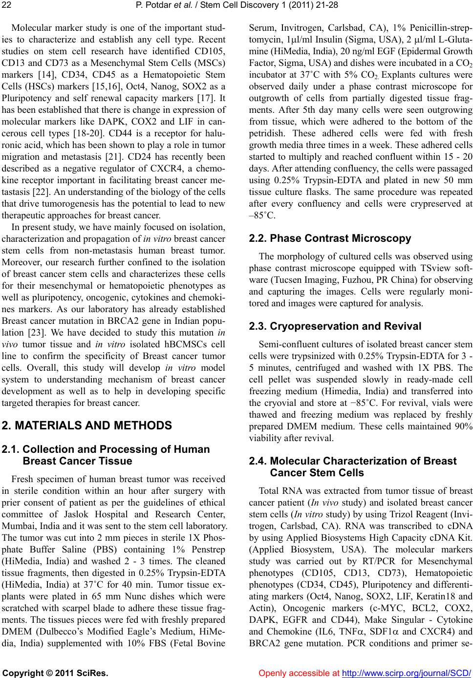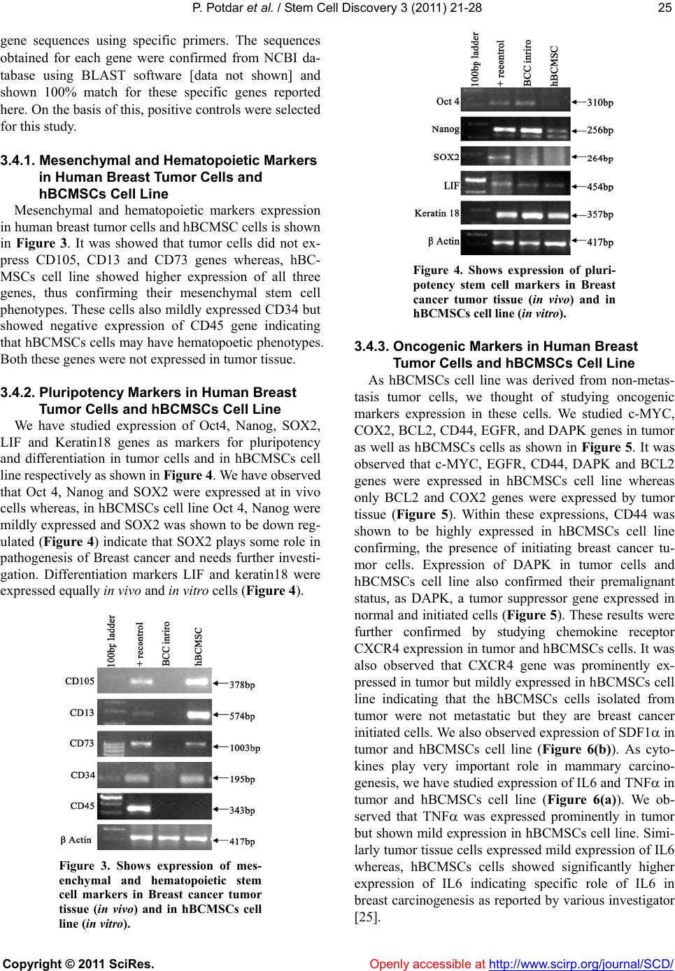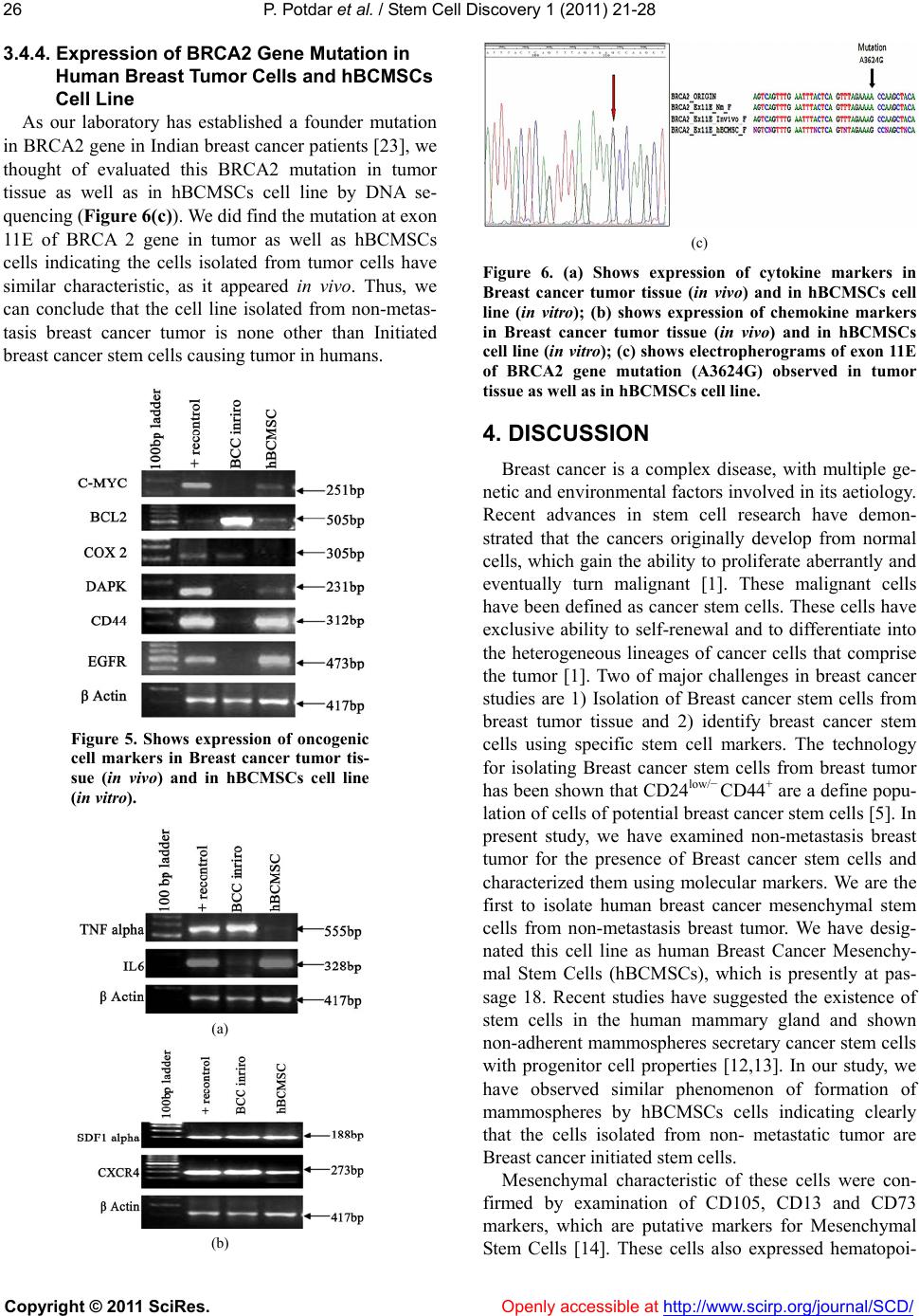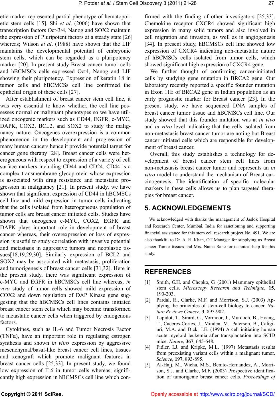
P. Potdar et al. / Stem Cell Discovery 1 (2011) 21-28
Copyright © 2011 SciRes. http://www.scirp.org/journal/SCD/Openly accessible at
28
the National Academy of Sciences, 100, 3983-3988.
[6] Bonnet, D. and Dick, J.E. (1997) Human acute myeloid
leukemia is organized as a hierarchy that originates from
a primitive hematopoietic cell. Nature Medicine, 3,
730-737.
[7] Collins, A.T., Berry, P.A., Hyde, C., Stower, M.J. and
Maitland, N.J. (2005) Prospective Identification of tu-
morigenic prostate cancer stem cells. Cancer Research,
65, 10946-10951.
[8] Hemmati, H.D., Nakano, I., Lazareff, J.A., Master-
man-Smith, M., Geschwind, D.H., Bronner-Fraser M.
and Komblurn, H.I. (2003) Cancerous stem cells can
arise from pediatric brain tumors. Proceedings of the Na-
tional Academy of Sciences, 100, 15178-15183.
[9] Kim, C.F., Jackson, E.L., Woolfenden, A.E., Lawrence,
S., Babar, I., Vogel, S., Crowley, D., Bronson, R.T. and
Jacks, T. (2005) Identification of bronchioalveolar stem
cells in normal lung and lung cancer. Cell, 121, 823-835.
[10] O’Brien, C.A., Pollett, A., Gallinger, S. and Dick, J.E.
(2007) A human colon cancer cell capable of initiating
tumor growth in immunodeficient mice. Nature, 445,
106-110.
[11] Potdar, P.D. and Subedi, R.P. (2011) Defining Molecular
Phenotypes of Mesenchymal and hematopoietic Stem
Cells derived from Peripheral blood of Acute Lympho-
cytic Leukemia patients for regenerative stem cell ther-
apy. Journal of Stem cells & Regenerative Medicine, 7,
29-40.
[12] Grimshaw, M.J., Cooper, L., Papazisis, K., Coleman, J.A.,
Bohnenkamp, H.R., Chiapero-Stanke, L., Taylor-Papa-
dimitriou, J. and Burchell, J.M. (2008) Mammosphere-
culture of metastatic breast cancer cells enriches for tu-
morigenic breast cancer cells. Breast Cancer Research,
10, 52.
[13] Wicha, M., Liu, S. and Dontu, G. (2006) Cancer stem
cells: an old idea – a paradigm shift. Cancer Research,
66, 1883-1890.
[14] Barry, F.P., Boynton, R.E., Haynesworth, S., Murphy,
J.M. and Zaia, J. (1999) The monoclonal antibody SH-2,
raised against human mesenchymal stem cells, recog-
nizes an epitope on endoglin (CD105). Biochemical and
Biophysical Resear ch Comm un i c a t io n s, 265, 134-139.
[15] Healy, L., May, G., Gale, K., Grosvel, F., Greaves, M.
and Enver, T. (1995) The stem cell antigen CD34 func-
tions as a regulator of hemopoietic cell adhesion. Pro-
ceedings of the National Academy of Sciences, 92,
12240-12244.
[16] Ogata, K., Satoh, C., Tachibana, M., Hyodo, H., Tamura,
H., Dan, K., Kimura, T., Sonoda, Y. and Tsuji, T. (2005)
Identification and hematopoietic potential of CD45-
clonal cells with very immature phenotypes (CD45-
Cd34-Cd38-Lin-) in patients with myelodysplastic syn-
dromes. Stem Cell, 23, 619-630.
[17] Olamura-Nakanishi, S., Saito, M., Niwa, H. and Isshil-
cawa, F. (2005) Oct-3/4 and Sox2 regulate Oct-3/4 gene
in embryonic stem cells. Journal of Biological Chemistry,
22, 5307-5317.
[18] Prescott, S.M. and Fitzpatrick, F.A. (2000) Cyclooxy-
genase-2 and carcinogenesis. Biochimica et Biophysica
Acta, 1470, M69-M78.
[19] Katzenellenbogen, R.A., Baylin, S.B. and Herman, J.G.
(1999) Hypermethylation of the DAP-Kinase CpG island
is a common alteration in B-cell malignancies. Blood, 93,
4347-4353.
[20] William, R.L., Hilton, D.J., Pease, S., Willson, T.A.,
Stewart, C.L., Gearing, D.P., Wagner, E.F., Metcalf, D.,
Nicola, N.A. and Gough, N.M. (1988) Myeloid leukae-
mia inhibitory factor maintains the developmental poten-
tial of embryonic stem cells. Nature, 336, 684-687.
[21] Draffin, J.E., McFarlane, S., Hill, A., Johnston, P.G. and
Waugh, D.J. (2004) CD44 potentiates the adherence of
metastatic prostate and breast cancer cells to bone mar-
row endothelial cells. Cancer Research, 64, 5702-5711.
[22] Schabeth, H., Runz, S., Joumaa, S. and Altevogt, P. (2006)
CD24 affects CXCR4 function in pre-β lymphocytes and
breast carcinoma cells. Journal of Cell Science, 119,
314-325.
[23] Potdar, P.D. and Bisht, S. L. (2009) Identification and
screening for novel mutations in exon 10 and exon 11 of
BRCA 1 and BRCA 2 genes in hereditary and sporadic
breast cancer patients in Indian population- concept for
biomarkers for early detection. Annals of Oncology, 20,
51.
[24] Potdar, P.D. and Sutar, J. P. (2010) Establishment and
molecular characterization of mesenchymal stem cell
lines derived from human visceral & subcutaneous adi-
pose tissues. Journal of Stem cells & Regenerative Medi-
cine, 6, 1-10.
[25] Knupfer, H. and Preiss, R. (2007) Significance of inter-
leukin-6 (IL-6) in breast cancer. Breast Cancer Res Treat,
102, 129-35.
[26] Shi, W., Wang, H., PanYijie, G., Yunqian, G., Pei, G.,
(2006) Regulation of the Pluripotency Marker Rex-1 by
NANOG and SOX2. Journal of Biological Chemistry,
281, 23319-23325.
[27] Ku, N., et al. (1997) Mutation of human keratin 18 in
association with cryptogenic cirrhosis. Journal of Clini-
cal Investigation, 99, 19-23.
[28] Malumbres, M. and Barbacid, M. (2001) To cycle or not
to cycle: a critical decision in cancer. Nature Reviews
Cancer, 1, 222-231.
[29] Nesbit, C.E., Tersak, J.M. and Prochownik, E.V. (1999)
MYC oncogenes and human neoplastic disease. Onco-
gene, 18, 3004-3016.
[30] Bhargava, R., Gerald, W.L., Li, A.R., Pan, Q., Lal, P.,
Ladanyi, M. and Chen, B. (2005) EGFR gene amplifica-
tion in breast cancer: Correlation with epidermal growth
factor receptor mRNA and protein expression and HER-2
status and absence of EGFR-activating mutations. Mod-
ern Pathology, 18, 1027-1033.
[31] Swellam, M., Ismail, M., Eissa, S., Hamdy, M. and Mok-
htar, N. (2004) Emerging role of P53, Bcl-2 and telom-
erase activity in egyptian breast cancer patients. IUBMB
Life, 56, 483-490.
[32] Chen, Y., Shi, L., Zhang, L., Li, R., Liang, J., Yu, W., Sun,
L., Yang, X., Wang, Y., Zhang, Y. and Shang, Y. (2008)
The molecular mechanism governing the oncogenic po-
tential of SOX2 in breast cancer. The Journal of Bio-
logical Chemistry, 283, 17969-17978.
[33] Charafe-Jauffret, E., et al., (2006) Gene expression pro-
filing of breast cell lines identifies potential new basal
markers. Oncogene, 25, 2273-2284.
[34] Smith, M.C.P., Luker, K.E., Garbow, J.R., et al., (2004)
CXCR4 regulates growth of both primary and metastatic
breast cancer. Cancer Research, 64, 8604-8612.