New Journal of Glass and Ceramics, 2011, 1, 49-52
doi:10.4236/njgc.2011 .12008 Published Online July 2011 (http://www.SciRP.org/journal/njgc)
Copyright © 2011 SciRes. NJGC
49
Mechanochemical Synthesis of Nano Calcium
Silica t e Particles a t Room Temperature
Shiv Prakash Singh, Basudeb Karmakar*
Glass Science and Tech nolo gy Section, Glass Division, Central Glass and Ceramic Res earch Inst itute, Ko lkata, India.
Email: basudebk@cgcri.res.in
Received March 24th, 2011; revised April 21st, 2011 ; accepted April 28th, 2011.
ABSTRACT
Nano-sized calcium silicate powders were synthesized at room temperature by the new mechanochemical method using
a high energy planetary ball mill. The formation of c alcium silicate from its ra w materials (calcium carbonate and de-
hydrated silica gel) was monitored by the XRD analysis with progression of ball milling. It is observed that the synthet-
ic process comes to an end through the following three sequential stages: comminution of raw materials, recombination
of comminuted raw materials to final product, and comminution of final product to smaller sizes. The nanostructure of
the synthesized powder was realized by the FESEM photomicrograph, TEM image and XRD analyses. These analytical
observations have revealed that the nano-sized polycrystalline calcium silicate particles are formed after about 6 h of
ball milling and they are spheroidal in shape. The average particle size of the as-genera ted calcium silicate nanocrys-
talline powders is found to be around 21 nm which decreases with increasing ball milling but increases with annealing
at elevated temperature.
Keywords: Mechanochemical Processing, Calcium Silicate Na noparticle, XRD, FES EM, TEM
1. Introduction
Calcium silicate has received significant attention be-
cause of their potential applications as host of phosphors
and ceramic insulators [1-3]. Nanosized calcium silicate
material has been studied over the past years because of
their significant properties which are different from their
bigger sizes. Conventionally, calcium silicate is prepared
by the solid state reaction of CaO or CaCO3 and quartz
(SiO2) at 1150˚C - 1200˚C for several hours [1-2]. It is
also prepared by the chemical methods such as combus-
tion, sol-gel, co-precipitation, etc. routes followed by
heat treatment at various temperatures [3-5]. All these
methods are limited by some of their inherent disadvan-
tages, for example, requirement of high temperature,
long processing time, low yield or hazardous to health
and environment.
Recently, synthesis of nanomaterials by mechano-
chemical route opens up a new page in the nanoscience
and nanotechnology. It is a quite simple, energy saving,
low-cost, industrially scalable and viable
non-conventional technique for the preparation of ho-
moge neou s and nano -sized (1 - 100 nm) multicomponent
materials [6-9]. It is an environmentally friendly sol-
vent-free and waste- free synthetic route as well unlike
chemical methods. The main advantage of the mechano-
chemical method is that the solid-state reactions are car-
ried out at room temperature instead of high temperature.
Practically, it is a high energy milling process that in-
volves repeated mixing, deformation, comminuting,
welding and re-welding of the reactant powder particles
in a closed vial of a planetary ball mill [10-13]. Here, the
solid-state reactions are progressed by the kinetic energy
transferred from the milling balls to the milled powders.
Its versatility gives the impressions that the mechano-
chemical method is a promising novel technique for the
synthesis of various advanced nanopowder materials. To
the best of our knowledge, the synthesis of calcium sili-
cate nanoparticles by the mechanochemical method has
not been explored previously.
In this letter, we demonstrate the mechanochemical
synthesis of calcium silicate nanoparticles from the cal-
cium carbonate and silica gel at room temperature with-
out applying additional heat treatment. Its synthesis is
recognized by the FESEM photomicrograph, TEM image
and X R D ana lyses.
Mechan ochemical Synthesis of Nano Calcium Silicate P ar ticles at Room Temperature
Copyright © 2011 SciRes. NJGC
50
2. Experimental Pro cedure
The starting raw materials were high purity calcium car-
bonate, CaCO3 (99%, Fluka, Switzerland) and dehy-
drated silica gel, SiO2 (99%, Fluka, Switzerland). A
stoichiometric mixture of these raw materials equivalent
to 5 g calcium s ilicate was ball milled in a 500 ml zirco-
nia jar with 100 numbers of 10 mm diameter zirconia
milling balls at 300 rpm in a high energy planetary ball
mill (PM 100, Retsch). Separate samples were prepared
by varyin g t he mil li n g d ura ti o n s uch as 1 , 3, 6 , 1 0 , 15, 2 0
and 26 h. One sample of 26 h duration was annealed at
750oC for 10 h in air to confirm the completion of syn-
thesis by the mechanochemical method and also to ob-
serve the effect of heating on the mechanochemically
synthesized calcium silicate at elevated temperature.
The XRD patterns of the staring raw materials mixture
and as-milled product samples were recorded at 25oC
with X’pert P ro MPD diffractometer (PANalytical) oper-
ating at 45 kV and 35 mA using Ni-filtered CuKα (λ =
1.5406 Å) radiation and the X’celerator with step size
0.05˚ (2θ) and step time 0.5 sec from 10˚ to 80˚. FESEM
photomicrographs were recorded with a Gemini Zeiss
Supra™ 35VP Model (Carl Zeiss) instrument using an
accelerating voltage of 4.9 kV. TEM images were taken
using a FEI instrument (TECHNAI G2) operating at the
accelerating voltage of 300 kV.
3. Results and Discussion
Calcium silicate (CaSiO3) is synthesized from the raw
materials, calcium carbonate (CaCO3) and dehydrated
silica (SiO2) gel, by the mechanochemical method using
a high ener gy planetar y ball mill. It s solid state s ynthesis
reaction which occurred in this method at room tempera-
ture could be represented by
3 232
CaCO SiOCaSiO CO+ =+↑
(1)
The synthesis of nano calcium silicate particles has
found to be progressed in this method through the
following three sequential stages. It is also more clearly
depicted in Figure 1.
Mixture of raw
materia ls
Commin ution of
raw materials
(Stage-I)
Recombination of comminuted
raw materials to
final pr oduc t
(Stage-II)
Commin ution of
final pr oduc t to
sma l ler sizes
(Stage-III)
The effects of milling time on the generation and
subsequent comminution of calcium silicate are moni-
tored by the XRD patterns by comparing with its standard
XRD pattern (JCPDS File Card No. 27-0088). These are
shown in Figure 2. The particle sizes were calculated
using the following Scher rer’s formula [14] :
D0.9/Bcos2 ( peak )
λθ
= (2)
where D is diameter of the calcium silicate particle, λ is
wavelength of CuK
α
radiation (
λ
= 1.5406 Å), B is full
width at half maximum (FWHM) of the intensity peak
and 2θ is diffraction angle at maxima of high intensity
peak in the patterns.
Figure 2 shows that during early stage of milling (up
to 3 h) there is no evidence of calcium silicate formation
but only the comminution of starting raw materials, e.g.,
calcium carbonate, down to 11 nm. Then after the particle
size instantl y increases (2 1 nm) up to 6 h of milling wit h
generation of calcium silicate. Between 3 and 6 h of mil-
ling the qualitative change of XRD patterns due to cal-
cium silicate formation is clearly visible. This observa-
tion sho ws that during th is per iod of milling the particles
are severely deformed, amorphized and re-combined due
to high e ner gy impacts of the millin g balls. At 6 h of mil-
ling, it clearly shows the calcium silicate peaks which
confirm the formation of calcium silicate crystalline
phase by the mechanochemical method. Subsequently,
the particle size of the as-generated calcium silicate de-
creases down to 13 nm and the peak intensities increases
with increase in milling time, that is, 6 h onward. Thus,
the synthesis of more calcium silicate gradually increases
along with reduction of its sizes with increasing milling
time.
Further qualitative changes are observed after 10 h of
milling in the XRD patterns. It shows more prominent
development of <320>, <–432> and <–921> hkl planes
of calcium silicate (JCPDS File Card No. 27-0088). The
information obtained from XRD patterns analyses agree
well with the field emission scanning electron microsco-
py (FESEM) photomicrograph as shown in Figure 3(a).
FESEM observations revealed the material morphology
with milling time by mechanochemical treatment. The
as-prepared sample after 10 h milling indicates that the
synthesized calcium silicate particles are joined with each
other due to recombination reactions thereby increases
the sizes as observed in the XRD. This is already shown
in the Figure 1. The transmission electron microscopy
(TEM) image of the neck of two particles and its SAED
pattern are shown in Figure 4(a). The formation of cal-
cium silicate was started around 6 h of milling. Further
milling, the calcium silicate gradually developed broad
XRD peaks without any characteristic changes of the
Mechan ochemical Synthesis of Nano Calcium Silicate P ar ticles at Room Temperature
Copyright © 2011 SciRes. NJGC
51
Figure 1 . The particle size as a function of milling time.
Figure 2. X-ray diffraction patterns of samples milled for
different duration and the 26 h milled sample annealed at
750oC (Top). The standard pattern (JCPDS File Card No.
27-0088) of calcium silicate (CS) is also shown for compari-
son (Bottom).
patterns which point towards the gradual reduction of its
particle size with increasing milling time, as realized by
evaluating using the Equation (2).
The main c ha nges o ccur aft er 2 6 h o f mill ing i s that it s
particle sizes reduced to about 13 nm as calculated from
XRD. The morphological changes of 26 h milled calcium
silicate particles are also clearly visible in the FESEM
photomicrograph as shown in Figure 3(b). The synthe-
sized calcium silicates separated from each other and get
their defined morphological shapes which are spheroidal
in nature. The transmission electron microscope (TEM)
image and the SAED pattern, as shown in Figure 4(b),
illustrate the polycrystallinity of the mechanochemically
synthesized calcium silicate. The TEM image also shows
the rough and irregular characteristics of the surfaces of
calcium silicate particles. It is very difficult to conclude
the same with only the FESEM photomicrograph as
shown in the Figure 3(b). However, the TEM image
gives the unambiguo us vie w.
We have annealed the synthesized calcium silicate af-
ter 26 h of milling at 750oC for 10 h in an electrical fur-
nace to confirm the completion of synthesis of calcium
silicate by the mechanochemical method at 6 h or on-
ward. Its XRD pattern is shown at the top of the Figure
2. It is seen that the width of peaks of the XRD pattern of
the annealed powder reduces with increase in their inten-
sities but without any additional peak. Thus, it indicates
only the growing of the calcium silicate particle size due
to fusion or sintering of smaller particles on annealing at
750oC. The heat treated powders do not exhibit any new
XRD peak which clearl y depicts tha t the solid state reac-
tion has completed in the mechanochemical process and
no further new compound is formed even though after
application of high temperature.
4. Conclusions
Synthesis of calcium silicate nanoparticles at room
temperature by the new mechanochemical method has
been demonstrated. The formation of calcium silicate ha s
been recognized by the FESEM photomicrograph, TEM
image and XRD analyses. It is observed that the syn
thetic process comes to an end through the following
(a)
(b)
Figure 3. FESEM photomicrographs of samples obtained
after (a) 10 and (b) 26 h of milling.
Mechan ochemical Synthesis of Nano Calcium Silicate P ar ticles at Room Temperature
Copyright © 2011 SciRes. NJGC
52
(a)
(b)
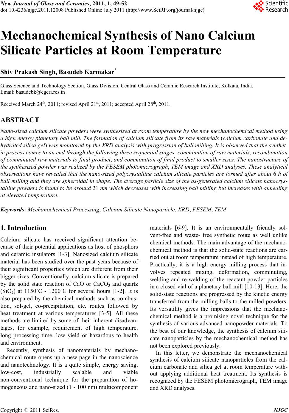
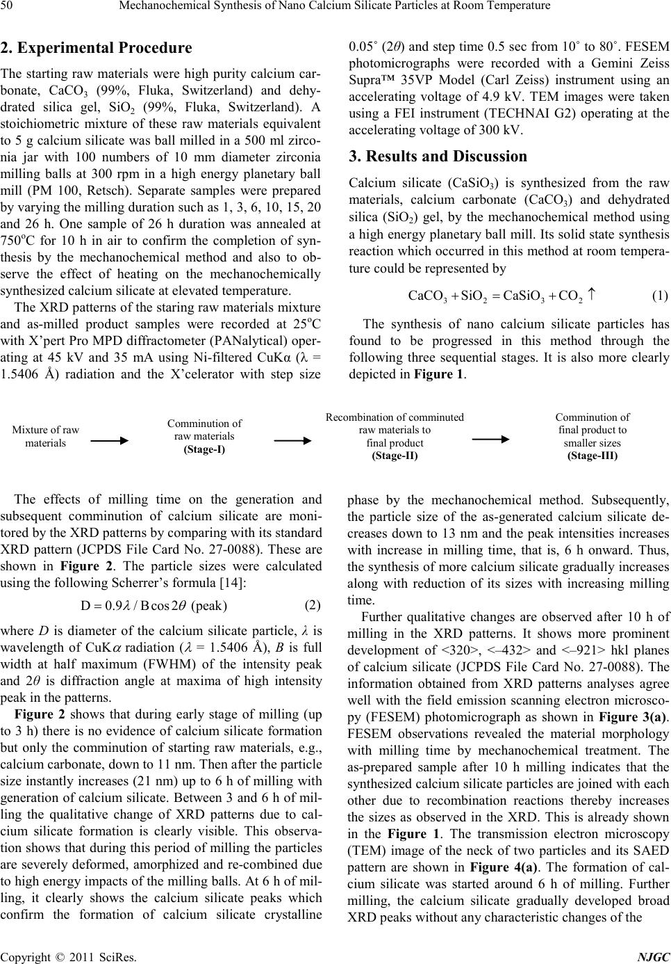
Figure 4. TEM images and SAEDs (insets) of samples ob-
tained after (a) 10 and (b) 26 h of milling .
three sequential stages: comminution of raw materials,
recombination of comminuted raw materials to final
product, and comminution of final product to smaller
sizes. The nano-sized polycrystalline calcium silicate
particles are found to generate after about 6 h of ball mil-
ling and they are spheroidal in shape. The average par-
ticle size of the as-generated particles is found to be
around 21 nm which decreases down to 13 nm with in-
creasing ball milling but increases up to 30 nm after an-
nealing at 750oC. The annealed sample does not show
any new XRD peaks, therefore, it authenticates the com-
pletion of the solid state synthesis reactions of calcium
silicate at room temperature by the mechanochemical
method using high energy planetary ball milling. We
beli eve tha t thi s work would op en a ne w vista in the area
of nanopar tic le research.
5. Acknowledg ements
The authors grateful ly ack nowledge the fina ncial suppo rt
of NMITLI, CSIR, New Delhi. They gratefully thank
Director o f the institute for his kind per missio n to p ublish
this paper. The technical supports obtained from the in-
frastructural facilities (XRD and Electron Microscope
Divisions) of this institute are also thankfully acknowl-
edged.
REFERENCES
[1] W. M. Yen and M. J. Weber, “Inorganic Phosphors:
Compositions, Preparation and Optical Properties,” CRC
Press, Boca Raton, 2004. doi:10.1201/9780203506325
[2] S. Kamiya and H. Mizuno, Phosphors for Lamps,” I n: S.
Shionoya and W. N. Yen, Eds., Phosphor Handbook,
CRC Press, Boca Raton, 1999, pp. 389-444.
[3] T. I. Khristov, N. V. Popovich and S. S. Galaktionov,
“Calcium Silicate Phosphors Obtained by the Sol-Gel
Method,” Glass and Ceramics, Vol. 51, No. 9-10, 1994,
pp. 7-12. doi:10.1007/BF00679827
[4] S. J. Dhoble, N. S. Dhoble and R. B. Pode, “Preparation
and Ch aracterizat ion of Eu 3+ Activat ed CaSiO3, (C aA)SiO3
[A = Ba or Sr] Phosphors,” Bulletin of Materials Scien ce,
Vol. 26, N o. 4, 20 03, pp. 37 7-382.
doi:10.1007/BF02711179
[5] Y. Nish isu, M. Kobayashi, H. Ohya and T. Akiya, “Prep-
aration of Eu-Doped Alkaline-Earth Silicate Phosphor
Particles by Using Liquid-Phase Synthesis Method,”
Journal of Alloys and Compounds, Vol. 408-412, 2006,
pp. 898-902. doi:10.1016/j.jallcom.2005.01.101
[6] C. L. Chew, A. Srinivas, T. Sritharan and F. Y. C. Boey,
“Mechanochemical Activation of Strontium Bismuth
Tantalate Synthesis,” Scripta Materialia, Vol. 53, 2005,
pp. 1197 -1199. doi:10.1016/j.scriptamat.2005.07.009
[7] Q. Zhang and F. Saito, “Mechanochemical Synthesis of
Lanthanum Aluminate by Grinding Lanthanum Oxide
with Transition Alumina,” Journal of the American Ce-
ramic Society, Vol. 83, No. 2, 2000, pp . 439-441.
doi:10.1111/j.1151-2916.2000.tb01215.x
[8] D. Domanski, G. Urretavizcaya, F. J. Castro and F. C.
Gennari, “Mechanochemical Synthesis of Magnesium
Aluminate Spinel Powder at Room Temperature,” Journal
of the American Ceramic Society, Vol. 87, No. 11, 2004,
pp. 2020 -2024. doi:10.1111/j.1151-2916.2004.tb06354.x
[9] I. Bergmann, V. Šepelák and K. D. Becker, “Preparation
of Nanoscale MgFe2O4 via Non-Conventional Mechano-
chemical Ro ute,” Solid State Ionics, Vol. 177, No. 19-25,
2006, pp . 1865-1868.
[10] T. Rojac, M. Kosec, B. Malič and J. Holc, “The Applica-
tion of a Milling Map in the Mechanochemical Synthesis
of Ceramic Oxides,” Journal of the European Ceramic
Soci ety, Vol. 26, No. 16, 2006, pp. 3711-3716.
doi:10.1016/j.jeurceramsoc.2005.11.013
[11] J. Lee, Q. Zhang and F. Saito, “Mechanochemical Syn-
thesis of Lanthanum Oxyfluoride from Lanthanum Oxide
and Lanthanum Fluoride,” Journal of the American Ce-
ramic Society, Vol . 84, No. 4, 20 01, pp . 863-865.
doi:10.1111/j.1151-2916.2001.tb00753.x
[12] T. Sritharan, F. Y. C. Boey, A. Srinivas, “Synthesis of
Complex Ceramics by Mechanochemical Activation,”
Journal of Materials Processing Technology, Vol. 192-
193, 2007, pp. 255-258.
[13] M. Shoji and K. Sakurai, A Versatile Scheme for Pre-
paring Single-Phase Yttrium Oxysulfate Phosphor,
Journal of Alloys and Compounds, Vol. 426, No. 1-2,
2006, pp . 244-246. doi:10.1016/j.jallcom.2005.12.110
[14] B. D. Cullity, Elements of XRay Diffraction,” 2nd Edi-
tion, Addison-Wesley, Massachusetts, 1978.