International Journal of Clinical Medicine, 2015, 6, 831-837
Published Online November 2015 in SciRes. http://www.scirp.org/journal/ijcm
http://dx.doi.org/10.4236/ijcm.2015.611109
How to cite this paper: Nallapati, S.N., Otikunta, A.N., Reddy, Y.V.S. and Srinivas, R. (2015) E/e’ as a Predictor of Short-
Term Survival Following ST-Elevation Myocardial Infarction. International Journal of Clinical Medicine, 6, 831-837.
http://dx.doi.org/10.4236/ijcm.2015.611109
E/e’ as a Predictor of Short-Term
Survival Following ST-Elevation
Myocardial Infarction
Sivaprasad Naidu Nallapati, Adikesava Naidu Otikunta, Y. V. Subba Reddy, Ravi Srinivas
Depart ment of Cardiology, Osmania General Hospital and Osmania Medical College, Hyderabad, India
Received 15 May 2015; accepted 16 November 2015; published 19 Novembe r 2015
Copyright © 2015 by authors and Scientific Research Publishing Inc.
This work is licensed under the Creative Commons Attribution International License (CC BY).
http://creativecommons.org/licenses/by/4.0/
Abstract
Background: We examined the usefulness of tissue Doppler imaging in evaluating the diastolic
dysfunction and assessed the prognostic value of ratio between early mitral inflow velocity and
mitral annular early diastolic velocity (E/e’) in unselected patients with acute ST-elevation myo-
cardial infarction (STEMI). Methods: Patients presenting with acute STEMI at Osmania General
Hospital, Hyderabad, India between January-2012 and June-2012 were examined in this study. All
patients underwent echocardiographic examination comprising Doppler assessment of transmi-
tral flow velocities (peak E-wave velocity) and Doppler tissue imaging of the medial mitral valve
annulus (e’). All patients were followed up for 6 months and all-cause mortality was measured as
the study endpoint. Role of E/e’ ratio as a predictor of survival after acute STEMI was evaluated by
a comparative analysis of patients with E/e’ ≤15 and >15. Results: A total of 50 patients with acute
STEMI (mean age: 52.2 ± 13.2 years; 80% males) were analyzed in this study. Of them, 23 (46%)
patients had an E/e’ >15. Clinical parameters such as Killip class ≥2 as well as left ventricular ejec-
tion fraction of <40% were significantly associated with E/e’ >15. Five (10%) patients died during
the 6-month follow-up period. All deaths occurred among patients from the E/e’ >15 group, indi-
cating that E/e’ is an effective predictor of overall survival. Conclusion: E/e’ ratio was identified as
a strong predictor of survival after acute myocardial infarction and can be suitable for risk-s tr ati-
fication of patients in this setting. Further studies are warranted to substantiate the findings.
Keywords
ST-Elevated Myocardial Infarction, Echoca rdiography, Transmitral Flow Velocity, Medial Mitral
Valve Annulus, E/e’ Ratio, Survival
S. N. Nallapati et al.
832
1. Introduction
Coronary artery disease is the leading cause of morbidity and mortality worldwide, and acute myocardial infarc-
tion is the most common mode of its presentation [1]. In patients with myocardial infarction, measurement of
left ventricular diastolic dysfunction is important in decision-making and predicting the mortality because it is
associated with progressive left ventricular dilatation, development of heart failure, and cardiac death [2]. Al-
though invasive cardiac catheterization is the gold standard to measure le ft ventric ular filling, it car ries the risk
of subsequent complications [3]. In this regard, echocardiography using tissue Doppler imaging has emerged as
a valuable non-invasive tool, with similar predictive value as compared to cardiac catheterization [3] [4]. Dopp-
ler tissue imaging of mitral valve annulus appears to be particularly useful, and reduced early mitral annulus ve-
locity (e’) indicates impaired myocardial relaxation [2]. In addition, the ratio of early transmitral flow velocity
(E) to early diastolic sep tal mitral annulus velo city (E/e’) correlate s well with left ventricular diastolic d ysfunc-
tion [5] [6]. The prognostic value of E/e’ >1 5 followi ng myocardial infarc t ion has been evaluated previously in a
study by Hillis et al. [2]. They indicated that E/e’ is a powerful predictor of survival after acute myocardial in-
farction. Subseq ue ntly, E/e’ ra tio has bec ome central in the guid elines for left ventric ular d iastolic e valuatio n [7].
However, till date no study from India has substantiated the prognostic value of E/e’ following myocardial in-
farction. In these lines, we examined the usefulness of tissue Doppler imaging in evaluating the diastolic dys-
function and a ssessed the prognos tic value o f E/e’ in unse lected patients with ac ute ST -elevation myocardial in-
farction (STEMI).
2. Methods
2.1. Study Population
In this prospective cohort study, patients presenting with acute STEMI with or without thrombolysis at Osmania
General Hospital, Hydera ba d , India betw een Janu ary-2012 and June-2012 were examined. Patients with NSTEMI,
unstable angina, or post-infarction angina and patients presenting after more than 7 days of onset of myocardial
infarction were excluded.
2.2. Data Collection Statistical Analyses
Baseline characteristics and details of clinical presentation including the Killip-Kimball classification based on
the first physical exa mination on admission, histor y and previous treat ments were note d. All patients underwent
echocardiographic examination comprising Doppler assessment of transmitral flow velocities and Doppler tissue
imagi ng o f the med ial mitral valve an nulu s (Figure 1) . Here, the left ventricular systolic function was measured
Figure 1. Tissue Doppler imaging of mitral annulus.
S. N. Nallapati et al.
833
semi-quantitatively using a visually estimated le ft ventricular ejection fraction (LVEF). In ad dition, left ventricle
dimensions, including and left ventricular end-systolic diameter (LVESD) and left ventricular end-diastolic di-
ameter (LVESDD) were estimated. Mitral regurgitation was graded using color flow imaging. Mitral inflow was
evaluated in the apical four-chamber view, using pulsed-wave Doppler echocardiography. Subsequently, peak
E-wave and peak A-wave velocities were estimated from the mitral inflow profile. Tissue Doppler imaging of
the mitral annulus was obtaine d fro m the apical four-c hamber view to measure e’ and a’. Further, the decision to
treat with thrombolytic therapy was at the discretion of treating cardiologist. Severity of coronary artery disease
was examined by elective coronary angiography. Subsequently, elective revascularization by PCI/CABG was
performed. All patients were prospectively followed -up for 6 months to analyze patients’ survival. All-cause
death was estimated as the study end point.
Since E/e’ ratio >15 is reported to be the best Doppler predictor of an elevated (>12 mm Hg) mean left ven-
tricular diastolic pressure (mLVDP), clinical characteristics, echocardiographic characteristics, and survival
outcomes were compared between patients with E/e’ ratio >15 and patients with E/e’ ratio ≤15.
2.3. Statistical Analyses
The Statistical Package for Social Sciences (SPSS; Chicago, IL, USA) program, version 15 was used for statis-
tical analysis. Continuous data are expressed as mean ± standard deviation and are compared using the Mann
Whitney U test. Categorical data are presented as frequencies and percentages and are compared using the Chi-
square test or Fisher exact test. Correlations were calculated using the Spearmen rho test and independent pre-
dictors of an elevated E/e’ ratios were identified by regression analyses. Survival was plotted according to the
Kaplan-Meier method, and mortality rates were compared using the log-rank test. P value of <0.05 was consi-
dered a s statistica lly significant.
3. Results
3.1. Clinical Characteristics
A total of 50 patients, 40 males and 10 females, with acute STEMI were analyzed in this study. Baseline cha-
racteristics of overall patients, along with a comparative analysis of patients with E/e’ ≤15 and >15 are given in
Table 1.
The mean age of overall patients was 52.2 ± 13.2 years. Of them, 23 (46%) patients had an E/e’ >15. Patients
with E/e’ >15 were significantly older than patients with E/e’ ≤15 (57.65 ± 12.7 vs. 47.6 ± 12.0 years, P =
0.012) . Further, diab etes mellitus (n = 12, 80%; P = 0.002), hypertension (n = 18, 58%; P = 0.029) were signifi-
cantl y more commo n amo ng p atients with E/e’ >1 5. In additio n, patients with E/e ’ >15 were s ignificantly more
Table 1. Baseline clini cal characteris tics.
Characteristics All patients
(n = 50)
Patients with E/e’ ≤15
(n = 27) Patients with E/e’ >15
(n = 23) P v alue
Age in years 52.2 ± 13 .2 47.6 ± 12.0 57.65 ± 12.7 0.012
Age >45 years 26 (52%) 11 (42%) 15 (58%) 0.084
Ma le g ender 40 (80%) 22 (55%) 18 (45%) 0.778
Hypertension 31 (62%) 13 (42%) 18 ( 58%) 0.029
Diabetes mellitus 15 (30%) 3 (20%) 12 (80%) 0.002
Smok er 23 (46%) 13 ( 56%) 10 (44%) 0.741
Killips class ≥2 14 (28%) 4 (28%) 10 (72%) 0.024
Anterior wall STEMI 33 (66%) 16 (48%) 17 (52% ) 0.276
Poster ior wall STEMI 3 (6%) 1 (33%) 2 ( 67%) 0.549
Thrombolytic therapy 24 (48%) 13 (54%) 11 (46%) 0.982
E, Early tr ansmitral flow velocit y; e’, Early diastolic septal mitral annulus velocity; STEM I, ST-elevati on myocardial infarction.
S. N. Nallapati et al.
834
likely to pre sent with worse clinical p rofile, a s determined by high frequency of the Killip class ≥2 on admission
(P = 0.024). There were no differences in occurrence of AWMI and requirement for inotropic support in both
groups. There were moderate correlations between Killip class ≥2 E/e’ ratio >15 (odds ratio: −2.93; P = 0.025).
Treatment with thrombolytic therapy was similar in both group s .
3.2. Echocardiographic Characteristics
The echocardiographic characteristics for the entire study cohort along with a comparison of patients with E/e’
ratio >15 and ≤15 are given in Ta ble 2. P atients with an E/ e’ ratio >1 5 exhibited worse systolic function, a s de-
termined by the LVEF. Accordingly, the frequency of patients with LVEF <55% as well as LVEF <40% was
significantly higher in E/e’ >15 group, indicating more severe heart failure in patients with E/e’ ratio >15.
LVEF <40% was also identified as an independent clinical predictor of E/e’ ratio >15 (odds ratio: 6.6, 95% con-
fidence interval: 1.1 to 20; P = 0.01). LVESD and LVEDD were comparable between two groups, while cases of
E/A ratio >2 were non-significantly higher in the E/e’ > 15 g roup .
3.3. Angiographic Characteristics and In-Hospital Revascularization
Table 3 represents the angiographic characteristics and in-hospital revascularization for all patients’ enrolle d in
Table 2. Echocardiographic characteri stics.
Characteristics All patients (n = 50) Patients with E/e’ ≤15
(n = 27) Patients with E/e’ >15
(n = 23) P v alue
LVEF (%) 51.4 ± 11.1 54.1 ± 11.2 48.3 10.3 0.045
LVEF < 55%
31 (62%)
13 (42%)
18 (58%)
0.029
LVEF < 40% 10 (20%) 2 (20%) 8 (80%) 0.016
LVESD (cm) 3.3 ± 0.7 3.2 ± 0.7 3.5 ± 0.7 0.068
LVEDD (cm) 4.7 ± 0.7 4.6 ± 0.7 4.8 ± 0.6 0.297
Peak E-wave velocity (cm/s)
93.2 ± 20.1
82.4 ± 19.8
106.0 ± 10.8
<0.001
Peak A-wave velocity (cm/s) 64.6 ± 21.7 64.7 ± 19.5 64.4 ± 24.5 0.823
E/A ratio 1.7 ± 0.9 1.4 ± 0.5 1.9 ± 1.1 0.086
E/A ratio >2 11 (22%) 4 (36%) 7 (64%) 0.184
e’ (cm/s)
7.2 ± 2. 6
8.8 ± 2. 4
5.3 ± 1. 1
<0.001
a’ (cm/ s ) 8.4 ± 2.1 9.2 ± 1.8 7.6 ± 2.5 0.076
E/e’ ratio 14.7 ± 7.1 9.5 ± 2.0 20.8 ± 5.9 <0.001
Mitral regurgitation 8 (16%) 2 (25%) 6 ( 75%) 0.072
E, Early tr ansmitra l flow velocit y; A, Late transmi tral flow velocity; e’, Early diastolic septal mitral annulus velocity; a’, Late diastolic septal mitral
annulus velocity LVEF, Left ventricular ejection fraction; LVESD, Left ventricular end-systolic diameter; LVESDD, Left ventricular end-diastolic
diameter.
Table 3. Angiograph ic characteristics, in-hosp ital revascularizat ion, and survival outcomes.
Characteristics All patients (n = 50) Patients with E/e’
≤15 (n = 27) Patients with
E/e ’ >15 (n = 23) P value
Angiog raphic chara ct eristics
45 (90%)
26 (58%)
19 (42%)
0.108
Sin gle-vessel disease 28 (56%) 18 (64%) 10 (36%) 0.099
Double-vessel diseas e 13 (26 %) 8 (61%) 5 (39%) 0.526
Triple-vessel dis ease 4 (10%) 0 (0%) 4 (100%) 0.024
In-hospital revascularization
PCI/CABG 9 (22%) 24 (% ) 17 (%) 0.169
Survival outco mes
Deat h 5 (12%) 0 (0%) 5 (100%) 0.011
E, Early trans mitra l flow velocity; e’, Ear ly diastoli c septal mitra l annulus veloci ty; PCI, Per cutaneous cor onary inter vention; CABG, coron ary artery
bypass grafting.
S. N. Nallapati et al.
835
the study. Four patients in E/e’ >15 group and one patient in the E/e’ ≤15 group did not undergo coronary angi-
ography. Single-vessel disease and double-vessel disease were non-significantly more frequent in the E/e’ ≤15
group while triple-vessel disease was significantly more frequent in the E/e’ >15 group. Six patients from the
E/e’ >15 group and three patients from the E/e’ ≤15 group did not undergo PCI/CABG.
3.4. Survival Outcomes
Five (10%) patients died during the 6-month follow-up period (Table 3). All deaths occurred among patients
from the E/e’ >15 group, indicating that E/e’ is an effective predictor of overall survival. The Kaplan-Meier
anal ysis also substa ntiated the fi nding (Figure 2) . The o verall sur vival was 78. 3% in E/e ’ >15 gro up co mpar ed
to 100% in the E/e’ ≤15 group (log-rank P = 0.011).
4. Discussion
The echoca rdiogra m is a stand ard too l in the manage ment of patie nts with ac ute myocardial infarc tion. It is val-
uable in e stablis hi ng the dia gno sis, locatio n, and e xte nt of m yocardial i n farction [8] [9]. In t he p r ese nt st udy, t he
role of echocardiographic characteristics in providing progno stic information of survival following acute STEMI
was assessed. We observed that the E/e’ ratio was a strong predictor of survival after acute STEMI.
Earlier, H illis et al. had co nducted a study in similar lines [2]. They had found that E/e’ was a powerful pre-
dictor of survival after acute myocardial infarction in 250 unselected patients followed up for a median duration
of 13 months (risk ratio: 4.8; p = 0.002). Further, the progno stic value of E/e’ was incremental to clinical factors
and conventional echocardiographic parameters of left ventricular systolic and diastolic function [2]. Findings of
the present study also substantiated the strong role E/e’ in predicting the short-term survival in patients with
acute STE MI. Notabl y, all deaths occurr ed exclusi vely in the E/e ’ >15 group. This can be interpreted as E/e’ ≤15
may have a strong association with survival, while the risk of all-cause death will be high in patients with
E/e’ >15 following STEMI. Obeidat et al. have also demonstrated the usefulness of echocardiographic predic-
tors in prognosis of patients afte r first ac ute myocard ia l infarction [10].
In present study, impaired LVEF was more common among patients with E/e’ ratio >15. Further, LVEF <40%
was identified as an independent clinical predictor of E/e’ ratio >15. Further, a high frequency of the Killips
class ≥2 on admission was also evident among patients with E/e’ >15. Similar to the observations of present
study, Hillis et al. also reported a significant association of Killip Calss ≥2 and left ventricular ejection frac-
tion ≤40% with E/e’ ratio >15 [2]. We also observed that all cases of triple-vessel diseases occurred in E/e’ >15
group, indicating a trend of severe coronary artery disease in patients with E/e’ >15.
Figure 2 . Kaplan -Meier p l ot of surviv a l i n ST EMI pat ie n ts strat if ie d b y E/e’ 15
and >15.
S. N. Nallapati et al.
836
Although the E/e’ ratio is a robust marker in the predictio n of left ventricular filling pressure, it is imperfect
and sho uld be interpr eted wit h conside ration o f many si tuatio ns such a s severe mitral annular calcification, sig-
nificant mitral stenosis, moderate-to-severe mitral regurgitation, severe left ventricular dysfunction, and hyper-
trophic cardiomyopathy. These conditions can be a source of misleading information in the setting of acute
myocardial infarction [3].
Study Limitations
Our study has certain limitations: 1) the sample size is inadequate to draw conclusions in whole population; 2)
echocardiographer was not blinded in this study, which might impact selection bias; 3) certain compounding
factors like PCI/CABG and compliance of drug intake were not considered in the analysis, which might influ-
ence t he study fi ndings; 4) Since E/e’ ratio reflects an instantaneous measure of left ventricular filling p ressure,
a single measurement may not convey maximal prognostic information as the left ventricular filling pressure
may change over the course of the peri-infarct period; and 5) the follow-up period was only 6 months, which is
not enough to draw significant co ncl us io ns of sur v iva l. We s ug ges t tha t st ud ie s i nvo l vi ng l ar ge r sample -size and
long-term follow-up would offer necessary insights regarding the prognostic value of E/e’ ratio. We are of opi-
nion that echocardiographic estimation of E/e’ can be used as a valuable bed-size tool in the risk-strati ficatio n of
the patients with acute myocardial infarction.
5. Conclusion
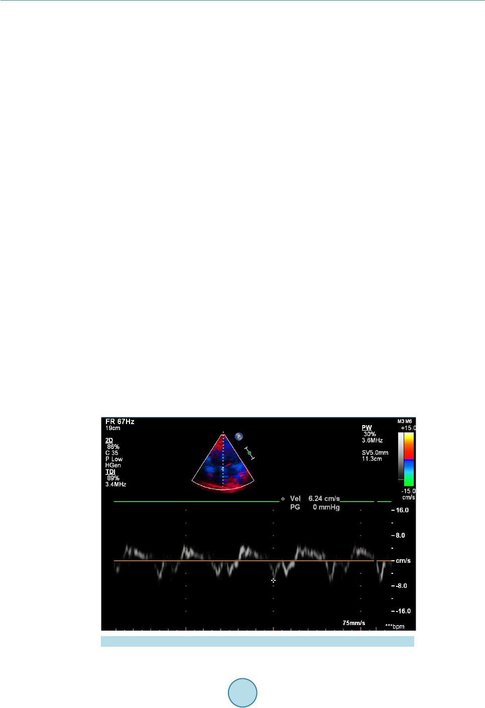
Findings of the present study suggest that an E/e’ ratio >15 is a powerful predictor of decreased survival after
acute STEMI. Further studies are warranted to substantiate the findings. However, we opine that measurement
of E/e’ may assist ris k-stratification of patie nts in this settin g.
References
[1] Gaziano, T.A., Bitton, A., Anand, S., Abrahams-Gessel, S. and Murphy, A. (2010) Growing Epidemic of Coronary
Heart Disease in Low- and Midd le-Income Countries. Current Problems in Cardiology, 35, 72-115.
http://dx.doi.org/10.1016/j.cpcardiol.2009.10.002
[2] Hillis, G.S., Moller, J.E., Pellikka, P.A., Gersh, B.J., Wright, R.S., Ommen, S.R., Reeder, G.S. and Oh, J.K. (2004)
Noninvasive Estimation of Left Ventricular Filling Pressure by E/e' Is a Powerful Predictor of Survival after Acute
Myocardial Infarction. Journal of the American College of Cardiology, 43, 360-367.
http://dx.doi.org/10.1016/j.jacc.2003.07.044
[3] Park, J.H. and Marwick, T.H. (2011) Use and Limitations of E/e' to Assess Left Ventri cular Fillin g Pressure b y Echo-
cardiography. Journal of Cardiovascular Ultrasound, 19, 169-173. http://dx.doi.org/10.4250/jcu.2011.19.4.169
[4] Liang, H.Y., Cauduro, S.A., Pellikka, P.A., Bailey, K.R., Grossardt, B.R., Yang, E.H., Rihal, C., Seward, J.B., Miller,
F.A. and Abraham, T.P. (2006) Comparison of Usefulness of Echocardiographic Doppler Variables to Left Ventricular
End-Diastolic Pressure in Predicting Futu r e H eart Failure Events. American Journal of Cardiology, 97, 866-871.
http://dx.doi.org/10.1016/j.amjcard.2005.09.136
[5] Ommen, S.R., Nishimura, R.A., Appleton, C.P., Miller, F.A., Oh, J.K., Redfield, M.M. and Tajik, A.J. (2000) Clinical
Utility of Doppler Echocardiography and Tissue Doppler Imaging in the Estimation of Left Ventricular Filling Pres-
sures: A Comparative Simul taneous Doppler-Catheterization Study. Circulation, 102, 1788-1794.
http://dx.doi.org/10.1161/01.CIR.102.15.1788
[6] Poulsen, S.H. (2001) Clinical Aspects of Left Ventricular Diastolic Function Assessed by Doppler Echocardiography
Following Acute Myocardial Infarction. Danish Medical Bulletin, 48, 199-210.
[7] Nagueh, S.F., Appleton, C.P., Gillebert, T.C., Marino, P.N., Oh, J.K., Smiseth, O.A., Waggoner, A.D., Flachskampf,
F.A., Pellikka, P.A. and Evangelisa, A. (2009) Recommendations for the Evaluation of Left Ventricular Diastolic
Function by Echocardiography. European Journal of Echocardiography, 10, 165-193.
http://dx.doi.org/10.1093/ejechocard/jep007
[8] Cerisano, G. and Bolognese, L. (2001) Echo-Doppler Evaluation of Left Ventricular Diastolic Dysfunction during
Acute Myocardial Infarct ion: Methodological, Clinical and Prognostic Implications. Italian Heart Journal, 2, 13-20.
[9] Shanks, M., Ng, A.C., van de Veire, N.R., Antoni, M.L., Bertini, M., Delgado, V., Nucifora, G., Holman, E.R., Choy,
J.B., Leung, D.Y., Schalij, M.J. and Bax, J.J. (2010) Incremental Prognostic Value of Novel Left Ventricular Diastolic
Indexes for P rediction of Clinical Outco me in Patients with ST-Elevation M yocardial Infarction . American Journal of
S. N. Nallapati et al.
837
Cardiology, 105, 592-597. http://dx.doi.org/10.1016/j.amjcard.2009.10.039
[10] Obeidat , O., Alam, M., Divine, G.W., Khaja, F., Goldstein, S. and Sabbah, H. (2004) Echocardiographic Predictors of
Prognosis after First Acute Myocardial Infarction. American Journal of Cardiology, 94, 1 278-1280.
http://dx.doi.org/10.1016/j.amjcard.2004.07.112