Surgical Science
Vol.5 No.1(2014), Article ID:42413,3 pages DOI:10.4236/ss.2014.51006
Musculoskeletal Manifestations of Atypical Mycobacterium: Case Reports
1Department of Orthopaedics, Our Lady of Lourdes Hospital, Drogheda, Ireland
2Our Lady of Lourdes Hospital, Drogheda, Ireland
Email: *mujeeb_rohilla@yahoo.com
Copyright © 2014 Mujeeeb Rohilla et al. This is an open access article distributed under the Creative Commons Attribution License, which permits unrestricted use, distribution, and reproduction in any medium, provided the original work is properly cited. In accordance of the Creative Commons Attribution License all Copyrights © 2014 are reserved for SCIRP and the owner of the intellectual property Mujeeeb Rohilla et al. All Copyright © 2014 are guarded by law and by SCIRP as a guardian.
Received December 17, 2013; revised January 8, 2014; accepted January 15, 2014
KEYWORDS
Musculoskeletal; Atypical Mycobacterium; Antiboitics
ABSTRACT
Musculoskeletal manifestations of atypical Mycobacterium are very rare and they can be easily missed by the junior doctors. We are presenting two cases of atypical Mycobacterium: the first one was infected by cleaning a fish tank and the other was immune-compromised patients. Both the cases were treated with broad spectrum antiboitics initially, with no results, unless the specimen cultures were obtained surgically and the specific cultures were asked. The treatment was started after obtaining the culture results. Both the patients were started on antituberculous treatment for 3 - 6 months with good functional results.
1. Introduction
Musculoskeletal atypical Mycobacterium infections in humans are rare and are frequently encountered. When they do arise, they can pose a diagnostic challenge for the clinicians. They can present as non-healing wounds or as skin leisions, tenosynovitis, osteomyelitis or septic arthritis. They are frequently present in the environment (soil, water) and in animal reservoirs. Atypical mycobacterium infections in adult immunocompetent hosts are uncommon, and the lungs are most commonly infected in 90% of the cases [1]. The hand and the wrist are the most commonly involved sites because of their abundance of synovial fluid and tissue combined with a higher probability of penetrating injuries at those sites. However, only a few cases have been reported so far of nontuberculous mycobaterial infections of the musculoskeletal system. Consequently, clinical awareness of this disease has been poor because of the difficulty of making the diagnosis, and the radiographs obtained in the early stage can be normal without clinical significance. Thus, this can cause delay in the treatment. Atypical mycobacterial infections are now being seen with increasing frequency as a cause of localised soft tissue infections [2].
Although they are generally of low pathogenecity to humans, more than 140 species have been reported in the literature, only 25 species have been strongly associated with atypical mycobacterial diseases and the rest are environmental organisims. Correct species identification is very important because all of them differ in their clinical relevance. Further, they differ in their growth rate, temperature tolerance, and drug susceptibility. The diagnosis of such infections is complex and requires good communication between clinicians and microbiologists. Because of limited sensitivity and specificity of symptoms, radiology and direct microscopy of clinical samples, culture remains the gold standard for the correct diagnosis [3].
2. Case Report 1
A 46-year-old (Right Hand Dominant) lady presented with a 3 cm × 5 cm cut wound on the dorsal aspect at the level of 2nd web space of her left hand from a metal skewer while cleaning the fish tank, which was cleaneddressed and treated with oral antibiotics as an outpatient. 10 days later she complained of a painful swelling on her left hand and pain in all her fingers Examination revealed a subcutaneous 3 × 3 cm tender swelling, with no distal neurovascular involvement, and restricted finger movements (Figure 1). She underwent surgical exploration and tendon sheaths were found to be not involved. A specimen was excised, microbiology cultures were sterile and histology showed non-specific chronic granulation tissue, and the wound healed uneventfully. 3 months later she developed a painful, 2 cm × 1.5 cm lump on dorsum of index finger over PIPJ, for which she underwent excisional biopsy, C&S and histology. Histology results should granulomatous infection, with multi-nucleated giant cells, and central necrosis, at that stage culture for mycobacterium were requested, which showed positive Zeil-Nelson staining, and Mycobacterium species were isolated 2 months later, (Mycobacterium Marinium), sensitive to rifampicin and ciprofloxacin. She was treated with rifampicin and ciprofloxacin for 3 months achieving complete cure of her lesions. The patient was asymptomatic at the time of discharge.
3. Case Report 2
A 56-year-old woman with a previous history of joint disease presented with an acutely swollen, painful right knee of 48 hours duration without any history of trauma. Examination revealed inflammation, effusion and decreased range of motion. Joint aspirate drained about 30 ml of serosanguineous fluid and she was started on broad spectrum IV antibiotics for the suspicion of septic arthritis. Gram stain and bacterial cultures of the aspirate were negative and clinically she did not showed any response to antibiotics. During this course of treatment she had MRI scan of right knee was obtained which showed central and marginal leisions and diffuse involvement of bone marrow (Figure 2). She underwent arthroscopic washout at 72 hours later, cultures of which were sent for mycobacterium, which revealed white patchy growth in the joint, and the culture showed mycobacterium avium. The patient was treated with 6 months of treatment with rifampicin, isonizide and ethambutol, and a good clinical response was achieved.
4. Discussion
Although atypical Mycobacterium related infections are quite rare, and manifest in variety of ways, the diagnosis should be kept in mind, especially in the presence of predisposing circumstances, which lead to atypical mycobacterial infections. These predisposing circumstances include immunocompromised patients, people with preexisting joint diseases, and those with a history showing exposure to potential sources. As atypical Mycobacterium

Figure 1. Showing a nodule at the level of index finger.
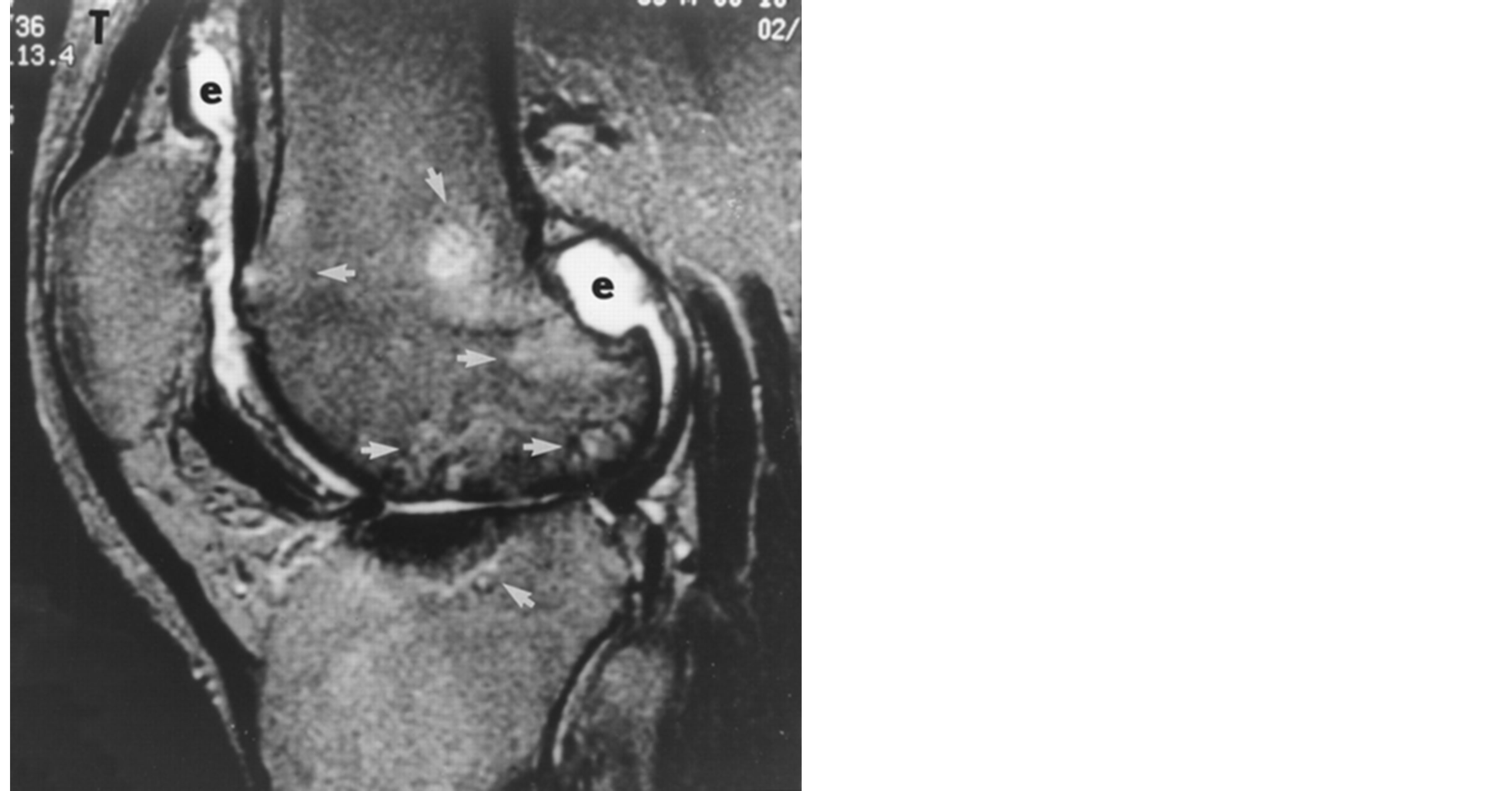
Figure 2. MRI of right knee showing central and marginal leisions with diffuse involvement of bone marrow.
does not grow under routine culture conditions, the diagnosis is easily missed resulting in delay of treatment. The microbiologist should be informed so that appropriate cultures could be performed.
Cutaneous exposure to fish tanks is the main source of M. Marinium infections, it usually acquired by direct inoculation through broken skin. Infections usually develop on fingers or hands, can be a solitary nodule or multiple, and in majority remains confined to skin, but can lead to tenosynovitis, arthritis, bursitis and osteomyelitis, especially in the immunocompromised. There are no set guidelines regarding duration, and number of antimicrobials, but 2 drug regimen including a macrolide ranging from 3 - 6 months is usually successful.
Mycobacterium related arthritis affects the knee joints commonly and up to 50% of these patients are immunocompromised or have received intrarticular injections. Diagnosis is based on culture of synovial fluid or culture of specimens obtained from exploration. Antimicrobial treatment involves use of 2 - 3 agents, over a period of 3 - 6 months, with or without use of surgical interventions.
The clinical manifestations and radiographic and histological features of bones, joints and soft infections by NTM are usually indistinguishable from those of tuberculosis. The imaging appearances of NTM tenosynovitis in the medical literature were frequently reported as exuberant tenosynovitis, which rarely involves the underlying muscles and bony structures, with preserved joint space and without tendon tear even in long standing phase [4]. All these deseases are characterized by a gradual and insidious onset with slowly progressive enlargement of the involved tendon, followed by pain and limitation of movements [5].
5. Conclusions
Although humans are routinely exposed to atypical mycobacterium, the rate of clinical Infection is low because the bacterias usually colonize rather than invade the host. Depending on the specific strain and host, however these bacterias can cause various infections [6].
Nevertheless, the imaging of NTM infections is nonspecific in the early stages and in the late stages, and the imaging resembles other granulomatous or mycobacterial infections. Thus, to determine the final diagnosis, it is useful to perform open biopsy and appropriate mycobacterium cultures so the optimal combination therapy and duration of treatment can be administered.
REFERENCES
- J. O. Falkinham 3rd, “Epidemiology of Infections by Nontuberculous Mycobacteria,” Clinical Microbiology Reviews, Vol. 9, No. 2, 1996, pp. 177-215.
- R. J. Wallace Jr., B. A. Brown and G. O. Onyi, “Skin, Soft Tissue, and Bone Infections Due to Mycobacterium Chelonaechelonae: Importance of Prior Corticosteroid Therapy, Frequency of Disseminated Infections, and Resistance to Oral Antimicrobials Other Than Clarithromycin,” The Journal of Infectious Diseases, Vol. 166, No. 2, 1992, pp. 405-412. http://dx.doi.org/10.1093/infdis/166.2.405
- J. Van Ingen, “Diagnosis of Nontuberculous Mycobacterial Infections,” Seminars in Respiratory and Critical Care Medicine, Vol. 34, No. 1, 2013, pp. 103-109. http://dx.doi.org/10.1055/s-0033-1333569
- S. Jaovisidha, C. Chen, K. N. Ryu, P. Siriwongparirat, P. Pekanan and D. J. Sartoris, “Tuberculous Tenosynovitis and Bursitis. Imaging Findings in 21 Cases,” Radiology, Vol. 201, No. 2, 1996, pp. 507-513.
- K. K. Amrani, M. Sundaram, A. Y. Shin and A. T. Bishop, “Mycobacterium Marinium Infections of Distal Upper Extremities: Clinical Course and Imaging Findings in Two Cases with Delayed Diagnosis,” Skeletal Radiology, Vol. 32, No. 9, 2003, pp. 546-549. http://dx.doi.org/10.1007/s00256-003-0643-z
- Z. Toossi and J. Ellner, “Mycobacterium Tuberculosis and Other Mycobacterium. Infectious Diseases,” 2nd Edition, Saunders, Philadelphia, 1998, pp. 2299-2307.
NOTES
*Corresponding author.