Natural Resources
Vol.4 No.2(2013), Article ID:32906,6 pages DOI:10.4236/nr.2013.42025
Forest Fairy Ring Fungi Clitocybe nebularis, Soil Bacillus spp., and Plant Extracts Exhibit in Vitro Antagonism on Dieback Phytophthora Species
![]()
1Grosvenor Grammar School, Belfast, UK; 2Applied Plant Science Division, Agri-Food & Biosciences Institute, Belfast, UK.
Email: *jr.rao@afbini.gov.uk
Copyright © 2013 Catherine Hearst et al. This is an open access article distributed under the Creative Commons Attribution License, which permits unrestricted use, distribution, and reproduction in any medium, provided the original work is properly cited.
Received February 23rd, 2013; revised April 26th, 2013; accepted May 9th, 2013
Keywords: Natural Forest Resources; Antifungal Extracts; Clitocybe nebularis; Bacillus licheniformis; Bacillus pumilis; Phytophthora Species; Japanese Larch Dieback Disease
ABSTRACT
In vitro Kirby-Bauer disc-diffusion assays coupled with bio-imaging software techniques were used to assess native forest dwelling “fairy ring” forming fungi (Clitocybe nebularis) and co-habitant forest tree-root colonising non pathogenic, antibiotic producing bacteria (Bacillus licheniformis, Bacillus pumilis) for their antagonism towards Japanese larch dieback oomycetes phytopathogens which also affects ornamental alternative hosts. The aqueous extracts of C. nebularis exhibited the highest clearance (inhibitory) zone of 21.4 × 105 pixels = 573%) against Phytophthora ramorum than growth/clear zone Cartesian integrates recorded in untreated (control) disc (3.7 × 105 pixels = 100%) over 3-day incubation. The fairy ring fungal extracts also exhibited substantive antagonism against P. kernoviae (147%), P. lateralis (347%) and a solanaceous crop infecting P. infestans (86%). Quite encouragingly, the soil oomycete phytopathogen P. ramorum was inhibited strongly (mean ~ 177%) by both forest bacilli. Aqueous extracts of non-forest antifungal herbaceous plants (garlic and elderberry) expressed similar inhibitory effects (mean ~ 70%). A seaweed fungal elicitor component fucoidan showed moderate levels (mean ~ 85%) of antagonism against P. ramorum, P. kernoviae, P. lateralis and P. infestans. The results in this in vitro study highlight the intensity and vigor of antagonistic forest microflora and non-forest herbaceous antifungal agents such as garlic and other plant extracts as serious candidates for suppression of the oomycete Phytophthora pathogenic fungi in forest soils. This study calls for urgent scoping and impact assessment studies in pot experiments and mini-plot forest trials to gauge the fitness of these natural resources for field level potential biotechnological applications to combat the devastating dieback disease in the native woodlands and horticulture.
1. Introduction
Climate change, trade of ornamental alternative hosts in circulation and air, water, soil, plant and plant products exacerbate the pathogen proliferation and epidemiology of the dieback disease caused by Phytophthora ramorum [1] in Japanese larch (Larix kaempferi) resulting in Sudden Oak Death [2] in the UK [3] and Europe [4]. Containment of the outbreaks of this disease normally involves early detection, removal and destruction of infected plants, clear-felling of the infected trees and fungicidal treatments; all such operations have environ mental and forest soil health implications. The life cycle of Phytophthora ramorum is complex depending on the host, and tend to persist in dead or dying tissues in soil for very long periods [5] and the soil phase itself is a critical mass in determining the survival and host root infections. New non-chemical measures to combat this devastating soil oomycetes are desirable worldwide for both woodland and horticulture industry. Recent reviews [6] summarise the current trends accentuating in alternative natural biocontrol methods of using medicinal extracts [7] or forest microbes for phytopathogens including Phytophthora spp. In natural habitats such as forests, field inoculated basidiomycetes [8] for mineralization purposes encounter limitations due to impact emanating from competing forest soil microorganisms. However, antimicrobial potentials of forest resources remain a hot pursuit and poorly understood.
Little is known on the efficacy of clusters of naturally evolved antagonists that may take on forest pathogens. Literature is scarce on natural resource expeditions seeking natural remedies for tree pathogens that may exist in their own habitat. Recently we have been actively scouting for natural resources comprising extracts of herbaceous plants including elderberry [9,10] exotic mushrooms such as shiitake [11,12] for antimicrobial complimentary therapies for human bacterial and fungal pathogens. In one of the expeditions, we stumbled across, a forest “fairy ring” forming mushroom—Clitocybe nebularis, and non-pathogenic tree-rhizospheric bacteria - Bacillus licheniformis, Bacillus pumilis producing unique enzymes, rare antibiotics [13] and exhibiting in vitro inhibition [14] of environmental pathogens. C. nebularis belongs to Tricholomatoid clade of Agaricales basidiomycetes, comprising ~50+ species including commercial Tricholoma matsutake [15] fairy ring forming mushrooms found in diverse ecological niches [16] as ectomychorrhizae or thrive as saprobic ground fungi [8]. Prompted in part by the prevalence of formidable natural cohabitant antagonists in the forest soil, we set out to examine if natural resources may be conservatively manipulated towards the suppression of invasive forest pathogens. We present in vitro scoping study examining antagonistic performance of forest resources comprising aqueous extract of (forest dwelling), ring forming fungi, Clitocybe nebularis, culture suspensions of (tree root/ rhizosphere colonising) bacteria (Bacillus licheniformis, Bacillus pumilis), together with non-forest prospective candidates such as garlic extract and aqueous extracts of native medicinal plant for their antifungal efficacy against dieback Phytophthora species.
2. Experimental
2.1. Microorganisms
Forest fungi Clitocybe nebularis, and bacteria Bacillus licheniformis, and Bacillus pumilis, all isolated from a local forest [14], Solanacea phytopathogen P. Infestans A1 were all held in culture collection archives at Plant Pathology, AFBI, Newforge Lane, Belfast, UK. The tree-infecting Phytophthora cultures (P. ramorum 3678, P. kernoviae 2444 & P. lateralis 44777 were obtained from LGC Standards, Teddington, Middlesex, UK. Soil bacterial isolates were grown in LB broth and the oomycete fungi on potato dextrose media.
2.2. Plant Extracts and Commercial Materials
Herbaceous specimens Sambucus nigra L. (elderberry), Filipendula spp. (meadowsweet), Buddliae (butterfly bush) were freshly collected from a local forest (kind permission of Ulster folk museum, Northern Ireland). Garlic (Allium sativum) (Sainsburys, UK), Fucoidan (SigmaAldrich) were purchased. Homogeneous aqueous extract preparations [9] were used in this study for bioassays.
2.3. Kirby-Bauer Disc Antagonism Assays
From stock Phytophthora cultures 1 cm2 plugs were excised and transferred individually to the Cartesian co-ordinate centre of four directional segments marked previously using a fine-tip marker pen of fresh plates of PDA, incubated for 3 days at ambient temperature to facilitate natural contours of hyphal growth to advance. The culture plates were examined using a binocular microscope and the outline of the perimeter of the hyphae was carefully traced by marker pen. Three sterile discs (Mast Group Ltd, Merseyside, UK) were saturated with a total of 60μls of an assay compound and the discs were stacked and placed at the 10mm distance (marked previously) to the “north” of the culture. At intervals of 3 days the extent of the hyphal growth (mm) was marked.
2.4. Bio-Imaging of Antifungal Inhibitory Zone
The area covered by each culture was measured and recorded using the bio-imaging technique [17] to assess inhibition/growth promoting properties of novel agents on moulds, using the Autochemisystem UVP Bioimaging system (UVP Products, Cambridge, UK), supported by LabWorks software package. Compared to recording clearance zones by bacterial lawn in the traditional antibiotic assays, the irregular contours of fungal growth is problematic in our bioassays. The emerging irregular progression of fungal hyphal growth from the culture plug at the centre of the Petri dish would resemble an undulated marine coastline. Initially the instrument was set on white light and to an exposure ratio of 490:500, with a constant focus of 47% calibration. Using the “Area Density” tool, (in pixels) the entire “area of individual plate” was measured first, recorded, and then using freehand “draw” tool followed on by the “area of irregular contours” of fungal growth co-integrated within the Cartesian coordinates estimated to reflect inhibition effects (clearance zones) as seen in culture plate.
2.5. Statistical Analyses
The arbitrary ratio, fungal growth, was calculated for each treatment and is the ratio of total pixels of surface area occupied by fungal growth/total pixels of surface area of the plate. Statistical analyses were performed employing the student t-test to compare fungal growth for each treatment against its control and where a probability value of greater than 0.05 (5%) was considered not significant.
3. Results and Discussion
3.1. Bio-Imaging of Antifungal Inhibitory Zone
Bio-imaging software facilitated the measurements (Figure 1) of the area of inhibition (zone of clearance) in pixels in (X-axis) which accurately maps the irregular contours of the phytopathogens tested (Y-axis) through the Cartesian co-ordinates marked in culture plates. Values (pixels) in excess of the control baseline (100%) highlights inhibitory activities i.e. higher the clearance zone, the greater the efficacy of antifungal activity. The bio-imaging software generated Cartesian calculus of area of the zone of clearance (inhibition), hitherto given in parantheses was estimated in pixels and corresponding equals in percentiles (Figure 1), roughly ca. ~3700 pixels of the area equalled one percent and variations thereupon were due to irregular fungal growth contours. The standard error (bars not shown) was ~ 2% (7465 pixels).
3.2. Antagonism of Native Forest Fairy Ring Mushroom Aqueous Extract towards Phytophthora
Aqueous extracts of the native fungal species Clitocybe nebularis, (Figure 1) gave the strongest inhibitory effects [14]. Aqueous extracts of C. nebularis (Figure 1) exhibited an inhibitory (clearance) zone of 21.4 × 10⁵ (pixels) = 573% against P. ramorum 3678 untreated (control) disc (3.7 × 105 = 100%) normal clear zone contour for Phytophthora growth over the 3 days of incubation. The ring fungi extracts also exhibited strong antagonism towards other tree-phytopathogens, P. kernoviae 2444 (17.6 × 105 = 147%) and P. lateralis 44777 (18.2 × 105 = 347%) and demonstrated its antifungal potency against solanaceous crop infecting P infestans A1 (6.9 × 105 = 86%).
3.3. Antagonism of Native Forest Bacteria towards Phytophthora sp.
In comparison to the fairy fungi extracts, the two native forest bacteria were the next strongest inhibitor of all tree infecting phytopathogens tested (Figure 1) except P. kernoviae. The soil oomycete phytopathogen P. ramorum was inhibited strongly by both non-pathogenic bacilli, B. licheniformis (6.61 × 105 = 177%), B. pumilis (6.64 × 105 = 178%). Interestingly, the antagonistic bacteria appear to share forest decaying litter or rhizosphere niche of the tree stands alongside the saprobic ectomycorhizal C. nebularis [16], whose fruiting bodies emerge as the fascinating and enigmatic fairy rings around the tree. In vitro antagonism of Bacillus spp. is widely known [6] and
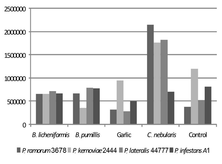
Figure 1. Antagonism of forest microflora towards Phytopthora sp.
linked to high microbial activity found in soils with high organic matter content such as in woodlands. Thus, introducing large populations of antagonistic “naturalised” bacteria used in this study as inoculants can be expected to be active suppressors of Phytophthora oomycete pathogens during their soil phase life cycle.
3.4. Chemical Constituents of Antagonistic Forest Microbes
Our results indicated that the inhibitory active principles in the aqueous extracts of the forest microflora would in part or fully be water soluble and may carry proteinaceous components. Our previous work with aqueous extracts indicated that aromatic and proteinaceous products from herbaceous plants [9,10] and forest mushrooms [11, 12] possessed synergistic antimicrobial activities. Interestingly, clitocypin and proteinaceous components in ring fungi exhibit mild antimicrobial activity [13]. Detailed chemical analyses [18] revealed phenyl acetic acid (PA), nebularine among other aromatics and nucleosidic compounds in C. nebularis fruiting bodies displaying antifungal activity against generic phytopathogens. The antibiotic production of the forest Bacillus spp. used in this study is well documented [6]. Options involving manipulation of specific antagonists/antibiotics isolated from Phytophthora suppressive soils for precise disease management [19] is a widely practised methodology for soil and water-borne oomycete infection containment.
3.5. Antifungal Efficacy of Herbaceous Medicinal Plant Extracts against Phytophthora sp.
Among the bio-resources that are not normally an integral part of a natural forest habitat, garlic (Allium sativum) extracts makes a strong candidate for forest patho-
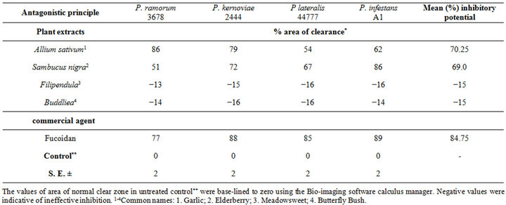
Table 1. In vitro growth inhibition* of Phytophthora sp challenged with herbaceous antagonistic agents.

Figure 2. Forest fairy ring fungi, and resources: in vitro antagonism of Phytophthora sp. The fungi Clitocybe nebularis is a notorious fairy ring forming mushrooms commonly found in woodlands (A) and become evident aboveground as a regular circle of fruiting bodies (B); often surrounded by dead vegetation. Kirby-Bauer Petri plate assay demonstrating antifungal activity against Phytophthora spp.; untreated control (C); garlic (D); Clitocybe nebularis (E); elderberry (F); forest bacteria, Bacillus licheniformis (G); Bacillus pumilis (H); and fucoidan (I). Disks were impregnated with aqueous extract (C)-(F), culture suspensions in G and H and aqueous solution (I).
gen suppression applications, partly due to their success with a number of disease control strategies in diverse crops [6]. In general, the garlic extracts were consistently inhibitory towards all 4 Phytopthora species challenged (Table 1), the mean percentage of clearance zones were ca. ~70% [P. ramorum (3.2 × 105 = 86%), P. kernoviae (2.8 × 105 = 79%), P. lateralis (1.2 × 105 = 54%) and Solanacea phytopathogen P. infestans (2.1 × 105 = 62%)]. Such broad spectrum potency of garlic was expected in the light of well established knowledge that the soluble volatiles in the Allium spp. inhibit several soil inhabiting phytopathogens in crops [20]. The in vitro antifungal activity of locally sourced herbaceous plants and known for their local medicinal values [9,12], such as Sambucus nigra L. (elderberry) was highly effective, whereas Filipendula spp. (meadowsweet), Buddliae (butterfly bush) were ineffective towards Phytophthora ramorum (Table 1) exhibiting negative values. The breach of the clear zone by test fungal growth culminates into net negative effect (−15%) on the area of inhibition (i.e. clearance zone). The elderberry extract treatment exhibited similar inhibitory (mean ca. ~69%) effects to those of garlic extracts, while fucoidan, a common component in seaweeds and a fungal elicitor exhibited a moderate inhibition (mean ca. ~85%) against the tree phytopathogens. Seaweeds are yet untapped natural resources in the sea-locked British Isles for biological means of forestry pathogen management; we intend to scout for its potential against tree pathogens in the future.
Figure 2 depicts a summary of key visual results of in vitro antagonism of a battery of antifungal agents against the dieback phytopathogens.
4. Conclusion
In conclusion, the present study mimics in vitro antagonism within a microcosm niche albeit demonstrating efficacies within a battery of charges and thus serves as a platform to encourage further considerations for natural combinatorial options for forest pathogen biocontrol. In vitro antagonistic performance of the forest co-habitants, viz., the fairy ring forming fungi C. nebularis, the two tree root colonising non-pathogenic bacilli tested in this study, is quite promising and complemented by antifungal activities of herbaceous medicinal plants elderberry, garlic towards dieback oomycete phytopathogens. In the light of sporadic incidents of Phytophthora ramorum in oak, and with the recent outbreaks of new dieback diseases such as Chalara fraxinea on ash trees (www.forestry.gov.uk), the development of a sustainable eco-friendly multiple disease control strategy is urgently needed. This scoping study offers a conceptual model of deploying habitat’s own antagonistic natural resources as group treatments that could potentially diminish the proliferation of dieback causal agents (e.g. Phytophthora ramorum oomycetes) in soils, offering an ecologically sustainable management of forest disease and warrants impact assessment in pot experiments and mini-plot woodland trials.
5. Acknowledgements
Authors thank Nuffield Science Foundation Bursary to CH, and the awards scheme administered by Sentinus, Northern Ireland (www.sentinus.co.uk).
REFERENCES
- D. Huberli and M. Garbelotto, “Phytophthora ramorum Is a Generalist Plant Pathogen with Differences in Virulence between Isolates from Infectious and Dead-End Hosts,” Forest Pathology, Vol. 42, No. 1, 2012, pp. 8-13. http://dx.doi.org/10.1111/j.1439-0329.2011. 00715.x
- C. M. Brasier and J. Webber, “Sudden Larch Death,” Nature Vol. 466, No. 7308, 2010, pp. 824-825. doi:10.1038/466824a
- V. Chadfield and M. Pautasso, “Phytophthora ramorum in England and Wales: Which Environmental Variables Predict County Disease Incidence?” Forest Pathology, Vol. 42, No. 2, 2012, pp. 150-159. doi:10.1111/j.1439-0329.2011.00735.x
- E. M. Goss, M. Larsen, A. Vercauteren, S. Werres, K. Heungens and N. J. Grünwald, “Phytophthora ramorum in Canada: Evidence for migration within North America and from Europe,” Phytopathology, Vol. 101, No. 1, 2011, pp. 166-171. doi:10.1094/PHYTO-05-10-0133
- N. Shishkoff, “Persistence of Phytophthora ramorum in Soil Mix and Roots of Nursery Ornamentals,” Plant Disease, Vol. 91, No. 10, 2007, pp. 1245-1249. doi:10.1094/PDIS-91-10-1245
- J. R. Stangarlin, O. J. Kuhn, L. Assi and K. R. F. SchwanEstrada, “Control of Plant Diseases Using Extracts from Medicinal Plants and Fungi,” Vol. 1, In: A. Méndez, Ed., Science Against Microbial Pathogens: Communicating Current Research and Technological Advances, Formatex Microbiology Series Publication, Formatex Research Center, Badajoz, 2011, pp. 1033-1042. http://www.formatex.org/microbiology3/
- I. K. Park, J. Kim, Y. S. Lee and S. L. Shin, “In Vivo Fungicidal Activity of Medicinal Plant Extracts against Six Phytopathogenic Fungi,” International Journal of Pest management, Vol. 54, No.1, 2008, pp. 63-68. doi:10.1080/09670870701549665
- G. Gramms, “The Universe of Basidiomycetous Ground Fungi” Vol. 1, In: Current Research Technology and Education Topics in Applied Microbiology and Microbial Biotechnology, A. Méndez, Ed., Formatex Microbiology Series Publication, Formatex Research Center, Badajoz, 2010, pp. 218-229. http://www.formatex.org/microbiology2/chapters1.html
- S. Woods-Panzaru, D. Nelson, G. McCollum, L. M. Ballard, Y. Maeda, C. E. Goldsmith, A. Loughrey, J. R. Rao and J. E. Moore, “An Examination of Antibacterial and Antifungal Properties of Constituents Described in Traditional Ulster Cures and Remedies,” Ulster Medical Journal, Vol. 78, No. 1, 2009, pp. 13-15.
- E. Efstratiou, A. I. Hussain, P. S. Nigam, J. E. Moore, M. A. Ayub and J. R. Rao, “Antimicrobial Activity of Callendula officinalis Petal Extracts against Fungi, as well as Gram-Negative and Gram-Positive Clinical Pathogens,” Complementary Therapies in Clinical Practice, Vol. 18, No. 3, 2012, pp. 173-176. doi:10.1016/j.ctcp.2012.02.003
- R. Hearst, D. Nelson, G. McCollum, B. C. Millar, Y. Maeda, C. E. Goldsmith, P. J. Rooney, A. Loughrey, J. R. Rao and J. E. Moore, “An Examination of the Antibacterial and Antifungal Properties of Constituents of Shiitake (Lentinula edodes) and oyster (Pleurotus ostreatus) Mushrooms,” Complementary Therapies in Clinical Practice, Vol. 15, No. 1, 2009, pp. 5-7. doi:10.1016/j.ctcp.2008.10.002
- J. R. Rao, T. J. Smyth, B. C. Millar and J. E. Moore, “Antimicrobial Properties of Shiitake Mushrooms (Lentinula edodes),” International Journal of Antimicrobial Agents, Vol. 33, No. 6, 2009, pp. 591-592. doi:10.1016/j.ijantimicag.2008.10.018
- J. Brizin, B. Rogelj, T. Popovic, B. Strukelj and A. Ritonja, “Clitocypin, a New Type of Cysteine Proteinase Inhibitor from Fruit Bodies of Mushroom Clitocybe nebularis,” Journal of Biological Chemistry, Vol. 275, No. 26, 2000, pp. 20104-20109. doi:10.1074/jbc.M001392200
- M. Hearst, D. Nelson, G. McCollum, L. M. Ballard, B. C. Millar, S. Moore, S. McClean, J. E. Moore and J. R. Rao, “An Investigation into Antimicrobial Properties of Protein Extractions from Native Fungal and Plant Species,” Journal of Pharmacognosy and Phytotherapy Vol. 2, No. 8, 2010, pp. 103-107.
- A. Yamada, H. Kobayashi, H. Murata, E. Kalmiş, F. Kalyoncu and M. Fukuda, “In Vitro Ectomycorrhizal Specificity between the Asian Red Pine Pinus densiflora and Tricholoma matsutake and Allied Species from Worldwide Pinaceae and Fagaceae Forests,” Mycorrhiza, Vol. 20, No. 5, 2010, pp. 333-339. doi:10.1007/s00572-009-0286-6
- C. G. Dowson, A. D. M. Rayner and L. Boddy, “Spatial Dynamics and Interactions of the Woodland Fairy Ring Fungus, Clitocybe nebularis,” New Phytologist, Vol. 111, No. 4, 1989, pp. 699-705. doi:10.1111/j.1469-8137.1989.tb02365.x
- J. E. Moore, G. McCollum, A. Murphy, B. C. Millar, D. Nelson, C. E. Goldsmith, P. J. Rooney, A. Loughrey and J. R. Rao, “Description of a Simple Bio-Imaging Technique to Assess Inhibition/Growth Promoting Properties of Novel Agents on Moulds,” British Journal of Biomedical Science, Vol. 67, No. 3, 2010, pp. 145-146.
- Y. S. Kim, I. K. Lee, S. J. Seok and B. S. Yun, “Chemical Constituents of the Fruiting Bodies of Clitocybe nebularis and Their Antifungal Activity,” Mycobiology, Vol. 36, No. 2, 2008, pp. 110-113. doi:10.4489/MYCO.2008.36.2.110
- B. Keen and T. Vancov, “Phytophthora cinnamomi Suppressive Soils,” Vol. 1, In: A. Méndez, Ed., Current Research Technology and Education Topics in Applied Microbiology and Microbial Biotechnology, Formatex Microbiology Series Publication, Formatex Research Center, Badajoz, 2010, pp. 239-250. http://www.formatex.org/microbiology2/chapters1.html
- H. Curtis, U. Noll, J. Stormann and A. J. Slusarenko, “Broad-Spectrum Activity of the Volatile Phytoanticipin Allicin in Extracts of Garlic (Allium sativum L.) against Plant Pathogenic Bacteria, Fungi and Oomycetes,” Physiological and Molecular Plant Pathology Vol. 65, No. 2, 2004, pp. 79-89. doi:10.1016/j.pmpp.2004.11.006
NOTES
*Corresponding author.