214 R. Wirestam et al. / J. Biomedical Science and Engineering 2 (2009) 210-215
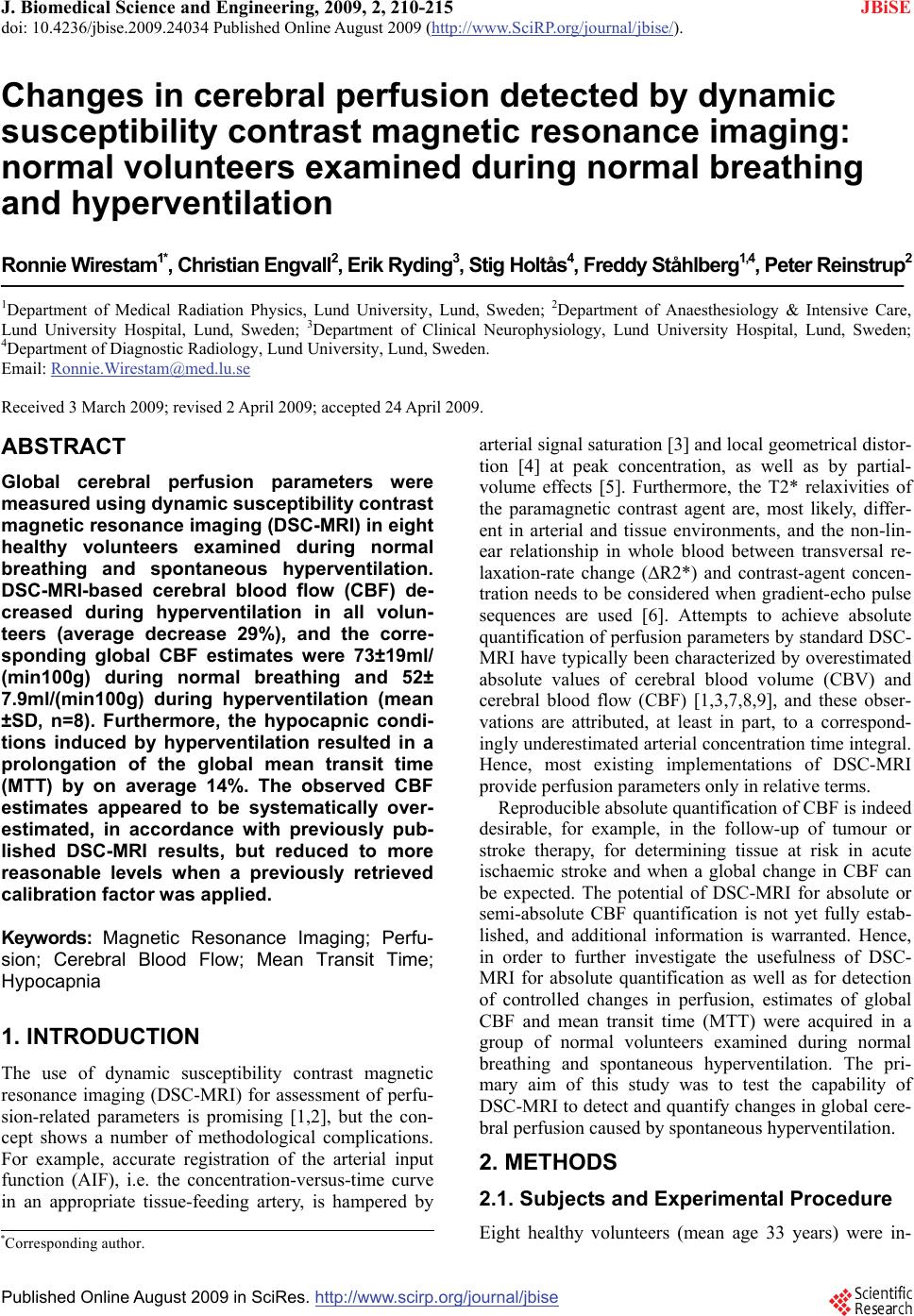
SciRes Copyright © 2009 JBiSE
were quite reasonable, and in accordance with previ-
ously published PET results from normal subjects. For
example, Kaneko et al. [20] observed MTT values of 6.1
s in grey matter and 8.1 s in white matter, and the large
study by Leenders et al. [21] showed CBV-to-CBF ratios
of 5.7 s in insular grey matter and 7.3 s in white matter.
Application of the calibration factor to the present data
resulted in a corrected whole-brain average CBF of ap-
proximately 42 ml/(min100g). Literature values of nor-
mal global CBF in humans at rest vary over a consider-
able range [22,23], but are typically between 40 and
50ml/(min100g) for the adult population. For example,
Knutsson et al. [9] obtained a whole-brain average CBF
of 40ml/(min100g) (in elderly normal subjects) by
Xe-133 SPECT, Slosman et al. [24] observed a global
CBF of 43ml/(min100g) in male volunteers (age interval
29-38 years), also by use of Xe-133 SPECT, Dörfler et
al. [22] reported a global CBF estimate of 48ml/
(min100g) based on extracranial sonography and Mat-
thew et al. [25] observed 40 ml/(min100g) using H2
15O
PET. Finally, Yonas et al. [26] employed stable xenon
computed tomography (Xe-CT) and extracted regional
CBF values of 92ml/(min100g) in the highest-flow
compartments, 54ml/(min 100g) in mixed-cortical re-
gions (calculated from linear-regression equations and
corresponding to the age of 33 years) and an age-inde-
pendent white-matter regional CBF of 20ml/(min100g).
In conclusion, DSC-MRI showed promising results in
the detection of controlled perfusion changes, induced
by spontaneous hyperventilation, in individual subjects.
In accordance with previously reported DSC-MRI ex-
periments, uncorrected absolute CBF values appeared to
be overestimated.
5. ACKNOWLEDGEMENTS
This study was supported by the Swedish Research Council (project no.
13514), the Swedish Cancer Society and the Crafoord Foundation,
Lund.
REFERENCES
[1] K. A. Rempp, G. Brix, F. Wenz, C. R. Becker , F. Gückel,
and W. J. Lorenz, (1994) Quantification of regional
cerebral blood flow and volume with dynamic suscepti-
bility contrast-enhanced MR imaging, Radiology, 193,
637-641.
[2] L. Østergaard, R. M. Weisskoff, D. A. Chesler, C.
Gyldensted, and B. R. Rosen, (1996) High resolution
measurement of cerebral blood flow using intravascular
tracer bolus passages, Part I: Mathematical Approach and
Statistical Analysis, Magnetic Resonance in Medicine,
36, 715-725.
[3] R. Ellinger, C. Kremser, M. F. Schocke, C. Kolbitsch, J.
Griebel, S. R. Felber, and F. T.Aichner, (2000) The
impact of peak saturation of the arterial input function on
quantitative evaluation of dynamic susceptibility con-
trast-enhanced MR studies, Journal of Computer Assisted
Tomography, 24, 942-948.
[4] M. Rausch, K. Scheffler, M. Rudin, and E. W. Radu,
(2000) Analysis of input functions from different arterial
branches with gamma variate functions and cluster
analysis for quantitative blood volume measurements,
Magnetic Resonance Imaging, 18, 1235-1243.
[5] J. J. Chen, M. R. Smith, and R. Frayne, (2005) The
impact of partial-volume effects in dynamic suscepti-
bility contrast magnetic resonance perfusion imaging,
Journal of Magnetic Resonance Imaging, 22, 390-399.
[6] B. F. Kjølby, L. Østergaard, and V. G. Kiselev, (2006)
Theoretical model of intravascular paramagnetic tracers
effect on tissue relaxation, Magnetic Resonance in
Medicine, 56, 187-197.
[7] C. B. Grandin, A. Bol, A. M. Smith, C. Michel, and G.
Cosnard, (2005) Absolute CBF and CBV measurements
by MRI bolus tracking before and after acetazolamide
challenge: Repeatability and comparison with PET in
humans, NeuroImage, 26, 525-535.
[8] K. E. Sakaie, W. Shin, K. R. Curtin, R. M. McCarthy, T.
A. Cashen, T. J. and Carroll, (2005) Method for
improving the accuracy of quantitative cerebral perfusion
imaging, Journal of Magnetic Resonance Imaging, 21,
512-519.
[9] L. Knutsson, S. Börjesson, E. M. Larsson, J. Risberg, L.
Gustafson, U. Passant, F. Ståhlberg, and R. Wirestam,
(2007) Absolute quantification of cerebral blood flow in
normal volunteers: Correlation between Xe-133 SPECT
and dynamic susceptibility contrast MRI, Journal of
Magnetic Resonance Imaging, 26, 913-920.
[10] P. Meier and K. L. Zierler, (1954) On the theory of
indicator-dilution method for measurement of blood flow
and volume, Journal of Applied Physiology, 6, 731-744.
[11] K. L. Zierler, (1965) Equations for measuring blood flow
by external monitoring of radioisotopes, Circulation
Research, 16, 309-321.
[12] T. Ernst, L. Chang, L. Itti, and O. Speck, (1999)
Correlation of regional cerebral blood flow from
perfusion MRI and SPECT in normal subjects, Magnetic
Resonance Imaging, 17, 349–354.
[13] J. B. Fortune, P. J. Feustel, C. deLuna, L. Graca, J.
Hasselbarth, and A. M. Kupinski, (1995) Cerebral blood
flow and blood volume in response to O2 and CO2
changes in normal humans, Journal of Trauma, 39,
463-472.
[14] H. Ito, I. Kanno, M. Ibaraki, J. Hatazawa, and S. Miura,
(2003) Changes in human cerebral blood flow and
cerebral blood volume during hypercapnia and
hypocapnia measured by positron emission tomography,
Journal of Cerebral Blood Flow and Metabolism, 23,
665-670.
[15] P. Reinstrup, E. Ryding, L. Algotsson, L. Berntman, and
T. Uski, (1994) Effects of nitrous oxide on human
regional cerebral blood flow and isolated pial arteries,
Anesthesiology, 81, 396–402.
[16] W. D. Obrist, T. W. Langfitt, J. L. Jaggi, J. Cruz, and T.
A. Gennarelli, (1984) Cerebral blood flow and meta-
bolism in comatose patients with acute head injury.
Relationship to intracranial hypertension, Journal of
Neurosurgery, 61, 241-253.
[17] H. Ito, M. Ibaraki, I. Kanno, H. Fukuda, and S. Miura,
(2005) Changes in the arterial fraction of human cerebral
blood volume during hypercapnia and hypocapnia