American Journal of Plant Sciences, 2011, 2, 262-267
doi:10.4236/ajps.2011.22028 Published Online June 2011 (http://www.SciRP.org/journal/ajps)
Copyright © 2011 SciRes. AJPS
Plant Regeneration through Indirect Somatic
Embryogenesis in Coelogyne Cristata Orchid
Aung Htay Naing, Jae Dong Chung, Ki Byung Lim
School of Applied Biosciences, College of Agriculture and Life Science, Kyungpook National University, Daegu, Republic of Korea.
Email: kblim@knu.ac.kr
Received April 7th, 2011; revised May 6th, 2011; accepted May 18th, 2011.
ABSTRACT
A newly efficient protocol has been established for high frequency somatic embryogenesis through callus culture of
Coelogyne cristata. The best frequency of callusing was obtained from leaf segments (3 - 5 mm) cultured on the MS
medium supplemented with 2 mg·L–1 2,4-D and 2 mg·L–1 BA combination. A negative effect of coconut water was ob-
served on the callus inductio n medium. When ca llus lines No. 4, 6 and 8 induced from leaf segments were sub-cultured
separately on 1/2 MS and MS media containing AC (1 - 3 g·L–1), formation of somatic embryos was found. However,
percentages of embryo formation and the number of embryos per explants were strongly affected by media and callus
lines used. The effect of 1/2 MS media is definitely be tter than MS medium for somatic embryogenesis from the selected
lines of leaf derived callus. Among the callus lin es, line no. 4 is the best for soma tic embryogenesis fo llowed by line no.
6 and 8. The somatic embryos converted into healthy plants with well developed shoots on the same media. The plant-
lets were transferred to 1/2 MS medium containing 1 g·L–1 AC for plant regenera tion until 8 weeks of culture and suc-
cessfully acclimatized in the greenhouse.
Keywords: Callus Formation, Embryo Formation, Leaf Segment, Plant Growth Regulators, Tropical Orchid
1. Introduction
Coelogyne cristata which belongs to the family Orchi-
daceae has high ornamental value as a cut flower. It pro-
duces its graceful racemes of white flowers with yellow
splotches on the throat, and has a long lasting and fra-
grant scent. In Myanmar, the pseudobulb of Coelogyne
cristata is used in making traditional medicine for dys-
entery and diarrhea. However, wild orchids including C.
cristata in Myanmar are nowadays steadily decreasing
due to over collection of orchid hunters, shifting cultiva-
tion, extension of crop cultivation and urban develop-
ment, which is causing loss of their natural habitats and
is leading to extinction. Thus, it is essential to take
measures to conserve their germplasm and to reveal effi-
cient propagation protocols to allow for the continuous
large supply of materials for medicine. However, there is
no report on plant regeneration through somatic em-
bryogenesis in C. cristata. The process has been induced
in tissue cultures of orchids either directly from the epi-
dermal cells of explants [1-3] or indirectly via interven-
ing callus [4].
The phenomenon of somatic embryogenesis via callus
is rather rare as success of callus formation in orchids has
limitations due to slow growth and a tendency to become
necrotic [5,6]. Especially, callus formation from leaf ex-
plants of orchids has been and is the rarest even now.
Even when callus induction was achieved, the frequency
of callusing might have been ultimately low and the cal-
lus was difficult to maintain and eventually failed to sur-
vive. Even when plant regeneration from callus of or-
chids was achieved, it usually occurred via protocorm-
like-bodies PLBs [6-8]. Therefore, success of somatic
embryogenesis via callus culture is interesting in plant
regeneration of orchids. This study presents optimal con-
centrations of 2,4-D and BA for callus formation and
high formation of somatic embryos from the culture of
leaf derived callus in Coelogyne cristata using different
kinds of media and AC
2. Materials and Methods
Mature capsules of Coelogyne cristata were collected
from Mingalardon Orchid Farm in Yangon, Myanmar.
The capsules were washed thoroughly under running tap
water. Then they were sterilized by immersion in 70%
ethanol for 10 seconds and flamed for a short time. After
they were dissected longitudinally, seeds were isolated
and sown on Hyponex (HP) basal media for in vitro ger-
Plant Regeneration through Indirect Somatic Embryogenesis in Coelogyne Cristata Orchid263
mination for 3 months. The germinated plantlets about
1.0 cm in height were transferred to Murashige and
Skoog (MS) basal media supplemented with NAA 1.0
mg·L–1 and BA 2.0 mg·L–1 for plant growth. After 2
months of culture, leaves of the in vitro plantlets become
the explants source to conduct further experiments.
2.1. Effect of PGRs and Coconut Water on
Callus Induction from Leaf Segments
Leaf segments of about 3 - 5 mm in length were used as
explants. For investigations on the effects of coconut
water (5% v/v) (MB cell, Seoul, Korea) and plant growth
regulators (PGRs) on callus induction, various combina-
tions of 2,4-dichlorophenoxyacetic acid (2,4-D; 0, 1.0,
2.0 and 3.0 mg·L–1) and benzyladenine (BA; 0, 1.0, 2.0
and 3.0 mg·L–1) were added to MS basal media. Each
experiment consisted of 10 explants with three replica-
tions. The culture vessels were placed in dark conditions
for 12 weeks and forming calli were recorded.
2.2. Effect of Media and AC on Somatic
Embryogenesis
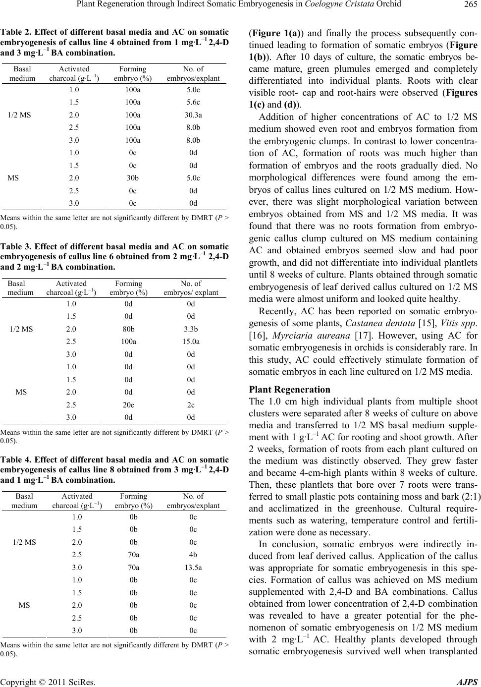
To examine the effect of media and AC on somatic em-
bryogenesis, the above leaves derived callus lines [line
No. 4 (26.7%), line No. 6 (40.0%), line No. 8 (36.7%)]
without CW, which showed better callus formation and
originated from the same tissue but cultured on the dif-
ferent media compositions, were chosen. And, each cal-
lus line was dissected into 2 - 3 mm sized callus pieces
and cultured on the different kinds of basal media (MS
and 1/2 MS) containing (1 - 3 g·L–1) activated charcoal
(AC). Each experiment consisted of 10 explants with
three replications. The culture vessels were placed at
room temperature under 16/8 hr day/night photoperiod at
a 20 - 50 μmolm²s¹ photon flux density. Data were
recorded after 8 weeks of culture.
2.3. Plant Regeneration
Single plants derived from the above experiments were
transferred to half-Murashige and Skoog 1/2 MS basal
medium supplemented with 1 g·L–1 AC for plant regen-
eration. Regenerated plants were transplanted and accli-
matized in the greenhouse.
For all cultures, percentage of sucrose and agar was
3.0 and 0.8% respectively. The pH of medium was ad-
justed to 5.8 before adding agar. Data were analyzed by
DMRT (P > 0.05).
3. Results and Discussion
Developing somatic embryogenic culture systems with
reliable regeneration capacity from ornamental plants is a
prerequisite for mass propagation and their genetic im-
provement. Studies were being intensively made to de-
velop a protocol for somatic embryogenesis. The process
can be induced in tissue cultures of orchids either directly
from the epidermal cells of explants [1-3] or indirectly via
intervening callus [4 ]. In this study, somatic embryo-
genesis was induced indirectly through callus culture.
In general, formation of callus in orchids is rather dif-
ficult due to its slow growth and necrotic tendency, espe-
cially, success of callus induction from leaf segments is
the rarest. In this study, when leaf segments were cul-
tured on the hormone free medium they became swollen,
and followed by initiation of a callus mass was visible
from the wound edges of leaf segment after 4 weeks of
culture. The calli were granular, whitish to light yellow
and comprised of isodiametric cells. Higher frequencies
of callus induction from explants were obtained on the
basal media supplemented with various concentrations of
2,4-D and BA (Table 1). However, ratios between con-
centrations of 2,4-D and BA were significantly associ-
ated with percentages of survival of explants and callus
formation. On the contrary, either the lowest concentra-
tion of 2,4-D and the highest concentration of BA or a
higher concentration of 2,4-D and lower concentration of
BA showed synergistic effects on callus induction from
leaf segments. Of the combinations of 2,4-D and BA
tested, 2 mg·L–1 2,4-D and 2 mg·L–1 BA were found as
optimal concentrations for the best callus induction
(40.0%). The color of callus induced from each combina-
tion was slightly changed from whitish to yellow and
most yellow callus showed granular type. However, cal-
lus mass obtained from highest concentrations of 3
mg·L–1 2,4-D was reddish yellow in color.
Addition of CW to the same media containing 2,4-D
and BA, frequencies of callusing were relatively low as
compared to those on the media devoid of CW. Almost
all of the leaf segments died on CW containing media.
However, percentage of callusing was stable on the me-
dia containing concentration of 3 mg·L–1 2,4-D and 1
mg·L–1 BA as devoid of CW.
Recently, combinations of 2,4-D and TDZ have been
reported for the callus induction of ornamental plants
including some orchid genera, Cymbidium [7], Vanda
coerulea [9], Cypripedium formosanum [10]. However,
BA alone or in combination with 2,4-D totally inhibited
callus induction in Paphiopedilum hybrid [11]. Similarly,
[12] mentioned that the combination of 2,4-D and BA
could not effectively induce callus from leaf segment in
Phalaenopsis. In the present study, combination of 2,4-D
and BA has successfully induced callus from leaf seg-
ments within a short period of time. However, the pres-
ence of CW inhibited callus induction from leaf segment
in this species. A similar result was observed in Pha-
laenopsis [12]. It is clear that CW has an inhibitory effect
on callus induction from leaf segments of orchids. Ac-
Copyright © 2011 SciRes. AJPS
Plant Regeneration through Indirect Somatic Embryogenesis in Coelogyne Cristata Orchid
264
cording to our findings in this study, exogenous hor-
mones are quite important for callus induction and no
effect of CW was observed on callus induction from leaf
segments in this species.
According to the results shown in Table 1, the lines
(No. 4, 6, 8) showed better callus formation. Thus, plant
regeneration through the lines should be critically con-
sidered. Although plant regeneration through organo-
genesis of callus culture has been fully developed in
some orchids, efficient protocol for embryogenesis
through callus culture of orchids still has been quite lim-
ited. Therefore, pieces of callus originating from the
above callus lines (4, 6, 8) without CW were separately
cultured on MS and 1/2 MS basal media supplemented
with different concentrations of AC (1 - 3 g·L–1) to ex-
Table 1. Effect of phytohormones on callus induction from
leaf segments of Coelogyne cristata after 12 weeks of culture
in dark conditions.
PGR (mg·L–1)
2,4-D BA
No. of
inoculated
explants
Survival
rate
Formation
of callus %
0 30 13g 3.3h
1 1 30 10h 5.7h
2 30 20f 13.3f
3 30 63a 26.7d*
2 1 30 43d 26.7d
2 30 46c 40.0a*
3 30 20f 5.3h
3 1 30 40e 36.7b*
2 30 50b 30c
3 30 40e 20e
With coconut water 5%
0 30 0j 0i
1 1 30 0j 0i
2 30 20f 0i
3 30 5i 5h
2 1 30 0j 0i
2 30 0j 0i
3 30 10h 10g
3 1 30 40e 40a
2 30 0j 0i
3 30 0j 0i
Means within the same letter are not significantly different by DMRT (P >
0.05).
amine somatic embryogenesis of the callus lines.
Degrees of somatic embryogenesis were totally af-
fected by callus lines and media. In general, explants
from all callus lines cultured on 1/2 MS basal media re-
sponded with embryo formation well. However, per-
centages of embryo formation and the number of em-
bryos were distinctly different along with the callus lines
used. Line 4 callus responded formation of embryos in all
treatments when they were cultured on 1/2 MS medium.
However, the highest number of embryos per explants
(30.3) was recorded on the media with 2.0 mg·L–1 AC.
When line 6 callus was used, the maximum average
number of embryos completely decreased (15.0/explant)
and the optimum concentration of AC was varied. Cul-
ture of line 8 callus on the same media responded with a
lower average number of embryos than that of line 6.
Although the callus lines originated from the same origin,
the line 8 attained from the highest concentration of
2,4-D (3 mg·L–1) was the lowest capacity for embryo-
genesis and also required the highest concentration of
AC (3 g·L–1) for optimum induction of somatic embryos.
It seems that the effect of higher concentration of 2,4-D
inhibited positively on somatic embryogenesis of later
experiments. Many workers also reported that the higher
concentrations of 2,4-D distinctly suppressed somatic
embryo formation in Oncidium [1,2] and in Phalaenopsis
[13]. The presence of 2,4-D in the culture medium
seemed to inhibit somatic embryogenesis in Citrus [14].
Thus, lower concentration of 2,4-D should be used in
callus induction to occupy higher somatic embryo forma-
tion capacity. As we discussed here, callus obtained from
hormone free medium might have the highest somatic
embryogenesis capacity as compared to those obtained
from 2,4-D containing media.
On MS medium, formation of embryos was rarely ob-
served from line 4 and line 6 calli and formation fre-
quency and number of embryos per callus were ex-
tremely low as compared to those on 1/2 MS medium.
Different capacities of somatic embryogenesis in callus
lines of the same origin were distinctly noticed in each
medium. On MS and 1/2 MS media, capacity of embryo
formation shows line 4 (30%, 5.0) > line 6 (20%, 2.0) >
line 8(0, 0), and line 4 (100%, 30.3) > line 6 (100%, 15.0)
> line 8 (70%, 12.0) (Tables 2, 3 and 4), respectively.
According to our findings herein, 1/2 MS medium in-
duced a higher number of somatic embryos (12 to 30 per
explant) as compared to MS media where the number of
embryos was 2 to 4 per explant. The obtained embryos
passed through the developmental stages, firstly the em-
bryogenic callus produced localized groups of cells, which
differentiated into embryogenic forms on the peripheral
region of the cluster old callus, showed induction of
rhizogenesis and trichome after 20 days of culture
Copyright © 2011 SciRes. AJPS
Plant Regeneration through Indirect Somatic Embryogenesis in Coelogyne Cristata Orchid265
Table 2. Effect of different basal media and AC on somatic
embryogenesis of callus line 4 obtained from 1 mg·L1 2,4-D
and 3 mg·L1 BA combination.
Basal
medium
Activated
charcoal (g·L–1)
Forming
embryo (%)
No. of
embryos/explant
1.0 100a 5.0c
1.5 100a 5.6c
2.0 100a 30.3a
2.5 100a 8.0b
1/2 MS
3.0 100a 8.0b
1.0 0c 0d
1.5 0c 0d
2.0 30b 5.0c
2.5 0c 0d
MS
3.0 0c 0d
Means within the same letter are not significantly different by DMRT (P >
0.05).
Table 3. Effect of different basal media and AC on somatic
embryogenesis of callus line 6 obtained from 2 mg·L1 2,4-D
and 2 mg·L1 BA combination.
Basal
medium Activated
charcoal (g·L–1) Forming
embryo (%) No. of
embryos/ explant
1.0 0d 0d
1.5 0d 0d
2.0 80b 3.3b
2.5 100a 15.0a
1/2 MS
3.0 0d 0d
1.0 0d 0d
1.5 0d 0d
2.0 0d 0d
2.5 20c 2c
MS
3.0 0d 0d
Means within the same letter are not significantly different by DMRT (P >
0.05).
Table 4. Effect of different basal media and AC on somatic
embryogenesis of callus line 8 obtained from 3 mg·L–1 2,4-D
and 1 mg·L–1 BA combination.
Basal
medium Activated
charcoal (g·L–1) Forming
embryo (%) No. of
embryos/explant
1.0 0b 0c
1.5 0b 0c
2.0 0b 0c
2.5 70a 4b
1/2 MS
3.0 70a 13.5a
1.0 0b 0c
1.5 0b 0c
2.0 0b 0c
2.5 0b 0c
MS
3.0 0b 0c
Means within the same letter are not significantly different by DMRT (P >
0.05).
(Figure 1(a)) and finally the process subsequently con-
tinued leading to formation of somatic embryos (Figure
1(b)). After 10 days of culture, the somatic embryos be-
came mature, green plumules emerged and completely
differentiated into individual plants. Roots with clear
visible root- cap and root-hairs were observed (Figures
1(c) and (d)).
Addition of higher concentrations of AC to 1/2 MS
medium showed even root and embryos formation from
the embryogenic clumps. In contrast to lower concentra-
tion of AC, formation of roots was much higher than
formation of embryos and the roots gradually died. No
morphological differences were found among the em-
bryos of callus lines cultured on 1/2 MS medium. How-
ever, there was slight morphological variation between
embryos obtained from MS and 1/2 MS media. It was
found that there was no roots formation from embryo-
genic callus clump cultured on MS medium containing
AC and obtained embryos seemed slow and had poor
growth, and did not differentiate into individual plantlets
until 8 weeks of culture. Plants obtained through somatic
embryogenesis of leaf derived callus cultured on 1/2 MS
media were almost uniform and looked quite healthy.
Recently, AC has been reported on somatic embryo-
genesis of some plants, Castanea dentata [15], Vitis spp.
[16], Myrciaria aureana [17]. However, using AC for
somatic embryogenesis in orchids is considerably rare. In
this study, AC could effectively stimulate formation of
somatic embryos in each line cultured on 1/2 MS media.
Plant Regeneration
The 1.0 cm high individual plants from multiple shoot
clusters were separated after 8 weeks of culture on above
media and transferred to 1/2 MS basal medium supple-
ment with 1 g·L–1 AC for rooting and shoot growth. After
2 weeks, formation of roots from each plant cultured on
the medium was distinctly observed. They grew faster
and became 4-cm-high plants within 8 weeks of culture.
Then, these plantlets that bore over 7 roots were trans-
ferred to small plastic pots containing moss and bark (2:1)
and acclimatized in the greenhouse. Cultural require-
ments such as watering, temperature control and fertili-
zation were done as necessary.
In conclusion, somatic embryos were indirectly in-
duced from leaf derived callus. Application of the callus
was appropriate for somatic embryogenesis in this spe-
cies. Formation of callus was achieved on MS medium
supplemented with 2,4-D and BA combinations. Callus
obtained from lower concentration of 2,4-D combination
was revealed to have a greater potential for the phe-
nomenon of somatic embryogenesis on 1/2 MS medium
with 2 mg·L–1 AC. Healthy plants developed through
somatic embryogenesis survived well when transplanted
Copyright © 2011 SciRes. AJPS
Plant Regeneration through Indirect Somatic Embryogenesis in Coelogyne Cristata Orchid
Copyright © 2011 SciRes. AJPS
266
Figure 1. Plant regeneration through somatic embryogenesis of leaf derived callus. (a) showing of clusters old callus, rhizogene-
sis and trichomes. (b) formation of somatic embryos. (c) fully developed somatic embryos. (d) conversion of somatic embryos
to shoots.
[2] J. T. Chen and W. C. Chang, “Effects of Auxins and Cy-
tokinins on Direct Somatic Embryogenesis on Leaf Ex-
plants of Oncidium ‘Gower Ramsey’,” Plant Growth
Regulation, Vol. 34, No. 2, June 2001, pp. 229-232.
doi:10.1023/A:1013304101647
in the greenhouse. This protocol is simple, easy to carry
out and can provide a large number of embryos and
plants for mass propagation in a short period of time. We
expect that this ability will also open up the prospect of
using biotechnological approaches for Coelogyne cristata
improvement. [3] H. H. Chung, J. T. Cheng and W. C. Chang, “Plant Re-
generation through Direct Somatic Embryogenesis from
Leaf Explants of Dendrobium,” Biologia Plantarum, Vol.
51, No. 2, June 2007, pp. 346-350.
doi:10.1007/s10535-007-0069-x
4. Acknowledgements
The authors acknowledge the financial support of Bio
Green 21 Program (code # 20070301034033), Rural De-
velopment Administration, Republic of Korea.
[4] J. F. Wu, J. T. Chen and W. C. Chang, “Effects of Auxins
and Cytokinins on Embryo Formation from Root-Derived
Callus of Oncidium ‘Gower Ramsey’,” Plant Cell, Tissue
and Organ Culture, Vol. 77, No. 1, April 2004, pp. 107-
109. doi:10.1023/B:TICU.0000016492.45075.a3 REFERENCES
[5] G. B. Kerbauy, “Plant Regeneration of Oncidium Vari-
cosum (Orchidaceae) by Means of Root Tip Culture,”
Plant Cell Reports, Vol. 3, No. 1, February 1984, pp.
27-29. doi:10.1007/BF00270224
[1] J. T. Chen, C. Chang and W. C. Chang, “Direct Somatic
Embryogenesis on Leaf Explants of Oncidium Gower
Ramsey and Subsequent Plant Regeneration,” Plant Cell
Report, Vol. 19, No. 2, December 1999, pp. 143-149.
doi:10.1007/s002990050724 [6] A. Begum, M. Tamaki and S. Kako, “Formation of Pro-
Plant Regeneration through Indirect Somatic Embryogenesis in Coelogyne Cristata Orchid267
tocorm-Like-Bodies (PLB) and Shoot Development
through in Vitro Culture of Outer Tissue of Cymbidium
PLB,” Journal of the Japanese Society for Horticultural
Science, Vol. 63, No. 3, 1994, pp. 663-673.
doi:10.2503/jjshs.63.663
[7] L. V. T. Huana, T. Takamura and M. Tanaka, “Callus
Formation and Plant Regeneration from Callus through
Somatic Embryo Structures in Cymbidium Orchid,” Plant
Science, Vol. 166, No. 6, June 2004, pp. 1443-1449.
doi:10.1016/j.plantsci.2004.01.023
[8] J. Roy and N. Banerjee, “Induction of Callus and Plant
Regeneration from Shoot Tip Explants of Dendrobium
Fimbriatum Lindl. var. Oculatum Hk.f,” Scientia Hor-
ticulturae, Vol. 97, No. 3, February 2003, pp. 333-340.
doi:10.1016/S0304-4238(02)00156-5
[9] N. T. Lang and N. T. Hang, “Using Biotechnological
Approaches for Vanda Orchid Improvement,” Omonrice,
Vol. 14, 2006, pp.140-143.
[10] Y. Lee and N. Lee, “Plant Regeneration from Proto-
corm-Derived Callus of Cypripedium Formosanmun,” In
Vitro Cellular & Developmental Biology—Plant, Vol. 39,
No. 5, September 2003, pp. 475-479.
doi:10.1079/IVP2003450
[11] Y. H. Lin, C. Chang and W. C. Chang, “Plant Regenera-
tion from Callus Culture of a Paphiopedilum Hybrid,”
Plant Cell, Tissue and Organ Culture, Vol. 62, No. 1, July
2000, pp. 21-25. doi:10.1023/A:1006425214471
[12] Y. Ishii, T. Takamura, M. Goi and M. Tanaka, “Callus
Induction and Somatic Embryogenesis of Phalaenopsis,”
Plant Cell Reports, Vol. 17, No. 6-7, April 1998, pp.
446-450. doi:10.1007/s002990050423
[13] H. L. Kuo, J. T. Chen and W. C. Chang, “Efficient Plant
Regeneration through Direct Somatic Embryogenesis
from Leaf Explants of Phalaenopsis ‘Little Steve’,” In
Vitro Cellular & Developmental Biology—Plant, Vol. 41,
No. 4, July 2005, pp. 453-456.
doi:10.1079/IVP2005644
[14] S. Fiore, F. D. Pasquale, F. Carimi and M. Sajeva, “Effect
of 2,4-D and 4-CPPU on Somatic Embryogenesis from
Stigma and Style Transverse Thin Cell Layers of Citrus,”
Plant Cell, Tissue and Organ Culture, Vol. 68, No. 1,
January 2002, pp. 57-63.
doi:10.1023/A:1012944100210
[15] G. M. Andrade and S. A. Merkle, “Enhancement of
American Chestnut Somatic Seedling Production,” Plant
Cell Reports, Vol. 24, No. 6, August 2005, pp. 326-334A.
doi:10.1007/s00299-005-0941-0
[16] G. Gambino, P. Ruffa, R. Vallania and I. Gribaudo, “So-
matic Embryogenesis from Whole Flowers, Anthers and
Ovaries of Grapevine (Vitis spp),” Plant Cell, Tissue and
Organ Culture, Vol. 90, No. 1, January 2007, pp. 79-83.
doi:10.1007/s11240-007-9256-x
[17] S. Y. Motoike, E. S. Saraiva, M. C. Ventrella, C. V. Silva
and L. C. C. Salomao, “Somatic Embryogenesis of Myr-
ciaria Aureana (Brazilian Grape Tree),” Plant Cell, Tissue
and Organ Culture, Vol. 89, No. 1, April 2007, pp. 75-81.
doi:10.1007/s11240-007-9210-y
Copyright © 2011 SciRes. AJPS