Journal of Water Resource and Protection
Vol.2 No.2(2010), Article ID:1297,6 pages DOI:10.4236/jwarp.2010.22015
Antibacterial Action and Physicochemical Properties of Stabilized Silver and Gold Nanostructures on the Surface of Disperse Silica
1Chuiko Institute of Surface Chemistry of NAS of Ukraine, Kyiv, Ukraine
2Marzeev Institute of Hygiene and Medical Ecology of AMS of Ukraine, Kyiv, Ukraine
E-mail: annaerem@ukr.net
Received November 10, 2009; revised December 4, 2009; accepted December 29, 2009
Keywords: silver, gold nanoparticles, binary stabilizer PVP and SDS, adsorption, high disperse silica, antimicrobial activity
ABSTRACT
This work is devoted to the synthesis and stabilization of nanosized Ag/SiO2 and Au/SiO2 disperse materials and investigation their morphology, optical and antimicrobial properties. First, Ag and Au nanoparticles (NPs) were produced in colloids via chemical (Ag) or photochemical (Au) reduction of appropriate ions. To prevent the oxidation of Ag NPs in colloid solution, external binary stabilizing agents PVP and SDS were used. Then, Ag and Au NPs (0.01-0.05% wt) were adsorbed from their colloid solutions on high disperse silica surface (Ssp=260m2/g) and samples prepared were dried. Materials obtained were studied by UV-vis, XRD, and TEM methods. Ag and Au NPs adsorbed on silica demonstrated a fair crystallinity in XRD. The surface plasmon resonance (SPR) band positions inherent to Ag and Au NPs on silica surface as well as the intensities of optical spectra were stable during 7 month and more. Obtained Ag NPs in colloids and Ag/SiO2 composites demonstrated excellent antimicrobial activity against a series of the microorganisms (Escherichia coli, Staphylococcus aurous, and Candida albicans). Au/SiO2 samples did not reveal any bactericide properties relative to the test microorganisms grown. The mechanisms of Ag(Au) NPs interaction with silica surface were analyzed.
1. Introduction
Water related infections belong to the one of the main health problems. The most dangerous water pathogenic pollutions such as Escherichia coli, Staphylococcus aurous are resistant to biodegradation. Silver nanoparticles (NPs) or silver ions have long been known to have strong inhibitory and bactericidal effect in solutions and composites with silica films and particles (Ag/SiO2) with developed surface area [1,2]. In case of Ag/SiO2 application, the specific surface area of silica is of great importance for effective adsorption of the species desirable. That is why developed silica surface covered with OH groups offers unique environment for the fixation and stabilization of nanosized silver and gold particles (Ag and Au NPs) for applications in optics, sensing, catalysis and as bactericides. However, Ag NPs are unstable on silica surface under ambient conditions due to fast oxidation [3]. It is important to prevent or inhibit the aggregation of NPs and oxidation process at the nanocluster formation stage, especially in case of silver. There are some methods to prevent the contact of Ag NPs with environment such as the protection with the inverse micelles, polymers, or their introduction into the solid materials, metal oxides or polymer films. Thus, stabilizing agents used for NPs surface modification control the particles formation, growth and aggregation rate [4,5].
Various methods are available for the NPs preparation. The most widely used techniques are photoor radiation method in presence of stabilizers, and chemical reduction using sodium tetrahydroborate, hydrazine, glucose, sodium citrate, and other reactants [6–10].
Previously we synthesized stable Ag and Au NPs within silica films prepared using sol-gel method via thermal reduction of appropriate metal ions [11]. These films were bright colored, transparent, stable and had characteristic SPR bands belonging to Ag or Au NPs. However, Ag clusters formed on disperse silica surface were found to be stable only for a few days [3]. The self-agglomeration and oxidation of Ag NPs were detected also within the porous silica films [12]. Photochemically generated Ag and Au NPs prepared in colloid solutions aggregated during 4-5 weeks, and Ag NPs were covered with oxide layer [9,10,13,14].
We suppose that the combination of the surfactant and polymer as the metal NPs stabilizers proposed for solution in [15,16] can be successfully used to prevent the oxidation and aggregation of Ag NPs in adsorbed state on high disperse silica surface in air.
The aim of this work was the synthesis of stable nanosized Ag/SiO2 and Au/SiO2 disperse materials using binary stabilizer SDS/PVP and investigation of their morphology, optical and antimicrobial properties.
2. Experimental Section
2.1. Initial Reagents
HAuCl4, AgNO3 (Меrck); Sodium dodecyl sulfate (SDS, Sigma Aldrich), Polyvinylpyrrolidone (PVP, grade: VFS 42-1491-85(Russia), FW=12000); Sodium tetrahydroborate (NaBH4, Fluka), Benzophenone (BP, Aldrich), i-propanol Sigma Aldrich); high-dispersed silica (HDS, brand А-300 with Ssp=260m2/g, “Khlorvinil”).
2.2. Ag and Au NPs Preparation
Ag/SiO2 and Au/SiO2 powders were obtained by adsorption of previously synthesized Me NPs colloids (total metal concentration 1,5·10-4 mol/l) on the surface of thermal treated HDS under stirring at room temperature. All Me colloids (0.01-0.05 wt % Me) were absorbed on silica surface.
Nanosized Ag particles in colloid solution were prepared via chemical reduction of AgNO3 with NaBH4 analogously to [9] in presence of SDS/PVP mixture previously dissolved in water. The reaction scheme is:
4Ag+ + nBH4- + 3nH2O →4Ag + nH2BO3- + 4nH+ + 2nH2
8Ag+ + nBH4- + 3nH2O →8Ag + nH2BO3- + 8nH+
Synthesized Ag NPs were adsorbed on the surface of HDS pretreated at 500 C. The Ag/SiO2 powders were dried at 85-95ºС during 4 hours.
Au colloids were synthesized via photochemical reduction of HAuCl4 [10] under UV-irradiation in water-ipropanol solution with mercury lamp irradiation (l=254 nm) in presence of mesoporous SiO2 powder with adsorbed benzophenone as the photocatalyst of Au3+ ions reduction and SDS as a stabilizer. Total reaction scheme including formation of ketyl-radicals from benzophenone and i-propanol is:
R2C•O– + Au3+ → R2C=O + Au2+ ( R = CH3, С6H5)
R2C•OH + Au3+ → R2C=O + Au2+ + H+
disproportionation: 2Au2+ → Au3+ + Au+
reduction of Au(I): R2C•O– + Au+ → R2C=O + Au0
Synthesized Au NPs were adsorbed on the surface of HDS pretreated at 500ºC. The Au/SiO2 powders were dried and treated at 500ºC during 5 hours.
2.3. Characterization
UV-VIS Absorption Spectroscopy studies were per-formed with a Perkin-Elmer Lambda 35 spectrophotometer in the wavelength range of 200–1000 nm. Measurements were made in 1 cm quartz cuvettes. Diffuse reflection spectra of Ме/SiO2 powders were recorded with PerkinElmer Lambda 35 spectrophotometer with a Labsphere RSA-PR-20 integrating sphere and handled using a Kubelka-Munk equation. The crystal structures of powders were determined with X-ray diffractometer (DRON-4-07, CuKα). The size and morphology of the Ag and Au nanoparticles in colloids and on silica surface were characterized with a JOEL JEM-100C transmission electron microscope (accelerating potential 100kV).
2.4. Antimicrobial Properties Study
Antimicrobial actions of NPs in colloids and on silica surface were determined using suspension tests according to the European standards EN 13727:2004 [17] and EN 13624:2002 [18]. Test microorganisms Staphylococcus aureus ATC C 6538, Escherіchіa colі K12 NCTC 10538 were used for evaluation of bactericidal activity and Candida albicans ATCC 10231 was used for evaluation of fungicidal activity of disinfectants.
Test microorganisms were grown in agar medium:
Tryptic soy agar for bacteria and Saburo agar for Candida albicans (HIMEDIA, India). Their specific quantities in test suspension (bacteria – up to 1.5·108 – 5.0·108 colony-forming units CFU/ml or 8lg, yeast-like fungi – up to 1.5·107 – 5.0·107 CFU/ml or 7lg) were monitored with a KFK-3 photoelectric colorimeter (λ = 620 nm, cuvette length – 10 mm).
Suspension method involved mixing of 1 ml of the test microorganisms with 1 ml of diluent and then adding 8 ml of the test disinfectant. The experimental mixture was maintained at (20.0±1.0)°С for the chosen contact times of 1; 2; 4; 24 h. After exposing for the required contact time, 1 ml was transferred to 9 ml of neutralizer (8.0 ml 0.1% Na2S + 1.0 ml H2O). After neutralizing for 5 min 1.0 ml of the test mixture was taken in duplicate and plated to detect survived test microorganisms. After incubation the numbers of survived bacteria (at 37.0±1.0°С for 24 to 48 h) or fungi (at 30.0±1.0°С for 48 h) in each sample were found.
The reduction values R=N0-Na were expressed in decimal logarithms: lgR= lgN0-lgNa, where N0 – number of cells per ml in the test mixtures at the beginning and Na – that at the end of the contact time t. An excellent antibacterial activity was in agreement with 99.9 percent reduction of microorganisms (R > 99.9 %). This related to 51g for bacteria and 41g for fungi.
3. Results and Discussion
SPR absorption band positions of the Ag and Au colloids obtained were at 417 and 530 nm respectively, and the average particle diameters were of 10-12 nm for Ag and 20-30 nm for Au (Figures 1(a), 1(b), 2(a), 2(b)).
When NPs from prepared colloids were adsorbed on the HDS surface adsorption equilibrium occurred in a few minutes. An irreversible binding of both types of NPs with silica was found. It should be noted, that a homogeneous distribution of the NPs was observed also after thermal treatment of bright-colored Ag/SiO2 at 85-90ºС and Au/SiO2 at 500ºС.
In the X-ray spectra of Me/SiO2 powders two clear reflections were noticed at 2q = 38° and 44°, more evident in case of Au/SiO2. These peaks were assigned to (111) and (200) planes of cubic Me respectively.
Sample color and characteristic optical spectra of Me NPs in colloids were stable for 7 month and more. Ag/SiO2 powders treated at 85-90°C were stable and oxidation-resistant on the developed HDS surface. Au/SiO2 powders were stable after treatment at 500ºС after destruction of protective shell.
The oxidation of Ag NPs on silica run after the thermal treatment of Ag/SiO2 powders in air at app. 200ºС due to destruction of organic protective shell and further interaction of Ag NPs with oxygen: 4Ag + O2 ↔ 2Ag2O.
Repeated treatment of Ag2O/SiO2 at 250 - 3000С resulted in the reduction of yellow color of Ag NPs which disappeared again when the samples were cooled to the room temperature. Obviously the presence of stabilizing shell was necessary to protect Ag NPs on HDS surface against oxidation.
Separately used PVP and SDS stabilizers provided the stability of Ag on the SiO2 during several days after drying. After that the oxidation of Ag on silica surface occurred and the color disappeared. The protection mechanism of Ag NPs in solution by binary PVP/SDS stabilizer consisted in capping of Ag NPs by SDS chains and absorption by PVP functional groups in accordance with [19]. Double shell of SDS micelles and polymer probably prevented Ag NPs against contact with oxygen. Binding of the metal nanoclusters by the surface of HDS is strong due to interaction between the functional groups of binary stabilizer of Ag NPs and OH-groups of silica surface [9] TEM image of Ag/SiO2 is shown in the Figure 1(d). According to [20], PVP in specified concentration range was adsorbed irreversibly. At the same time the binary shell did not prevent biocide action of Ag NPs as it is shown later.
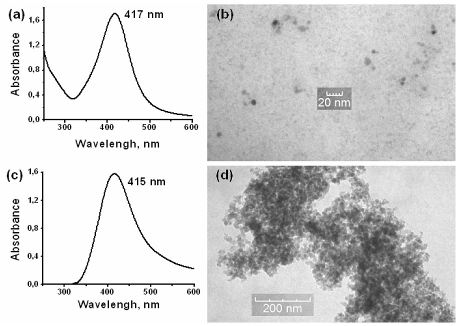
Figure 1. Optical spectra (a.c) and TEM images (b,d) of Ag NPs colloid and Ag/SiO2 powder respectively.
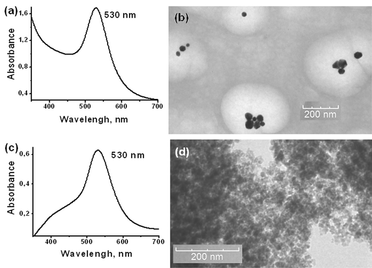
Figure 2. Optical spectra (a.c) and TEM images (b,d) of Au NPs colloid and Au/SiO2 powder respectively.
In accordance with [21,22], colloid silica particles in presence of SDS micelles after drying formed 3D structures with developed porosity. We suggested that Au NPs produced within colloid using only SDS stabilizer were localized within these “secondary” pores of HDS. TEM image of Au/SiO2 is shown in the Figure 2(d). Such location can provide an explanation for strong binding of Au NPs by silica after 500 C treatment.
Absorption maxima in the DRS of Ag/SiO2 and Au/SiO2 powders calculated via Kubelka-Munk equation were equal to those in colloid solutions. This is an evidence of nanoscale dimensionality of metal particles after absorption (Figure 1(c), 2(c)).
3.1. Ме/SiO2 Composites Antimicrobial Activity
To explain the inhibitor effects of silver on bacteria it was suggested that silver reacted with proteins by combining the thiol (-SH) groups leading to the bacteria inactivation [23]. In this work we examined the antibacterial activity of Ag/SiO2 and Au/SiO2 suspensions and of Ag (Au) colloids. Ag/SiO2 powders were tested after treating at 85-90°С, Au/SiO2 – after 500°C. The probes were diluted in distilled water with the concentration of 0.0016% wt. Me and of 3.13% wt. HDS in suspension. Colloids were tested with the concentration of 0.0016% wt. Me.
AgNO3 solution was used in the control experiments in the equal concentration as Ag NPs. Binary stabilizer PVP/SDS and NaBH4 were studied in the control with the same amount as in the colloids. Initial HDS was tested also at the concentration 3.13 % wt. in presence of the stabilizers and NaBH4.
The results of antimicrobial activity of Ag NPs in colloid and suspension against E. coli, S. aureus и C. albicans are presented in the Table 1.
Essential reduction values for bacteria E. coli (5 lg) and fungi C. albicans (4 lg) in colloids were achieved after 1 hour of exposure of microbial cells with Ag NPs. Staphylococcus were more Ag NPs-resistant, particularly 4.35 lg reduction was achieved only after 4 hour of exposure. Decrease in Ag concentration allowed to reveal the phenomenon mentioned. C. albicans bacteria were the most sensitive among of objects studied.
The control AgNO3 solution showed no antimicrobial action. The same result was also revealed for PVP/SDS and NaBH4 mixture. Thus experimental data presented indicated a high antimicrobial activity of silver colloids to all microorganisms.
Ag NPs embedding into SiO2 surface slightly decreased activity of Ag/SiO2 suspension. The exposure time increased and changes in interaction character of Ag NPs with the microbial cells appeared.
The contact time for 4 lg reduction reached for C. albicans remained the same as that in colloid (1 h). At the same time, Ag/SiO2 resistance of E.coli rose. The reduction value was only 3.58 lg after 4 hour contact time. On the contrary, S.aureus was more sensitive to Ag/SiO2 then to Ag in colloid.
Nevertheless the antimicrobial activity of Ag NPs/SiO2 complex remained high in general.
Nanosized gold at the same concentration range showed no antibacterial action on the indicated microorganisms. As it was shown in [24], Au NPs suppressed bacteria
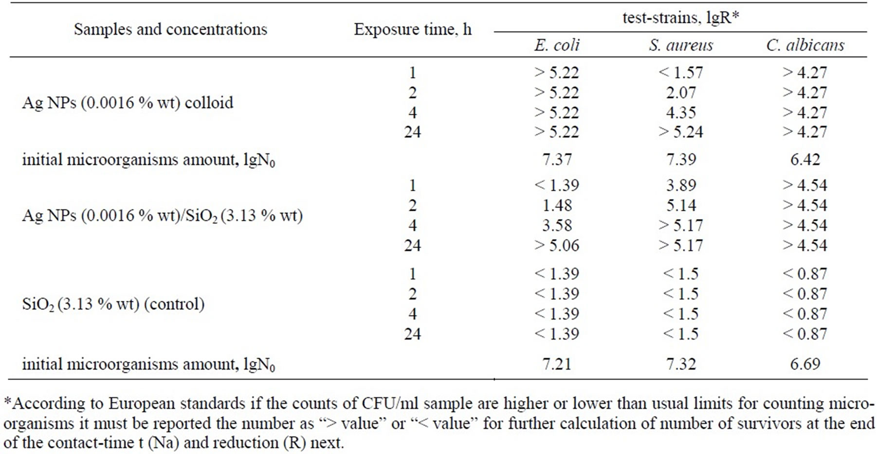
Table 1. The antimicrobial activity of Ag NPs in colloids and suspensions.
growth for the first stage of their reproduction.
Experimental data presented in Table 1 indicated almost total growth inhibition of all studied microbes when the extremely low concentrations of nanosized Ag were used. It should be noted that antimicrobial rates were over 1–2 orders of magnitude than those given in literature.
4. Conclusions
Stable Au and Ag nanoparticles on SiO2 surface were obtained by adsorption of previously synthesized Me NPs colloids in presence of SDS and binary SDS/PVP stabilizing agents.
The fixation of the metal nanoclusters on silica surface was a result of: 1) interaction between the functional groups of stabilizer shell of NPs and OH-groups of silica; 2) location of Me NPs within secondary pores of HDS globules.
Composite systems obtained containing strongly bound and homogeneously distributed Ag NPs on silica surface served as potential wide-spectrum effective antimicrobial materials for water cleaning as well as for medical and pharmaceutical application.
REFERENCES
- D. K. Tiwari, J. Behari, P. Sen, “Time and dosedependent antimicrobial potential of Ag nanoparticles synthesized by top-down approach,” Current Science, Vol. 95, No. 5, pp. 647–655, September 2008.
- M. Kawashita, S. Toda, H-M. Kim, T. Kokubo, N. Masuda, “Preparation of antibacterial silver-doped silica glass microspheres,” J.Biomedical Materials research, Part A, Vol. 66, No. 2, pp. 266–274, 2003.
- M. Hillenkamp, G. D. Domenicantonio, O. Eugster, C. Félix, “Instability of Ag nanoparticles in SiO2 at ambient conditions,” Nanotechnology, Vol. 18, pp. 015702, January 2007.
- E. D. Goddard, J. V. Gruber, “Principle of polymer science and technology in cosmetics and personal care,” M. Dekker, New York, 1999.
- R. Patakfalvi, Z. Viranyi, I. Dekany, “Kinetics of silver nanoparticle growth in aqueous polymer solutions,” Coll. Polym. Sci., Vol. 283, pp. 299–305, June 2004.
- L. Bois, F. Bessueille, E. Chassagneux, Y. Battie, N. Destouches, C. Hubert, A. Boukenter, S. Parola, “Silver nanoparticles growth in a mesoporous silica film templated with the F127 triblock copolymer,” Coll. Surf. A: Physicochem. Eng. Aspects., Vol. 325, No. 1–2, pp. 86–92, 2008.
- M. P. Pileni, I. Lisiecki, L. Motte, C. Petit, I. Cizeron, N. Moumen, P. Lixon, “Nanoparticles synthesized in reverse micelles,” Prog. Col. Polym. Sci., Vol. 93, No. 1, 1993.
- N. Toshima, T. Yonezava, K. Kushihashi, “Polymerprotected palladium – platinum bimetallic clusters: preparation, catalytic properties and structural considerations,” J.Chem. Soc., Faraday Trans., Vol. 89, pp. 2537–2543, 1993.
- I. Mukha, А. Eremenko, N. Smirnova, G. Korchak, A. Mikhiyenkova, I. Chekman, “Formation, physical – chemical and bactericide properties of stabilized silver nanostructures on the surface of disperse silica,” (Russian), Chemistry, Physics and Technology of Surface, Kyiv: Naukova Dumka, Vol. 15, pp. 255–266, 2009.
- G. Krylova, A. Eremenko, N. Smirnova, S. Eustis, “Photogeneration of nanosized gold on the surface of mesoporous silica modified by benzophenone,” Theor. and Experim. Chemistry. (Russian, Transl. English), Vol. 41, No. 6, pp. 365–370, November 2005.
- S. Eustis, G. Krylova, A. Eremenko, N. Smirnova, C. Tabor, W. Huang, M. El-Sayed, “Using silica films and powders modified with benzophenone to photoreduce silver nanoparticles,” J. Photochem. Photobiol. A: Chem., Vol. 181, pp. 385–393, 2006.
- O. Akhavan, R. Azimirad, A. Z. Moshfegh, J. Phys. D: Appl. Phys., Vol. 41, No. 19, pp. 195305, 2008.
- G. V. Krylova, A. M. Eremenko, N. P. Smirnova, S Eustis, “Photochemical preparation of nanoparticles of Ag in aqueous-alcoholic solutions and on the surface of mesoporous silica,” Theor. and Experim. Chemistry (Russian, Transl. English), Vol. 41, No. 2, pp. 100–104, March–April 2005.
- L. G. Grechko, A. M. Eremenko, G. V. Krylova, L. B. Lerman, N. P. Smirnova, N. G. Shkoda, “Optical properties of small silver particles within colloid solutions,” (Ukr), Proceedings of Kyiv University, Series: Physics & Mathematics, Vol. 4, pp. 450, 2004.
- J. P. Wilcoxon, R. L. Williamson, and R. Baughman, “Optical properties of gold colloids formed in inverse micelles,” J. Chem. Phys., Vol. 98, pp. 9933–9950, 1993.
- Y. N. Cheong Chan, R. R. Schrock, and R. E. Cohen, “Synthesis of silver and gold nanoclusters within microphase-separated diblock copolymers”, Chem.Mater., Vol. 4, pp. 24–27, 1992.
- EN 13727, “Chemical disinfectants and antiseptics – Quantitative suspension test for the evaluation of bactericidal activity in the medical area –Test method and requirements (phase 2, step 1),” Brussels, European Committee for Standardization, September 2004.
- EN 13624, “Chemical disinfectants and antiseptics - Quantitative suspension test for the evaluation of fungicidal or yeasticidal activity for instruments used in the medical area – Test method and requirements (phase 2, step 1),” Brussels, European Committee for Standardization, October 2002.
- Ch. Chen, L. Wang, G. Jiang, H. Yu, “Chemical preparation of special-shaped metal nanomaterials through encapsulation or inducement in soft solution,” Research Adv. Mater., Vol. 11, pp. 1–18, 2006.
- Yu. S. Lipatov and L. M. Sergeeva, “Adsorption of Polymers,” (Russian), Naukova Dumka, Kiev. 1972.
- S. R. Kline, E. W. Kaler, “Aggregation of colloidal silica by n-alkyl sulfates,” Langmuir, Vol. 12, No. 10, pp. 2402–2407, March 1996.
- Y. Guo, A. Guadalupe, “Functional silica aerogel from metastable lamellar composite,” Chem. Commun., pp. 315–316, January 1999.
- A. Lehninger, D. Nelson, M. Cox, “Principle of biochemistry,” second ed., New York: Worth Publishers, 1993.
- G. Yashan, G. Krylova, A. Eremenko, N. Smirnova, V. Zhalko-Tytarenko, V. Marievskiy, I. Chekman, “Bactericide properties of gold and silver nanoparticles in solution and on high disperse silica surface,” (Russian), Chemistry, Physics and Technology of Surface. Kyiv: Naukova Dumka, Vol. 14, pp. 524, 2008.