Surgical Science, 2011, 2, 137-139
doi:10.4236/ss.2011.23028 Published Online May 2011 (http://www.scirp.org/journal/ss)
Copyright © 2011 SciRes. SS
Primary Malignant Peripheral Nerve Sheath Tumor
of the Breast—A Case Report
Mushtaq Chalkoo, Shahnawaz Ahangar, Asim Rafiq Laharwal,
Aasim Mushtaq Patloo, Abbass Mohd, Shabir Ahmed Dar
Department of Surgery, Government Medical College, Srin agar, India
E-mail: Mushtaq_chalkoo@rediffmail.com; shanz101@gmail.com
Received February 19, 2011; revised April 3, 2011; accepte d April 8, 2011
Abstract
Malignant peripheral nerve sheath tumor is a rare soft tissue sarcoma. Breast is an extremely rare location of
this lesion and presentation as a breast lump in the absence of pain or previous benign neural tumor is even
rarer. We report such a lesion in a 60-year-old female who presented with hard and painless breast lump for
2 years. Histopathology revealed a malignant spindle cell tumor of low grade potential. It was subsequently
confirmed to be malignant peripheral nerve sheath tumor (MPNST) on the basis of immunopositivity for
vimentin, neurone specific enolase and S-100.
Keywords: Breast Lump; Malignant Peripheral Nerve Sheath Tumor
1. Introduction
Breast tumors are predominantly of epithelial origin, and
non-epithelial tumors arising from indigenous structures in
the breast are rare [1]. Prim ary pure brea st s arcoma is a rare
disease and constitutes 0.2% - 1.0% of all mammary ma-
lignancies [2]. The exact etiology is still unknown but these
tumors frequently occur in patients suffering with neurofi-
bromatosis Type I [NF 1]. The mean age at the time of
diagnosis of MPNST is in the thirties, but the patients with
NF 1 are about 10 yr younger than th e patients without NF
1 [3]. MPNSTs are also known to have an association with
previous irradiation. MPNSTs are known to arise within
the field of irradiation, 9 to 36 yr after radiation therapy
administered for treating previous malignancies [4]. Im-
munohistochemical analysis proves to be helpful in indis-
tinguishable cases. Though several cases of Malignant Pe-
ripheral Nerve Sheath Tumor (MPNST) of breast have
been reported [5]. We report the first case with a one year
follow up.
2. Case Report
A 60 years old female presented with a giant lump in he r
right breast for two years. The patient was concealing the
lump by hiding it under her pajamas and tying a belt
around it. It was only after the lump d eveloped ulcer ation
over its surface that th e patient sought medical help. She
had no previously significant medical or surgical history.
Her general physical examination was essentially no rmal.
Breast examination revealed a giant lump involving
whole of the right breast which had gravitated to the
groin (Figure 1). Overlying skin was ulcerated along the
inferio-medial aspect and the rest of the skin was hyper
pigmented. It was firm in consistency and free from the
underlying pectoral muscles. A solitary lymph node
about 1 × 1 cm was palpable in axilla with tenderness on
palpation. Fine needle aspiration cytology [FNAC] of the
lymph node revealed inflammatory changes. Patient was
put on antibiotic treatment for a period of 5 days as per
culture and sensitivity rep or t of the wou nd swab from the
Figure 1. Preoperative photograph of the patient.
M. CHALKOO ET AL.
138
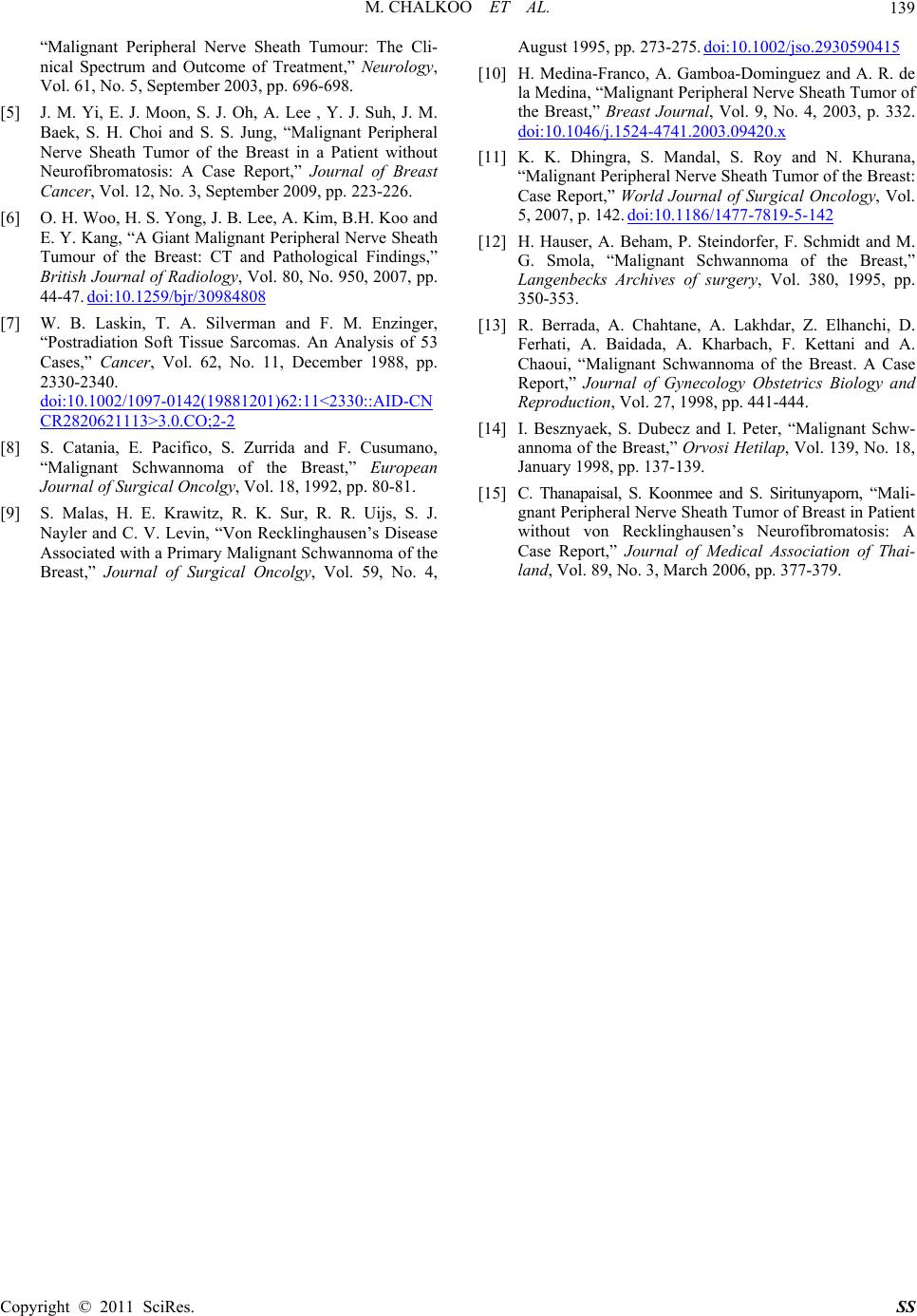
Figure 2. S-100 protein staining of the tumour tissue.
ulcer. Then simple mastectomy with excision of axillary
node was contemplated. The excised surgical specimen
consisted of a well-circumscribed, firm, solid mass
measuring 26 × 24 × 18 cm covered with skin ellipse
including nipple and areola. It weighed about 5 kg. The
cut section revealed grayish-white, nodular and myxoid
mass with prominent fibrous strands. No necrosis, hem-
orrhage or cystic degeneration was visible. Histopa-
thological examination revealed a malignant nerve
sheath tumor of low grade potential, with focal infiltra-
tion by mixed inflammatory cell infiltrate. It was sur-
rounded by fibrous tissue and spindle cells with numer-
ous vascular channels. The mitotic count was 16 mitosis
per 10 high-power fields. Immunohistochemical analysis
showed positivity for neuron specific enolase (NSE) and
vimentin and S-100 and expressed negativity for CD31,
CD34, FACTOR VIII, PLAP, EMA, SMA and calponin
(Figure 2). The excised lymph node revealed inflamma-
tory changes only.
3. Discussion
Malignant peripheral nerve sheath tumour [MPNST] is a
term that is used to describe tumours arising from the
peripheral nerves or their sheaths and it has replaced the
old terminology of schwanomas, malignant neurilem-
momas and the like. Overall they represent about 10% of
soft tissue sarcomas [6]. The incidence of MPNST in the
general population is 0.001% however it can increase to
5% - 42% in patients with neurofibromatosis type 1.
Peak incidence of these tumours is around 20 to 50 years
[5]. Laskinet et al., reported pointed out to irradiation of
peripheral nerves as one of the possible etiologies [7].
Such tumours develop after a long latent period (10 - 20
years) following irradiation and account for 11% of
MPNST. Histologically, spindle-shaped cells with con-
tours with curved cores and a slightly coloured cyto-
plasm are present. Cellular density and organization are
variable and the palisade provision of the cores is found.
On immunohistochemical staining, tumour cells show
reactivity to neuron sp ecific enolase (NSE) an d Vimentin
and S-100 [6]. The common sites of origin include the
extremities and trunk usually sciatic nerve, brachial
plexus and the sacral plexus. This tumour very rarely
affects breast. To the best of our knowledge only 9 such
cases have been reported with some of them being re-
ported in association with neurofibromatosis-1 and post
radiation [8-15]. In our case there was no clinical evi-
dence of neurofibromatosis type 1. Woo et al. noted
similar presentation in their case [6]. The diagnosis of
MPNST can only be done on the basis of immunohisto-
chemistry otherwise it can be easily mistaken for malig-
nant phylloides tumour, fibrosarcoma and leiomyosar-
coma [5]. Owing to the rarity of the tumour there are no
clear cut guidelines for its management. We performed a
simple mastectomy in our patient. Catania et al. and
Malas et al. have also recommended the same for the
treatment of MPNST of breast [8,9]. Role of radiother-
apy remains controversial. Yi et al. have tried adjuvant
therapy in their case. However our patient did not un-
dergo any radiation therapy [5]. One year into follow up
the patient is doing well with no local recurrence or any
distant metastasis. We have been following our patient
with 6 monthly ultrasound abdomen and with a detailed
history and physical examination every two months.
There are no reports available in the literature on the
median survival or the prognosis of MPNST of breast.
4. Conclusions
MPNST of breast is a very rare tumour. The optimum
therapy need s to be charted out but we believ e that it can
be managed by simple mastectomy.
5. References
[1] B. Majmudar, “Neurilemoma Presenting as a Lump in the
Breast,” Southern Medical Journal, Vol. 69, No. 4, April
1976, pp. 463-464.
doi:10.1097/00007611-197604000-00025
[2] T. Cil, A. Altintas, S. Pasa and A. Isikdogan, “Primary
Spindle Cell Sarcoma of the Breast,” Breast Care, Vol. 3,
No. 3, 2008, pp. 197-199. doi:10.1159/000121469
[3] B. S. Ducatman, B. W. Scheithauer, D. G. Piepgras, H. M.
Reiman and D. M. Ilstrup, “Malignant Peripheral Nerve
Sheath Tumours: A Clinicopathologic Study of 120
Cases,” Cancer, Vol. 57, No. 10, May 1986, pp.
2006-2021.
doi:10.1002/1097-0142(19860515)57:10<2006::AID-CN
CR2820571022>3.0.CO;2-6
[4] J. M. Baehring, R. A. Betensky and T. T. Batchelor,
Copyright © 2011 SciRes. SS
M. CHALKOO ET AL.
Copyright © 2011 SciRes. SS
139
“Malignant Peripheral Nerve Sheath Tumour: The Cli-
nical Spectrum and Outcome of Treatment,” Neurology,
Vol. 61, No. 5, September 2003, pp. 696-698.
[5] J. M. Yi, E. J. Moon, S. J. Oh, A. Lee , Y. J. Suh, J. M.
Baek, S. H. Choi and S. S. Jung, “Malignant Peripheral
Nerve Sheath Tumor of the Breast in a Patient without
Neurofibromatosis: A Case Report,” Journal of Breast
Cancer, Vol. 12, No. 3, September 2009, pp. 223-226.
[6] O. H. Woo, H. S. Yong, J. B. Lee, A. Kim, B.H. Koo and
E. Y. Kang, “A Giant Malignant Peripheral Nerve Sheath
Tumour of the Breast: CT and Pathological Findings,”
British Journal of Radiology, Vol. 80, No. 950, 2007, pp.
44-47. doi:10.1259/bjr/30984808
[7] W. B. Laskin, T. A. Silverman and F. M. Enzinger,
“Postradiation Soft Tissue Sarcomas. An Analysis of 53
Cases,” Cancer, Vol. 62, No. 11, December 1988, pp.
2330-2340.
doi:10.1002/1097-0142(19881201)62:11<2330::AID-CN
CR2820621113>3.0.CO;2-2
[8] S. Catania, E. Pacifico, S. Zurrida and F. Cusumano,
“Malignant Schwannoma of the Breast,” European
Journal of Surgical Oncolgy, Vol. 18, 1992, pp. 80-81.
[9] S. Malas, H. E. Krawitz, R. K. Sur, R. R. Uijs, S. J.
Nayler and C. V. Levin, “Von Recklinghausen’s Disease
Associated with a Primary Malignant Schwannoma of the
Breast,” Journal of Surgical Oncolgy, Vol. 59, No. 4,
August 1995, pp. 273-275. doi:10.1002/jso.2930590415
[10] H. Medina-Franco, A. Gamboa-Dominguez and A. R. de
la Medina, “Malignant Peripheral Nerve Sheath Tumor of
the Breast,” Breast Journal, Vol. 9, No. 4, 2003, p. 332.
doi:10.1046/j.1524-4741.2003.09420.x
[11] K. K. Dhingra, S. Mandal, S. Roy and N. Khurana,
“Malignant Peripheral Nerve Sheath Tumor of the Breast:
Case Report,” World Journal of Surgical Oncology, Vol.
5, 2007, p. 142. doi:10.1186/1477-7819-5-142
[12] H. Hauser, A. Beham, P. Steindorfer, F. Schmidt and M.
G. Smola, “Malignant Schwannoma of the Breast,”
Langenbecks Archives of surgery, Vol. 380, 1995, pp.
350-353.
[13] R. Berrada, A. Chahtane, A. Lakhdar, Z. Elhanchi, D.
Ferhati, A. Baidada, A. Kharbach, F. Kettani and A.
Chaoui, “Malignant Schwannoma of the Breast. A Case
Report,” Journal of Gynecology Obstetrics Biology and
Reproduction, Vol. 27, 1998, pp. 441-444.
[14] I. Besznyaek, S. Dubecz and I. Peter, “Malignant Schw-
annoma of the Breast,” Orvosi Hetilap, Vol. 139, No. 18,
January 1998, pp. 137-139.
[15] C. Thanapaisal, S. Koonmee and S. Siritunyaporn, “Mali-
gnant Peripheral Nerve Sheath Tumor of Breast in Patient
without von Recklinghausen’s Neurofibromatosis: A
Case Report,” Journal of Medical Association of Thai-
land, Vol. 89, No. 3, March 2006, pp. 377-379.