J. Biomedical Science and Engineering, 2011, 4, 352-356
doi:10.4236/jbise.2011.45044 Published Online May 2011 (http://www.SciRP.org/journal/jbise/
JBiSE
).
Published Online May 2011 in SciRes. http://www.scirp.org/journal/JBiSE
Enhanced stability of nano-emulsified paclitaxel
Ju Young Lee1a, Da Yeon Kim1a, Gyeong Hae Kim1, Kkot Nim Kang1, Byoung Hyun Min1, Bong Lee2,
Jae Ho Kim1, Moon Suk Kim1
1Department of Molecular Science and Technology, Ajou University, Suwon, Korea;
2Department of Polymer Engineering, Pukyong National University, Busan, Korea.
Email: moonskim@ajou.ac.kr
Received 9 February 2011; revised 28 March 2011; accepted 4 April 2011.
ABSTRACT
The main goal of this work was to develop an optimal
self-microemulsifying paclitaxel prepared with PLGA
and solubilizer such as tetraglycol, Cremophor ELP,
and Labrasol. The prepared PTx-loaded SMES showed
the size of the range of 80-130 nm by dynamic light
scattering and a spherical shape by atomic force mi-
croscopy. In experiment of storage stability in deion-
ized water (DW) or blood condition, PTx-loaded
SMES showed good stability in DW and comparable
stability in blood condition at 37˚C for 7 days. In ad-
dition, PTx-loaded SMES showed a significant in-
hibitory effect on B16F10 melanoma proliferation. In
conclusion, we confirmed that the formulations tried
in this study could be used as administration form for
animal trials of PTx.
Keywords: Self-Microemulsifying; Paclitaxel; Stability;
Anti-Tumor Activity
1. INTRODUCTION
Paclitaxel (PTx), a major anticancer drug isolated from
the bark of Taxus brevifolia, has significant activity in
clinical trials against a variety of tumors such as breast
cancer, advanced ovarian carcinoma, lung cancer, head
and neck carcinoma [1,2]. PTx is a hydrophobic drug
with poor aqueous solubility. To enhance its solubility
and allow parenteral administration, PTx is currently
formulated with solution of Cremophor® EL and ethanol
as dosage-form of PTx (Taxol®) for clinical application
[3,4].
Recently, numerous investigations have been focused
on the development of various PTx delivery systems
such as liposomes, emulsions, micelles, microspheres,
and polymeric nanoparticles [5-9]. Among these, self-
microemulsifying systems (SMES) may be a promising
way to load PTx in delivery system because it provides
high concentration of PTx in the aqueous media system
[10,11]. SMES are isotropic mixtures of oil, a surfactant,
and possibly one or more hydrophilic solvents or cosur-
factants, which form fine oil-in-water emulsions when
exposed to aqueous media under condition of gentle
agitation [12].
At present, many studies have highlighted the devel-
opment of PTx-loaded SMES with optimal condition for
blood circulation in effective concentration [13]. How-
ever, the major obstacle that limits the use of SMES is
due to the physical and/or to the chemical instability by
absorption of biological compounds such as protein dur-
ing drug circulation time in blood after intravenous ad-
ministration [14].
To improve stability of SMES, various formulations
of SMES is needed that can extend PTx circulation time
in blood. Our understanding is that the formulations of
SMES could examine under the blood condition. Thus,
the aim of this study was to examine various formula-
tions of PTx-loaded SMES to increase systemic clear-
ance through extending of the circulation time of PTx.
2. MATERIALS AND METHODS
2.1. Materials
Poly(d,l-lactide-co-glycolide) (PLGA, molecular weight,
8000, 20,000 and 90,000 g/mole) were purchased from
Boerhinger Ingelheim (Ingelheim, Germany). Paclitaxel
was purchased from Samyang Genex Co. (Seoul, Korea).
Caprylocaproyl macrogol-8 glyceride (Labrasol®) was
obtained from Gattefosse (Westwood, NJ, USA). Cre-
mophor ELP was purchased from BASF (Germany).
Tetraglycol was purchased from Sigma Chemical Co. (St.
Louis, MO, USA). All other chemicals were of reagent
grade. Deionized water (DW) was prepared by a Milli-Q
purification system from Millipore (Molsheim, France).
2.2. Preparation of PTx-Loaded SMES
A series of SMES was prepared in each of the various
formulas with various ratios of PLGA, paclitaxel, solu-
aJu Young Lee and Da Yeon Kim are equal first authors in this work.
J. Y. Lee et al . / J. Biomedical Science and Engineering 4 (2011) 352-356 353
bilizer, surfactant, and cosurfactant. Briefly, PLGA and
PTx were dissolved, respectively, by solubilizer such as
tetraglycol in glass vials. Surfactant and cosurfactant
were accurately weighed into glass vials. Then the com-
ponents were mixed by gentle stirring and vortex mixing
until PTx had perfectly dissolved. The mixture was
stored at room temperature until used. Before using,
PTx-loaded SMES was formed by contacting to aqueous
phase of the prepared mixture.
2.3. Size Analysis of PTx-Loaded SMES
For particle size analysis, formulation (50 μl) of PTx-
loaded SMES was diluted with DW to 50 ml in a volu-
metric flask and gently mixed by inverting the flask. The
droplet sizes of resultant emulsions were determined by
dynamic light scattering (DLS, ELS-8000, Photal, Japan)
at room temperature. The droplet size was individually
measured for three PTx-loaded SMES samples and then
calculated as average value. For atomic force micros-
copy (AFM), one drop of PTx-loaded SMES was trans-
ferred onto silicone wafer which washed with MeOH.
The wafer was quickly placed in liquid nitrogen, fol-
lowed by the freeze-drying for 2 days. AFM measure-
ments were carried out in the tapping mode with a
Nanoscope IV instrument (Digital Instruments Inc.).
2.4. Stability of PTx-Loaded SMES
PTx-loaded SMES was prepared with DW or a solution
of 0.9% NaCl and 5% bovine serum albumin (Sigma,
Germany) and individually placed in 10 ml tube. The
tube was constantly shaken at 100 rpm and 37˚C for 7
days. At the set time, the droplet size was individually
measured for three PTx-loaded SMES samples and then
calculated as average value.
2.5. Cell Culture and Cytotoxicity Tests
B16F10 melanoma cell line was obtained by KCLB
(Korea Cell Line Bank) and cultured in MEM (Mini-
mum Essential Medium, Gibco BRL, USA) supple-
mented with 10% fetal bovine serum (Gibco BRL, USA)
The cells were seeded into 75 cm2 flasks, cultured and
changed medium every 2 days. For cytotoxicity tests,
B16F10 cell suspension (2 × 10⁴ cells/well) was seed in
a 48-well plate. The cells were incubated overnight to
allow for cell attachment. Ptx (1 μg) of SMES solution
was added to each well for 7 days. Cell viability was
determined by using water-soluble enzyme substrate
MTT which was converted to purple water-insoluble
product formazan accumulated in the cytoplasm of vi-
able cells. Cell viability of each well performed indi-
vidually and then calculated as average value. In brief,
100 μl of PBS solution of the MTT tetrazolium substrate
(5 mg/ml) was added after 1, 4 and 7 days. After incuba-
tion for 4 h at 37˚C, the resulting purple formazan pre-
cipitate was solubilized by the addition of 1 ml of
DMSO and shaken for 30 min. An aliquot from each
well (100 μl) was transferred to 96-well plates and then
read using a plate reader of an ELISA (E-max, Molecu-
lar Device, USA). The optical density of each well de-
termined at 590 nm.
3. RESULTS AND DISCUSSION
3.1. Preparation of PTx-Loaded SMES
PLGA of different molecular weights used to compare
the stability of the self-microemulsifying PTx. To pre-
pare self-microemulsifying PTx consisting of mixtures
of Ptx, oil, a surfactant and PLGA, the formulation is
summarized in Ta b l e 1 . PLGA and PTx were dissolved
by solubilizers which were a mixture of tetraglycol,
Cremophor ELP, and Labrasol.
Firstly, the prepared PTx-loaded SMES were observed
visually. As shown in Figure 1(a), PTx-loaded SMES
(F1-0 - F10-0) showed the emulsion solution from
transparent to semitransparent according to various for-
mulations. The droplet size for all formulations of
PTx-loaded SMES was found in the range of 80 - 130
nm. The droplet size distribution is comparatively nar-
row for all formulations.
The morphology of PTx-loaded SMES was measured
by AFM as shown in Figure 2. PTx-loaded SMES (F10)
showed the spherical shape with smooth surface. A
comparatively uniform droplet size of PTx-loaded SMES
was also observed at AFM, indicating no aggregation or
adhesion among SMES.
3.2. Stability of PTx-Loaded SMES
The storage stability of PTx-loaded SMES is important
to maintain the therapeutic concentration of PTx. There-
fore, PTx-loaded SMES was firstly prepared with DW or
a solution of 0.9% NaCl and 5% bovine serum albumin.
The prepared PTx-loaded SMES was constantly shaken
at 100 rpm and 37˚C for 7 days to examine the storage
stability. After 4 and 7 days, there is no change of parti-
cle size for F1-F5 as shown in Figure 1(b), indicating
the storage stability of PTx-loaded SMES in DW. Mean-
while, F6 and F7 with PTx content < 0.05 wt showed the
precipitation of PTx. Hence we investigated the SMES
with PTx content of 0.003 wt according to different mo-
lecular weights of PLGA (F8-F10). F8, F9 and F10 in
DW showed no change of particle size at 37˚C for 4 and
7 days (Table 2).
Next, PTx-loaded SMES was prepared with a solution
of 0.9% NaCl and 5% bovine serum albumin as a model
blood condition. The haze color of PTx-loaded SMES is
deeply changed according to increasing incubation time
(Figure 3). The particle size f F8 and F10 increased to o
C
opyright © 2011 SciRes. JBiSE
J. Y. Lee et al . / J. Biomedical Science and Engineering 4 (2011) 352-356
Copyright © 2011 SciRes.
354
Table 1. Formulation for the preparation of PTx-loaded SMES.
Formulation
Composition (g, w/w)
F1 F2 F3 F4 F5 F6 F7 F8 F9 F10
Drug (Paclitaxel) 0.001 0.0010.0030.050.010.003
Solubilizer (Tetraglycol) 0.5 0.5 0.5
PLGA 8 kg/mol 0.0050.010.0150.005 0.005
PLGA 20 kg/mol 0.005
PLGA 90 kg/mol 0.005
Cosurfactant (Labrasol) 0.14 0.14 0.14
Surfactant (Cremophor ELP) 0.16 0.16 0.16
(a) (b)
Figure 1. Pictures (a) before and (b) after incubation at 37˚C for 7 days of PTx-loaded SMES pre-
pared in DW with different formulations F1-F10.
Ta ble 2 . The changes of particle size by incubation for 0 - 7 days at 37˚C of PTx-loaded SMES pre-
pared in DW and blood condition with different formulations F8-F10.
Condition In DW In blood condition a
Formulation F8 F9 F10 F8 F9 F10
Initial 80 ± 1 81 ± 1 128 ± 3 1483 ± 21970 ± 1 162 ± 4
4 days 84 ± 3 85 ± 3 129 ± 4 1920 ± 518301 ± 152 1399 ± 791
Particle size
(nm)b
7 days 80 ± 2 104 ± 2 124 ± 1 1970 ± 709773 ± 288 1005 ± 140
aA solution of 0.9% NaCl and 5% bovine serum albumin; bThe mean and standard deviation of particle size for each for-
mulation was calculated by individual measurement of three formulations.
JBiSE
J. Y. Lee et al . / J. Biomedical Science and Engineering 4 (2011) 352-356 355
Figure 2. AFM image of PTx-loaded SMES (F10).
1000 - 2000 nm after incubation, while that of formulation
F9 increased to 800 nm (Table 2). Even though these
formulations showed the increasing in particle size, there
is no precipitation under even blood condition. This
study does not provide sufficient data to allow acomplete
description of the stability to be proposed, however, it
indicated that the stability of PTx-loaded SMES de-
pended on the molecular weights of PLGA and concen-
tration of PTx.
3.3. Anti-Tumor Activity of PTx-Loaded SMES
The formulations F8-F10 were examined for their anti-
proliferative activities against B16F10 melanoma cell
line (Figure 4). The population of B16F10 cells shapely
increased as a function of culture time after addition of
saline (control). The PTx-loaded SMES (F8-F10) ex-
erted a significant inhibitory effect on cell proliferation.
The cell viability was approximately 40% and 20% at 4
days and 7 days, respectively. This indicated that PTx-
loaded SMES (F8-F10) displayed marked inhibition of
B16F10 cell proliferation.
3.4. Preparation of PTx-Loaded SMES
PLGA of different molecular weights used to compare
the stability of the self-microemulsifying PTx. To pre-
pare self-microemulsifying PTx consisting of mixtures
of Ptx, oil, a surfactant and PLGA, the formulation is
summarized in Ta b l e 1 . PLGA and PTx were dissolved
by solubilizers which were a mixture of tetraglycol,
Cremophor ELP, and Labrasol.
Firstly, the prepared PTx-loaded SMES were observed
visually. As shown in Figure 1(a), PTx-loaded SMES
(F1-0 - F10-0) showed the emulsion solution from
transparent to semitransparent according to various for-
mulations. The droplet size for all formulations of
PTx-loaded SMES was found in the range of 80 - 130 nm.
(a) (b) (c)
Figure 3. Pictures (a) before and after incubation for (b) 4 days and (c) 7 days at 37˚C of PTx-loaded SMES prepared in 5% BSA
ith different formulations F8-F10. w
C
opyright © 2011 SciRes. JBiSE
J. Y. Lee et al . / J. Biomedical Science and Engineering 4 (2011) 352-356
356
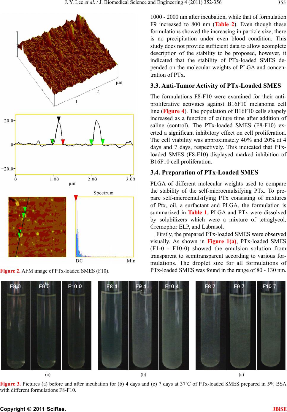
Figure 4. In vitro cytotoxicity of B16F10 melanoma cell
against PTx-loaded SMES of formulations F8, F9 and F10 for
1, 4 and 7 days. The cells grown on a culture plate without
PTx-loaded SMES were used as the control.
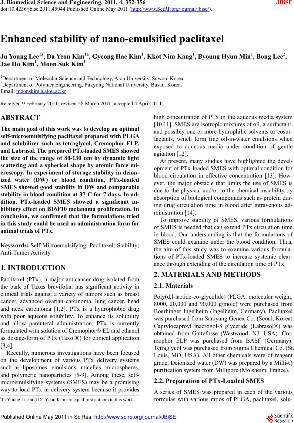
The droplet size distribution is comparatively narrow for
all formulations.
The morphology of PTx-loaded SMES was measured
by AFM as shown in Figure 2. PTx-loaded SMES (F10)
showed the spherical shape with smooth surface. A com-
paratively uniform droplet size of PTx-loaded SMES
was also observed at AFM, indicating no aggregation or
adhesion among SMES.
4. CONCLUSIONS
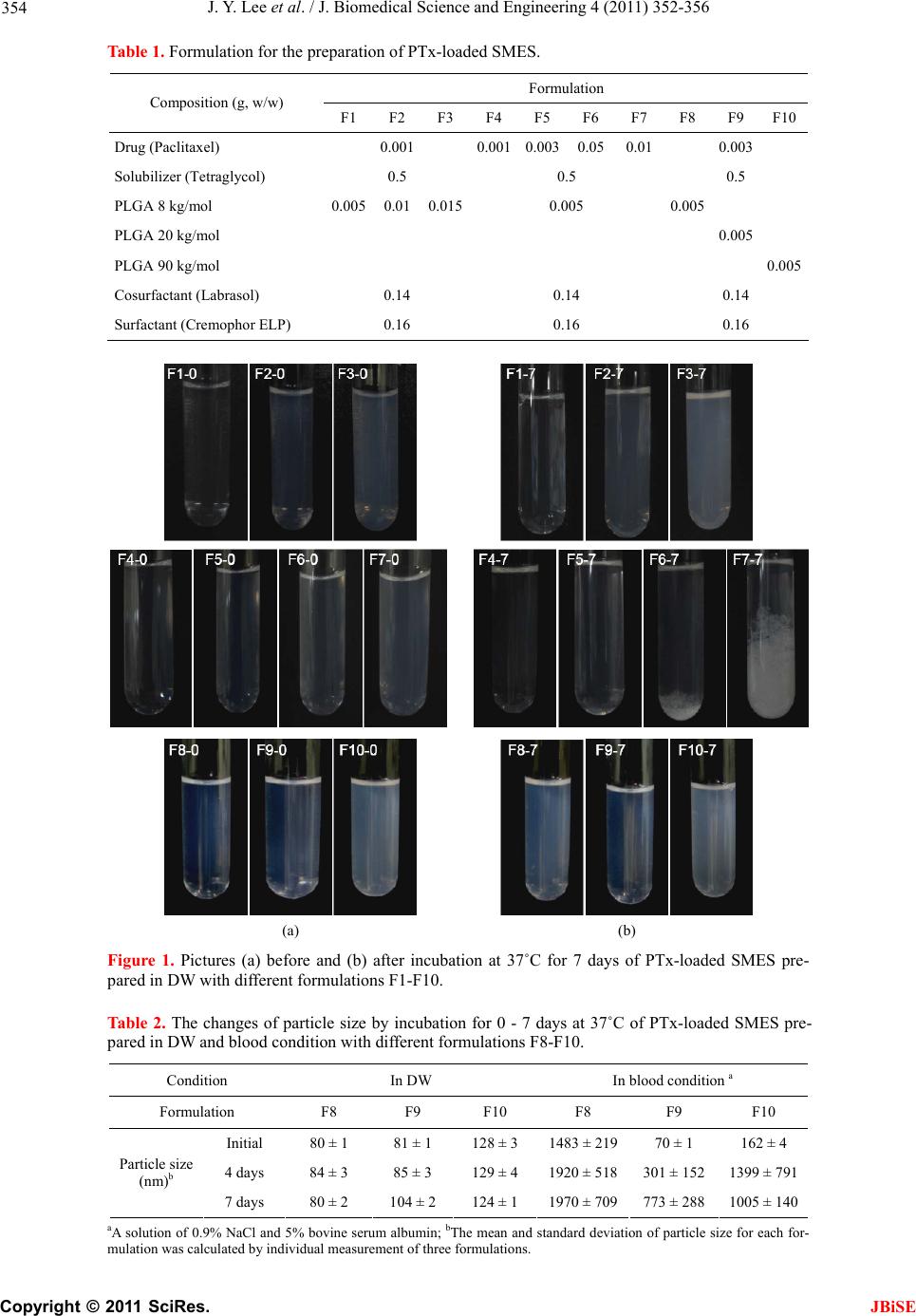
We prepared the PTx-loaded SMES with different for-
mulations to examine the storage stability. The prepared
PTx-loaded SMES showed a spherical shape with rang-
ing of 100 nm. We found the formulation of the PTx-
loaded SMES with stability for 7 days. The formulation
in this work could be used as administration form for
animal trials. Thus, further research on the animal model
using PTx-loaded SMES prepared in this work is now in
progress.
5. ACKNOWLEDGEMENTS
This work was supported by a grant from the new faculty research fund
of Ajou University, KMOHW (grant no A050082) and Priority Research
Centers Program through NRF funded by the Ministry of Education,
Science and Technology (2010-0028294).
REFERENCES
[1] Li, J.Y., Strobel, G., Sidhu, R., Hess, W.M. and Ford, E.J.
(1996) Endophytic taxol-producing fungi from bald cy-
press, Taxodium distichum. Microbiology, 142, 2223-
2226. doi:10.1099/13500872-142-8-2223
[2] Spencer, C.M. and Faulds, D. (1994) Paclitaxel: A review
of its pharmacodynamic and pharmacokinetic properties
and therapeutic potential in the treatment of cancer.
Drugs, 48, 794-847.
[3] Scripture, C.D., Szebeni, J., Loos, W.J., Figg, W.D. and
Sparreboom, A. (2005) Comparative in vitro properties
and clinical pharmacokinetics of Paclitaxel following the
administration of taxol(r) and paxene(r). Cancer Biology
& Ther apy, 4, 555-560.
[4] Cordes, N. and Plasswilm, L. (1998) Cell line and sched-
ule-dependent cytotoxicity of paclitaxel (Taxol): Role of
the solvent Cremophor EL/ethanol. Anticancer Research,
18, 1851-1857.
[5] Pang, K.S., Maeng, H.J. and Fan, J. (2009) Interplay of
transporters and enzymes in drug and metabolite proc-
essing. Molecular Pharmaceutics, 6, 1734-1755.
doi:10.1021/mp900258z
[6] Zhang, C., Qu, G., Sun, Y., Wu, X., Yao, Z., Guo, Q.,
Ding, Q., Yuan, S., Shen, Z., Ping, Q. and Zhou, H. (2008)
Pharmacokinetics, biodistribution, efficacy and safety of
N-octyl-O-sulfate chitosan micelles loaded with pacli-
taxel. Biomaterials, 29, 1233-1241.
doi:10.1016/j.biomaterials.2007.11.029
[7] Wei, H., Cheng, S.X., Zhang, X.Z. and Zhuo, R.X. (2009)
Thermo-sensitive polymeric micelles based on poly
(N-isopropylacrylamide) as drug carriers. Progress in
Polymer Science, 34, 893-910.
doi:10.1016/j.progpolymsci.2009.05.002
[8] Lee, H.B., Jeong, J.K., Sohn, S.I., Byun, Y., Ki, M.H. and
Seo, J.K. (2009) Drug delivery and regenerative medi-
cine technology. Tissue Engineering Regenerative Medi-
cine, 4, 663-667.
[9] Yu, W. and Zhang, N. (2009) Surface modification of
nanocarriers for cancer therapy. Current Nanoscience, 5,
123-134. doi:10.2174/157341309788185370
[10] Feng, S. and Huang, G. (2001) Effects of emulsifiers on
the controlled release of paclitaxel (Taxol) from nano-
spheres of biodegradable polymers. Journal of Con-
trolled Release, 71, 53-69.
doi:10.1016/S0168-3659(00)00364-3
[11] Gao, P
., Rush, B.D., Pfund, W.P., Huang, T., Bauer, J.M.,
Morozowich, W., Kuo, M.S. and Hageman, M.J. (2003)
Development of a supersaturable SEDDS (S-SEDDS)
formulation of paclitaxel with improved oral bioavail-
ability. Journal of Pharmaceutical Sciences, 92, 2386-
2398. doi:10.1002/jps.10511
[12] Gursoy, N., Garrigue, J.S., Razafindratsita, A., Lambert,
G. and Benita, S. (2003) Excipient effects on in vitro cy-
totoxicity of a novel paclitaxel self-emulsifying drug de-
livery system. Journal of Pharmaceutical Sciences, 92,
2411-2418. doi:10.1002/jps.10501
[13] Ten Tije, A.J., Verweij, J., Loos, W.J. and Sparreboom, A.
(2003) Pharmacological effects of formulation vehicles:
implications for cancer chemotherapy. Clinical Pharma-
cokinetics, 42, 665-685.
[14] Kan, P
., Chen, Z.B., Lee, C.J. and Chu, I.M. (1999) De-
velopment of nonionic surfactant/phospholipid o/w emul-
sion as a paclitaxel delivery system. Journal of Con-
trolled Release, 58, 271-278.
doi:10.1016/S0168-3659(98)00164-3
C
opyright © 2011 SciRes. JBiSE