 Journal of Geoscience and Environment Protection, 2014, 2, 129-133 Published Online April 2014 in SciRes. http://www.scirp.org/journal/gep http://dx.doi.org/10.4236/gep.2014.22018 How to cite this paper: Wang, M., & Lei, Y. X. (2014). Expression of Telomerase Reverse Transcriptase during the Malignant Transformation of Cadmium-Induced Cells. Journal of Geoscience and Environment Protection, 2, 129-133. http://dx.doi.org/10.4236/gep.2014.22018 Expression of Telomerase Reverse Transcriptase during the Malignant Transformation of Cadmium-Induced Cells Min Wang, Yixiong Lei* School of Public Health, Guangzhou Medical University, Guangzhou, China Email: *gz-leizeng@163.com Received February 2014 Abstract The objective of the present study was to investigate human telomerase reverse transcriptase (hTERT) mRNA and protein expres sion s during the cadmium chloride-induced malignant trans- formation of human bronchial epithelial (16HBE) cells. Fluorescence quantitative PCR (FQ-PCR) and Western blot analyses were performed to detect the hTERT mRNA and protein expressions in normal 16HBE cells, cadmium chloride-transformed 16HBE cells, and tumorigenic cells from nude mice inoculated with cadmium chloride-transformed 16HBE cells. Under the inner standard of GAPDH, the hTERT mRNA expression was significantly higher at different stages of malignant transformation (cadmium chloride-transformed 16HBE cells at passages 15 and 35 and tumori- genic cells from nude mice) than in normal 16HBE cells, and increased with the development of malignancy (P < 0.01). In addition, hTERT protein expression increased with the development of malignancy. These findings demonstrate that hTERT expression is related to cadmium chloride- induced malignant transformation. Cadmium chloride-induced malignant transformation is in- volved in changes in the hTERT activity, and might be an early event in cadmium chloride-induced malignant transformation. Keywords Cadmium Chloride; Human Bronchial Epithelial Cells; Malignant Transformation; Telomerase Reverse Transcriptase 1. Introduction The telomere is a cap-like structure of eukaryotic chromosomes. Human telomerase is one of the ribonucleo- *Corresponding author. This work was supported by the grants from the National Natural Science Foundation of China (No. 81373038), and by Science and Tec h- nology Planning Project of Guangzhou Municipality, China (No.2013J410037).  M. Wang, Y. X. Lei protein enzymes and consists of human telomerase reverse transcriptase (hTERT), the telomerase RNA compo- nent, and human telomerase-associated protein I (hTEPI). hTERT is a protein with reverse transcriptase activity that belongs to the catalytic subunit of telomerase and is a determinant of telomerase activation. Studies have shown that the hTERT gene is closely related to the formation of malignant tumors (Masutomi, 2003). Cadmium (Cd) is widely distributed in nature. Cd and its compounds are widely used in industry and belong to industrial poisons and environmental pollutants. In recent years, with the rapid development of the electronics industry, the requirement for electronic materials and products (such as nickel-cadmium batteries, transistors, and semi- conductors) has been increasing and, consequently, the requirement for and production of the corresponding raw materials (such as Cd) have been growing over the years. Thus, the chance of occupational exposure to Cd is also increasing. With the rapid development of industrial production, environmental pollution due to Cd and its compounds are increasing severely and may be long-standing in the environment ( Li , 2014; Wei, 2012). Ac- cording to statistics from the International Agency for Research on Cancer (IARC), Cd and its compounds are confirmed carcinogens and have been one of the metal poisons threatening human health (IARC, 1993). To further investigate the molecular mechanisms underlying the Cd-induced malignant transformation of hu- man bronchial epithelial (16HBE) cells, hTERT mRNA and protein expressions were investigated in normal 16HBE cells, 16HBE cells with Cd-induced malignant transformation, and tumorigenic cells from nude mice inoculated with 16HBE cells that underwent Cd-induced malignant transformation. 2. Material and Methods 2.1. Cells and Reagents Cadmium chloride-induced malignant transformation of 16HBE cells was reported in our previous study (Le i, 2008). Normal 16HBE cells, 16HBE cells with Cd-induced malignant transformation (passages 15 and 35), and tumorigenic cells from nude mice inoculated with Cd-induced malignantly transformed 16HBE cells were thawed and cultured. MEM powder (GIBCO-BRL, USA), calf serum (Guangzhou farm, China), trypsin, EDTA (HycloneE, USA), Taqman RNA quantitative PCR kit and probes (Ambion, USA), iScript Advanced cDNA synthesis kit (Bio-rad, USA), Espect spectrophotometer (Malcom, Japan), protein extraction kit, and BCA Pro- tein Assay kit (KeyGEN Biotech, Co., Ltd, China) were used in the present study. Primers were synthesized by the Invitrogen Corporation (Carlsbad, USA). The hTERT primers used in our study were 5'-TGTGCACC A AC ATC TAC AAG AT C-3' (forward primer) and 5'-CTGATGAAA TGGGAGCTGACG -3' (reverse primer). 2.2. Cell Culture and RNA Ext ract i on Cells were maintained in MEM containing 10% calf serum at 37˚C in a humidified environment with 5% CO2. Cells were digested with 0.25% trypsin in 0.02 EDTA (1:1, V/V). Cells (2.0 × 10⁶) were incubated with 1 ml of Trizol, followed by total RNA extraction according to the manufacturer’s instructions (GIBCO BRC). RNA pur- ity and concentration were determined by UV spectrophotometry and subjected to 1.0% agarose gel electropho- resis to detect RNA integrity. 2.3. RNA Reverse Transcription The RNA-Primer mixture (20 μL) was prepared with 1 μg of total RNA, 2 μl of 0.5 μM RT primer, and DEPC treated water (up to 15 μL), and then incubated at 65˚C for 5 min. The mixture was kept on ice for at least 2 min to produce the specific RNA-Primer Mix. Subsequently, reverse transcription was performed with 15 μL of RNA-Primer Mix, 4 μL of 5×iScript reaction Mix, and 1 μL of RTase (total volume: 20 μL) at 25˚C for 5 min, 42˚C for 30 min, and inactivated at 85˚C for 5 min. The products were diluted five-fold in ddH2O (20 μL of products in 80 μL of ddH2O) and then stored at -20˚C for use. 2.4. Detection of hTERT mRNA Expression wit h FQ-PCR Fluorescence quantitative PCR (FQ-PCR) was performed with the SYBR method. The mixture (10.0 μl) used for PCR included 5.0 μl of 2×SYBR Premix DimerEraser, 0.5 μl of 20 μM PCR RreversePr i mer, 0.5 μl of 20 μM PCR Forward Primer, 0.2 μl of ROX Reference Dye II, 1.0 μl of cDNA template, and 2.8 μl of ddH2O. PCR was performed under the following conditions: pre-denaturation at 95˚C for 30 s, 45 cycles of denaturation at  M. Wang, Y. X. Lei 95˚C for 5 s, annealing at 60˚C for 30 s, and extension at 72˚C for 30 s. Data were acquired and analyzed. 2.5. Analysis of hTERT Protein Expression by Western Blot First, total protein was extracted. In brief, the medium was removed and cells were washed with cold PBS twice and then transferred into a pre-chilled tube. After addition of pre-chilled lysis buffer, lysates were centrifuged at 12000 rpm for 15 min at 4˚C. The supernatant was harvested, protein concentration was determined with the BCA method and, based on this, the protein solution volume needed for loading was determined. The protein was boiled at 100˚C for 5 min, and 50 μg of protein was loaded and subjected to 10% SDS-PAGE analysis (40 mA, 90 min). Proteins were transferred onto a PVDF membrane that was then blocked in blocking buffer for 60 min at 37˚C. The membrane was treated with primary antibody (1:1000) at 37˚C for 1 h and then at 4˚C over - night. Following three washes in PBST (5 min each), the membrane was treated with secondary antibody (1:2000) at 37˚C for 1 h. Following three PBST washes (5 min each), visualization with DAB was performed. Gel image analysis was performed to determine the molecular weight of the target protein and the optical densi- ty of protein bands and, based on this, the relative protein expression was calculated. 2.6. Statistical Analysis Experiments were done in triplicate, and data were expressed as means ± standard deviation. One-way analysis of variance (ANOVA) was employed for comparisons among groups. Statistical analysis was performed with the SPSS version 15.0 (SPSS Inc, Chicago, IL, USA) for Windows. A value of P < 0.05 was considered statisti- cally significant. 3. Results 3.1. Detection of RNA integrity RNA extracted from different groups of cells was subjected to UV spectrophotometry. The results showed that the A260/A280 was 2.09-2.12 and the A260/A230 was 2.23-2.31, suggesting that RNA had a high purity and presented little contamination by DNA and/or proteins. 3.2. Detection of hTERT mRNA Expression In the 4 groups (normal 16HBE cells, 16HBE cells with Cd-induced malignant transformation at passage 15, 16HBE cells with Cd-induced malignant transformation at passage 35, and tumorigenic cells from nude mice), hTERT mRNA expression is shown in Table 1. The results show that hTERT mRNA expression was signifi- cantly higher at different stages of malignant transformation than in normal 16HBE cells under the inner stan- dard of GAPDH, and the hTERT mRNA expression increased with the development of malignant transforma- tion (P < 0.01). 3.3. Detection of hTERT Protein Expression GAPDH served as an internal reference. Western blotting was performed to detect the relative hTERT protein expression in 16HBE cells from the different groups (Figure 1). As shown in Figure 1, when compared with the control group, the hTERT protein expression showed an in- creasing tendency for the development of malignant transformation, and increased hTERT protein expression was obvious in malignant cells. Table 1. hTERT mRNA expression in the 16HBE cells from the 4 groups. Normal 16HBE cells 15th passage of Cd-treated cells 15th passage of Cd-treated cells Tumorigenic cells from nude mice GAPDH 2 8.93 ± 0.07 29.48 ± 0.09 30.96 ± 0.03 32.07 ± 0.17 hTE RT 27.29 ± 0.13 27.66 ± 0.08 26.56 ± 0.28 25.89 ± 0.07 2-ΔΔCt 1.00 1.13 6.77 23.26 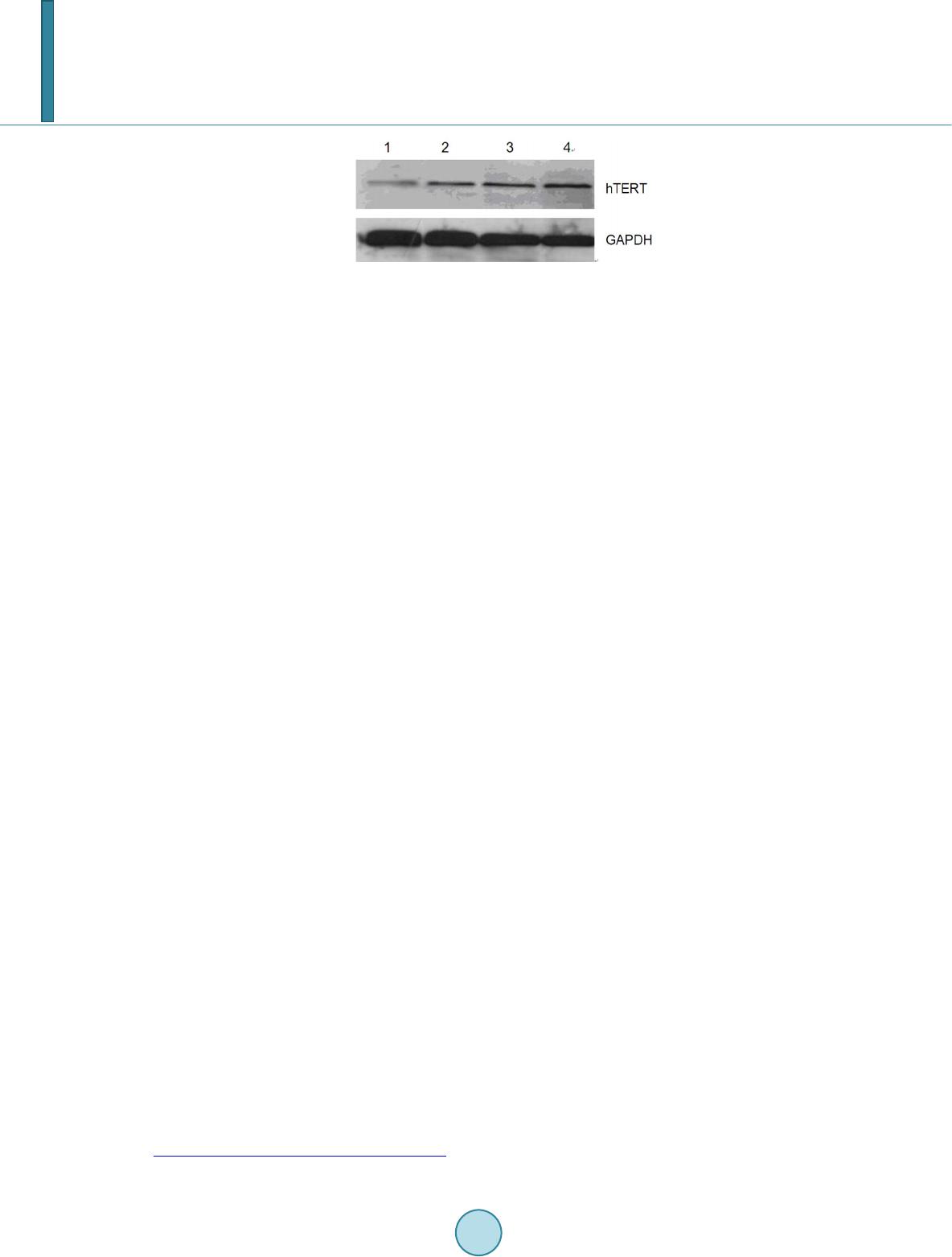 M. Wang, Y. X. Lei Figure 1. hTERT protein expression in 16HBE cells from the 4 groups. 1. Normal 16HBE cells; 2. 16HBE cells undergoing Cd-induced malignant trans- formation at passage 15; 3. 16HBE cells under- going Cd-induced malignant transformation at pas- sage 35; 4. Tumorigenic cells from nude mice un- dergo ing inoculation of 16HBE cells with Cd-induc- ed malignant transformation. 4. Discussion The telomere is a cap-like structure at the end of eukaryotic chromosomes that can bind G-rich nucleotide hex- amers and related proteins to form DNA-binding protein complexes. Telomerase is a DNA polymerase with re- verse transcriptase activity and is composed of RNA and proteins. During cell division, the telomerase can use its own RNA as a template to reverse transcribe the TTAGGG repeat fragments, which are then added to the end of the shortened and linear chromosomes, a process that may lead to the indefinite proliferation of cells and the occurrence of cancers (Neidle & Parkinson, 2003). In human somatic cells, telomerase expression is not ob- served, and thus the length of telomeres is not increased. The telomeres are shortened as cells divide. Finally, cell division and proliferation stop and cells start developing aging (Sara & Victoria, 2001). Studies have shown that hTERT is the catalytic subunit of telomerase and also a determinant of telomerase activation. The hTERT gene is associated with the formation and the malignancy of tumors. Under the catalysis of hTERT, telomerase can use its own RNA as a template for the synthesis of 5′-TTAGGG-3′ repeat sequences, which may offset te- lomere shortening during replication, leading to cell proliferation (Harrington, 2001). Our results showed that, at moderate concentrations (10 µmol/L), cadmium chloride could increase hTERT protein and mRNA expression in 16HBE cells at passage 15. The mRNA and protein expression further in- creased in 16HBE cells at independent growth stages and in malignant cells. This suggests that the increase in hTERT activity might be an early molecular event during the Cd-ind uced malignant transformation of cells, which may persist for the entire duration of the malignant transformation. In vitro, the malignant transformation of cells is involved in different stages of cancer formation and progression, including immortalization, morpho- logical changes, non-growth factor-dependent growth, growth on semi-solid medium, and tumorigenicity (Ross, 2003). Cells may acquire different malignant phenotypes via the sequential selective expansion, with a clonal growth advantage (Jetten, 1989 ) . Our findings demonstrated that the change in hTERT activity became evident over the development of malignant transformation, which might increase telomerase activity and play an impor- tant role in the Cd-induced malignant transformation of cells. The mechanisms underlying Cd-induced carcinogenesis are complex and have not been elucidated to date. Studies have shown that DNA injury, impaired DNA repair, changes in gene expression and cell cycle regula- tion, and abnormal DNA methylation are closely associated with Cd-induced carcinogenesis (Koizumi & Ya- mada , 2003; Zhou, 2012). Our findings showed that a change in hTERT activity occurred during the Cd-induced malignant transformation of cells, suggesting that hTERT expression is correlated with malignant transformation and carcinogenicity. Our findings may provide theoretical evidence for elucidating the mechanisms underlying Cd-induced carcinogenesis and present an indicator for the early diagnosis of carcinogenesis in Cd-exposed populations. In addition, our results provide a clue for detecting hTERT protein expression in malignant tumors (especially in populations with Cd exposure) and open potential avenues for hTERT mRNA-targeting therapy. References Harrington, L. (2003). Biochemical Aspects of Telomerase Function. Cancer Letters, 19 4 , 139-154. http://dx.doi.org/10.1016/S0304-3835(02)00701-2  M. Wang, Y. X. Lei IARC (1993). Beryllium, Cadmium, Mercury and Exposures in the Glass Manufacturing Industry. International Agency for Research on Cancer, 58, 119-238. Jetten, A. M. (1989). Multistep Process of Squamous Differentiation in Tracheobronchial Epithelial Cells in Vitro: Analogy with Epidermal Differentiation. Environmental Health Perspectives, 80, 149-160. http://dx.doi.org/10.1289/ehp.8980149 Koizumi, S., & Yamada, H. (2003). DNA Microarray Analysis of Altered Gene Expression in C admi um-Exposed Human Cells. Journal of Occupational Health, 45, 331 -334. http://dx.doi.org/10.1539/joh.45.331 Lei, Y. X., Wei , L., Wang, M., Wu, G. R., & Li , M. (2008). Malignant Transformation and Abnormal Expression of Euka- ryotic Initiation Factor in Bronchial Epithelial Cells Induced by Cadmium Chloride. Biomedical and Environmental Sciences , 21, 332-338. http://dx.doi.org/10.1016/S0895-3988(08) 600 51-3 Li, Z., Ma, Z., van der Kuijp, T. J., Yuan, Z., & Huang, L. (2014). A Review of Soil Heavy Metal Pollution from Mines in China: Pollution and Health Risk Assessment. Science of the Total Environment, 468-46 9, 843 -853. http://dx.doi.org/10.1016/j.scitotenv.2013.08.090 Masutomi, K., Yu, E. Y., Khurts, S., Ben-Po r ath , I., Currier, J. L., Metz, G. B., Brooks, M. W., Kaneko, S., Murakami, S., DeCaprio , J. A., Weinberg, R. A., Stewart, S. A., & Hahn, W. C. (2003). Telomerase Maintains Telomere Structure in Normal Human Cells. Cel l, 1 14, 241-253. http://dx.doi.org/10.1016/S0092-8674(03) 0055 0 -6 Neidle, S., & Parkinson, G. N. (2003). The Structure of Telomeric DNA. Current Opinion in Structural Biology, 13, 275- 283. http://dx.doi.org/10.1016/S0959-440X( 03) 0007 2 -1 Ross, R. A., Biedler, J. L., & Spengler, B. A. (2003). A Role for Distinct Cell Types in Determining Malignancy in Human Neuroblastoma Cell Lines and Tumor s. Cancer Letters, 197 , 35-39. http://dx.doi.org/10.1016/S0304-3835(03)00079-X Sara, K., & Victoria, L. (2001). Positive and Negative Regulation of Telomerase Access to the Telo mere. Cell Science, 113, 3357-3364 Wei, L., Lei, Y. X., Wu, L., Wang, M., Lu, Q., & He, C. C. (2012). Alterations in the Expression of Translation Factors as Molecular Markers in Cadmium-Exposed Workers. Biomarkers, 17, 78 -84. http://dx.doi.org/10.3109/1354750X.2011.639463 Zhou , Z. H., Lei, Y. X., & Wang , C. X. (2012). Analysis of Aberrant Methylation in DNA Repair Genes during Malignant Transformation of Human Bronchial Epithelial Cells Induced by Ca dmi um. Toxicological Sciences, 125, 412-4 17. http://dx.doi.org/10.1093/toxsci/kfr320
|