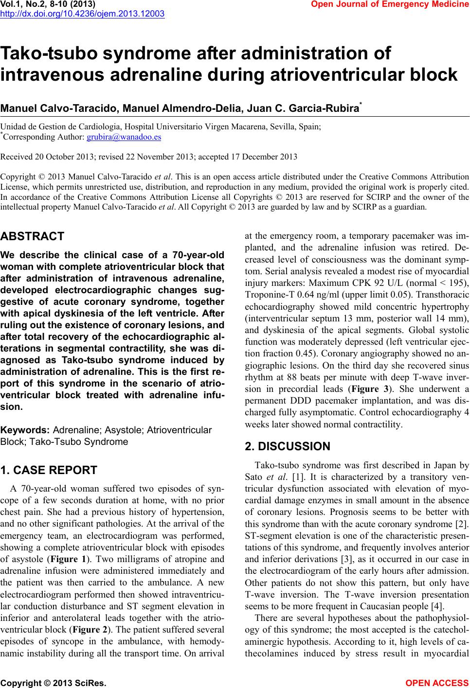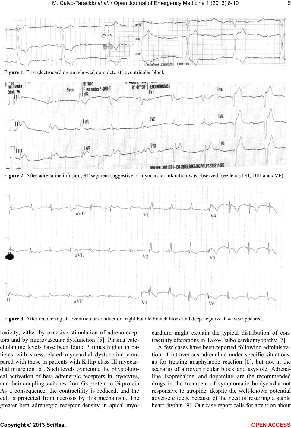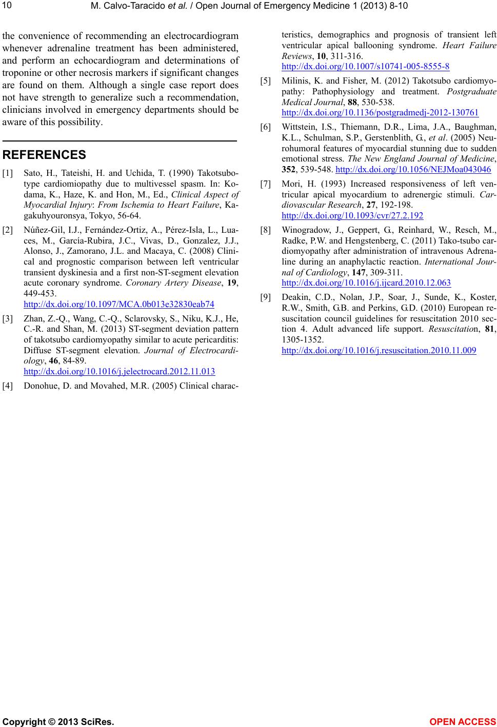
M. Calvo-Taracido et al. / Open Journal of Emergency Medicine 1 (2013) 8-10
10
the convenience of recommending an electrocardiogram
whenever adrenaline treatment has been administered,
and perform an echocardiogram and determinations of
troponine or othe r necros is mark ers if significant ch an g es
are found on them. Although a single case report does
not have strength to generalize such a recommendation,
clinicians involved in emergency departments should be
aware of this possibility.
REFERENCES
[1] Sato, H., Tateishi, H. and Uchida, T. (1990) Takotsubo-
type cardiomiopathy due to multivessel spasm. In: Ko-
dama, K., Haze, K. and Hon, M., Ed., Clinical Aspect of
Myocardial Injury: From Ischemia to Heart Failure, Ka-
gakuhyouronsya, Tokyo, 56-64.
[2] Núñez-Gil, I.J., Fernández-Ortiz, A., Pérez-Isla, L., Lua-
ces, M., García-Rubira, J.C., Vivas, D., Gonzalez, J.J.,
Alonso, J., Zamorano, J.L. and Macaya, C. (2008) Clini-
cal and prognostic comparison between left ventricular
transient dyskinesia and a first non-ST-segment elevation
acute coronary syndrome. Coronary Artery Disease, 19,
449-453.
http://dx.doi.org/10.1097/MCA.0b013e32830eab74
[3] Zhan, Z.-Q., Wang, C. -Q., Sclarovsky, S., Ni ku, K.J., He,
C.-R. and Shan, M. (2013) ST-segment deviation pattern
of takotsubo cardiomyopathy similar to acute pericarditis:
Diffuse ST-segment elevation. Journal of Electrocardi-
ology, 46, 84-89.
http://dx.doi.org/10.1016/j.jelectrocard.2012.11.013
[4] Donohue, D. and Movahed, M.R. (2005) Clinical charac-
teristics, demographics and prognosis of transient left
ventricular apical ballooning syndrome. Heart Failure
Reviews, 10, 311-316.
http://dx.doi.org/10.1007/s10741-005-8555-8
[5] Milinis, K. and Fisher, M. (2012) Takotsubo cardiomyo-
pathy: Pathophysiology and treatment. Postgraduate
Medical Journal, 88, 530-538.
http://dx.doi.org/10.1136/postgradmedj-2012-130761
[6] Wittstein, I.S., Thiemann, D.R., Lima, J.A., Baughman,
K.L., Schulman, S.P., Gerstenblith, G., et al. (2005) Neu-
rohumoral features of myocardial stunning due to sudden
emotional stress. The New England Journal of Medicine,
352, 539-548. http://dx.doi.org/10.1056/NEJMoa043046
[7] Mori, H. (1993) Increased responsiveness of left ven-
tricular apical myocardium to adrenergic stimuli. Car-
diovascular Research, 27, 192-198.
http://dx.doi.org/10.1093/cvr/27.2.192
[8] Winogradow, J., Geppert, G., Reinhard, W., Resch, M.,
Radke, P.W. and Hengstenberg, C. (2011) Tako-tsubo car-
diomyopathy after administration of intravenous Adrena-
line during an anaphylactic reaction. International Jour-
nal of Cardiology, 147, 309-311.
http://dx.doi.org/10.1016/j.ijcard.2010.12.063
[9] Deakin, C.D., Nolan, J.P., Soar, J., Sunde, K., Koster,
R.W., Smith, G.B. and Perkins, G.D. (2010) European re-
suscitation council guidelines for resuscitation 2010 sec-
tion 4. Adult advanced life support. Resuscitation, 81,
1305-1352.
http://dx.doi.org/10.1016/j.resuscitation.2010.11.009
Copyright © 2013 SciRes. OPEN A CCESS