Open Journal of Stomatology, 2013, 3, 515-519 OJST
http://dx.doi.org/10.4236/ojst.2013.39085 Published Online December 2013 (http://www.scirp.org/journal/ojst/)
Mandibular changes associated with maxillary impaction
and molar intrusion
Sondos Abuzinada, Fahad Alsulaimani
King Abdulaziz University, Jeddah, Saudi Arabia
Email: sondoz@hotmail.com
Received 29 October 2013; revised 29 November 2013; accepted 11 December 2013
Copyright © 2013 Sondos Abuzinada, Fahad Alsulaimani. This is an open access article distributed under the Creative Commons
Attribution License, which permits unrestricted use, distribution, and reproduction in any medium, provided the original work is
properly cited.
ABSTRACT
We report a case series of ten patients who presented
with anterior open bite. The initial evaluation in-
cluded clinical pictures, lateral cephalometric radio-
graphs and dental casts. They were assessed accord-
ing to the severity of the open bite and the cause
(skeletal or dental). They all underwent orthodontic
treatment as an initial step. Five patients with dental
open bite underwent molar intrusion using titanium
screws and five patients underwent maxillary Le Fort
I impaction. We report the mandibular changes asso-
ciated with these different treatment modalities with
improved esthetics.
Keywords: Maxillary Impaction; Molar Intrusion; Open
Bite; Le Fort I; Titanium Screws
1. INTRODUCTION
Vertical Maxillary Excess (VME) is one of the most fre-
quently encountered dentofacial deformities. This can
result in an anterior open bite or an unesthetic gummy
smile. The correction of vertical problems with or with-
out open bite usually includes maxillary Le Fort I im-
paction [1]. On the other hand, the correction of an open
bite can be managed orthodontically by molar intrusion
[2].
The mandible in such cases is either retrognathic or
within normal position and will respond to the superior
positioning of the maxilla by autorotation. In some cases
this autorotation will add the need to perform mandibular
surgery to either advance or setback the mandible and is
case-dependent [3,4]. With the mandibular autorotation,
the chin position advances. This can be estimated in the
presurgical prediction, but requires great accuracy as this
will decide if only maxillary Le Fort I impaction will
need or two jaw surgeries. Some studies have been made
for predicting mandibular autorotation following manxil-
lary impaction [5,6]. However, controversies in locating
the center of mandibular autorotation revealed the inabil-
ity to accurately predict the final position of the mandible
after maxillary impaction [6-8]. On the other hand, it has
been documented that the mandible will move forward
following maxillary impaction with a chin advancement
in 1:1 ratio [9].
Molar intrusion is used to manage an open bite. A
number of patients do not wish to perform orthognathic
surgery because of the involved risk. For such patients,
various alternatives can be used, including multibrackets
in conjunction with high-pull headgear therapy (2), mul-
tiple-loop edgewise arch wire (MEAW) therapy (3), re-
versed curve nickel-titanium wire with intermaxillary
elastics (4) and extraction therapy (5). These treatment
modalities can achieve acceptable overbite and interin-
cisal relationship which also guide the mandible and chin
into a new position leading to a more esthetic appearance
[10].
In this article we present a series of cases. Some un-
derwent orthodontic molar intrusion and others under-
went maxillary Le Fort I impaction. We compare the
changes in mandibular position and the resulting esthetic
improvement for each technique.
2. PATIENTS AND METHODS
10 patients presented to our joint maxillofacial surgery
and orthodontics clinic. Their age group varied between
15 and 27 years. Their chief complaint included a com-
bination of anterior open bite and “a gummy smile”.
They all underwent a detailed clinical examination which
included, radiographs (lateral cephalograms, orthopanto-
grams OPG), dental models and photographs. Cepha-
lometric analysis was done for all 10 patients using
Stiner analysis. The treatment plan was established after
correlating cephalometric analysis with clinical presenta-
OPEN ACCESS
S. Abuzinada, F. Alsulaimani / Open Journal of Stomatology 3 (2013) 515-519
516
tion and the chief complaint. The treatment plan for pa-
tients diagnosed with vertical maxillary excess and ret-
rognathia included presurgical orthodontic treatment
(leveling, alignment and decompensation) followed by
maxillary Le fort l osteotomy with impaction and fixa-
tion using four 1.5 mm miniplates. This was followed by
an advancement genioplasty according to patients need.
However, patients diagnosed with anterior open bite
without vertical maxillary excess underwent orthodontic
treatment. The cause of the anterior open bite in these
cases was suggested to have been extrusion of both upper
molars, based on cephalometric readings, therefore, the
plan was to intrude both upper molars using implanted
titanium screws to deliver the force.
3. ORTHODONTIC TREATMENT FOR
OPEN BITE
Bonding and banding of all teeth, using 0.022-inch slot,
preadjusted edgewise appliances were placed in both
arches (Victory SeriesTM, Roth Rx, 3M Unitek, Monrovia,
CA, USA) and leveling and aligning phase using pro-
gressive sequencing of arch wires. was performed. Cor-
rection of the two-step occlusal plane was done during the
leveling and aligning phase by extrusion of anterior teeth.
Once a heavy stainless steel arch wire (0.019" × 0.025")
was reached, Titanium screws (1.6-mm diameter, 8-mm
length; (RMO Co. Ltd., Denver, Colorado, USA) were
inserted bilaterally in the alveolar bone of the maxilla
through the buccal mucosa between the second bicuspid
and the first molar from both the labial and palatal area in
some patients as needed. All the screws were implanted at
the same visit under local anesthesia was administered.
Analgesics were prescribed to the patients for 3 days after
the implantation. One week after implantation of the ti-
tanium screws, intrusion of posterior teeth began using
elastic chains. The total active treatment period was 19
months. The implant screw anchorage was stable for the
entire duration of the treatment, and the screws were
removed during the retention phase. Another cepha-
lometric radiograph was taken upon completion of or-
thodontic treatment and Stiner analysis was repeated.
4. RESULTS
Five patients with anterior open bite and VME under-
went orthognathic surgery (Le fort I maxillary impaction
with more posterior impaction. An advancement genio-
plasty was also done in three patients. Five patients with
anterior open bite underwent orthodontic treatment only.
The results of the cephalometric analysis are listed in
(Ta ble 1). The counter clockwise rotation of mandibular
plane improved the chin position in three patients who
underwent orthodontic treatment (Figure 1) and in all
five patients who underwent maxillary impaction this
was demonstrated by a decrease in mandibular plane
angle and an increase in SNA (Figure 2). Two patients
who underwent orthodontic treatment for closure of an-
terior open bite showed clockwise rotation of the man-
dibular plane with an increase in mandibular plane angle
and a minimal decrease in SNB. However, all patients
showed improvement in facial profile and function. The
Ta b l e 1 . Cephalometric values for patients who underwent molar intrusion and patients who underwent maxillary impaction (DX:
diagnosis; DOB: dental anterior open bite).
Patient number Dx Incisal show at rest Open bite (mm)Man plane Occlusal planePoint B to NFH SNB
1 DOB 3 mm 5 mm 30
25
14
8
0
2
70
73
2 DOB 3 mm 4 mm 47
45
16
20
10
10
75
72
3 DOB 2 4 mm 38
36
17
16
9
10
85
84
4 DOB 3 5 mm 35
33
15
17
6
6
82
80
5 DOB 4 mm 5 mm 39
36
18
15
6
8
81
81
6 VME 5 mm 4 mm 52
50
19
18
13
9
80
84
7 VME 7 mm 4 mm 49
44
17
14
10
10
72
73
8 VME 8 mm - 42
33
13
9
55
52
80
85
9 VME 6 mm 3 mm 35
28
13
11
11
7
69
72
10 VME 6 mm 7 mm 33
30
15
15
26
22
68
70
Copyright © 2013 SciRes. OPEN ACCESS
S. Abuzinada, F. Alsulaimani / Open Journal of Stomatology 3 (2013) 515-519 517
(a) (b) (c)
Figure 1. (a) Lateral cephalometric radiograph before molar intrusion; (b) Lateral cephalometric radiograph after molar
intrusion; (c) Lateral cephalometric tracing before molar intrusion (blue) after molar intrusion (red).
(a) (b)
Figure 2. (a) Lateral cephalometric radiograph before maxillary impaction; (b) Lateral
cephalometric radiograph after maxillary impaction.
retrognathic chin and convex profiles were corrected,
resulting in a straight profile. The facial proportions were
also improved due to of the decrease in the lower facial
height. The strain in the circumoral musculature during
lip closure was improved.
5. DISCUSSION
Many studies in the literature mention the esthetic effects
of maxillary Le Fort I impaction related to the resulting
mandibular counter clockwise rotation [1,3,4]. However,
not much is mentioned regarding the effect of molar in-
trusion on the mandibular plane angle and the resulting
esthetic effect. The results of our study showed that mo-
lar intrusion used to close anterior open bite gives some
esthetic results as seen in maxillary Le Fort I impaction.
However, this was not noted in all orthodontically treated
cases with anterior open bite.
The esthetic changes resulting from maxillary impac-
tion are mostly related to the degree of mandibular auto-
rotation. The published data on this subject focused
mainly on prediction analysis of mandibular autorotation
following maxillary impaction [3,4]. Chin position was
advanced in all patients who underwent Le Fort I impac-
tion and this was noted in the literature by counter clock-
wise rotation of the mandible and an increase in SNB
and NB-Pog. However, we only used SNB to demon-
strate the effect of maxillary impaction on mandible po-
sition and NB-Pog was not used to demonstrate the
amount of postoperative mandibular advancement in pa-
tients who underwent maxillary impaction due to the fact
that three patients required genioplasty to further im-
prove esthetics.
Copyright © 2013 SciRes. OPEN ACCESS
S. Abuzinada, F. Alsulaimani / Open Journal of Stomatology 3 (2013) 515-519
518
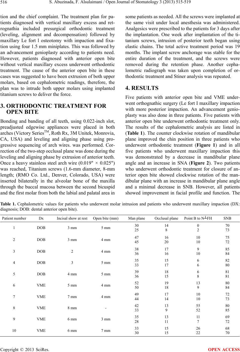
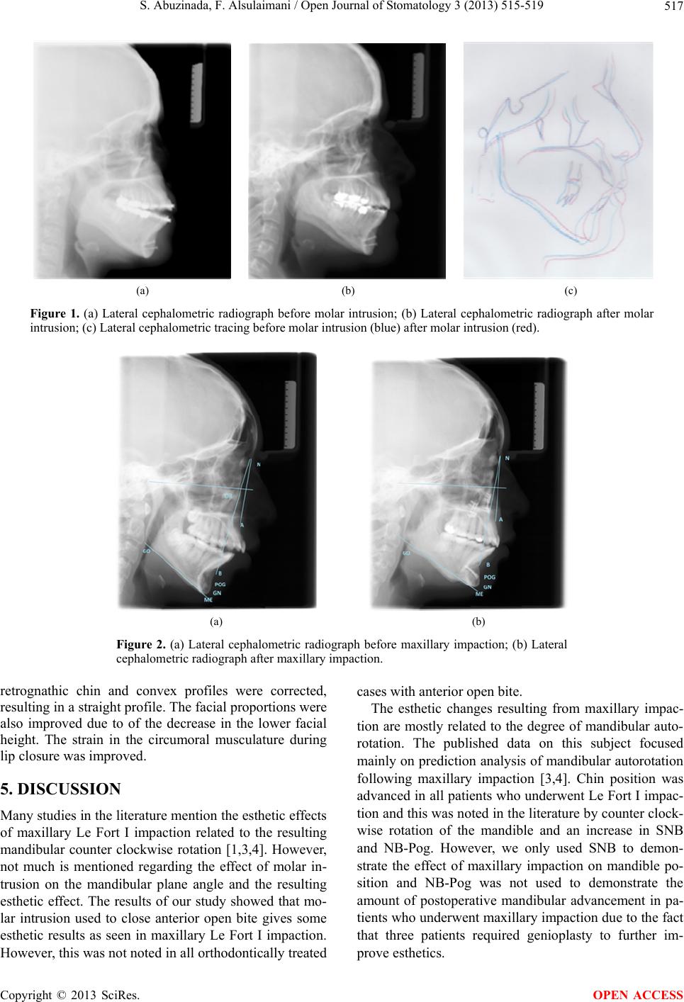
Patients who underwent orthodontic treatment for
open bite closure showed improved esthetics, however,
not all patients showed mandibular counter clockwise
rotation as seen in Le Fort I impaction. Some patients
showed clockwise mandibular rotation. This was ex-
plained by the orthodontic treatment, since two of the
patients have constricted maxilla. Expansion appliance
such as Quad-Helix was used to expand the maxillary
teeth. It is well documented with expansion clock wise
rotation of the mandibular plane which has taken place
due to extrusion of the palatal cusp. The clock wise rota-
tion point pog will also rotate backward. The difference
between pre and post treatment was in average 2 degrees
which could be due to the occlusal plane measured at
functional occlusion with no anterior teeth contacting a
measurement error that was possible. The most important
is the reduction of the mandibular plane which showed a
counter clock wise rotation in all the patients with es-
thetic improvement.
On the other hand, the skeletal improvement is poor
due to failure to establish absolute anchorage during mo-
lar intrusion. To obtain absolute anchorage, several de-
vises have been used such as dental implant [11-14],
screws [15,16] and miniplates [17,18]. The advantages of
these devices are by providing absolute anchorage dif-
ferent teeth movement without the need for patient’s co-
operation. Several reports have been reported on the use
of screw for anchorage in teeth movement, intrusion or
retraction of anterior teeth [10,11], and protraction of
posterior teeth in the mandible. In addition, few papers
have reported the use of titanium screws for orthodontic
anchorage to intrude upper and/or lower molars of an
adult patient with severe skeletal anterior open bite.
The mandible will follow any changes in occlusion
resulting from maxillary impaction or molar intrusion.
The noted changes in mandibular and chin position were
quite variable and less predictable following molar intru-
sion, however, the improved esthetics can be appreciated
in all patients. Future standardized studies will help us
make accurate predictions following molar intrusion or
maxillary impaction.
REFERENCES
[1] Schendel, S., Eisenfeld, J., Bell, W., et al. (1976) Supe-
rior repositioning of the maxilla: stability and soft tissue
osseous relations. American Journal of Orthodontics, 70,
663-674.
http://dx.doi.org/10.1016/0002-9416(76)90226-8
[2] Sherwood, K. (2007) Correction of skeletal open bite
with implant anchored molar/bicuspid intrusion. Oral &
Maxillofacial Surgery Clinics of North America, 19, 339-
350. http://dx.doi.org/10.1016/0002-9416(76)90226-8
[3] Nadjmi, N., Mommaerts, M., Abeloos, J. and Clercq, C.
(1998) Prediction of mandibular autorotation. Journal of
Oral and Maxillofacial Surgery, 56, 1241-1247.
http://dx.doi.org/10.1016/S0278-2391(98)90599-7
[4] Y.-C. Wang, Ko, E.W.-C., Huang, C.-S. and Chen, Y.-R.
(2006) The inter-relationship between mandibular auto-
rotation and maxillary LeFort I impaction osteotomies.
Journal of Craniofacial Surgery, 17, 898-904.
http://dx.doi.org/10.1097/01.scs.0000234985.99863.97
[5] Sperry, T.P., Steinberg, M.J. and Gans, B.J. (1982) Man-
dibular movement during autorotation as a result of max-
illary impaction surgery. American Journal of Orthodon-
tics, 81, 116-123.
http://dx.doi.org/10.1016/0002-9416(82)90035-5
[6] Bryan, D.C. (1994) An investigation into the accuracy
and validity of three points used in the assessment of
autorotation in orthognathic surgery. British Journal of
Oral and Maxillofacial Surgery, 32, 363-272.
http://dx.doi.org/10.1016/0266-4356(94)90026-4
[7] Nattestad, A. and Vedtofte, P. (1992) Mandibular autoro-
tation in orthognathic surgery: A new method of locating
the centre of mandibular rotation and determining its
consequence in orthognathic surgery. Journal of Cranio-
facial Surgery, 20, 163-170.
[8] Rekow, E.D., Speidel, T.M. and Koening, R.A. (1993)
Location of the mandibular center of autorotation in max-
illary impaction surgery. American Journal of Orthodon-
tics and Dentofacial Orthopedics, 103, 530-536.
http://dx.doi.org/10.1016/0889-5406(93)70093-4
[9] Wang, Y., Ko, E., Huang, C. and Chen, Y. (2006) The
inter-relationship between mandibular autorotation and
maxillary LeFort I impaction osteotomies. Journal of
Craniofacial Surgery, 17, 898-904.
http://dx.doi.org/10.1097/01.scs.0000234985.99863.97
[10] Alsulaimani, F. (2012) Severe anterior open-bite case
treated using miniscrew anchorage: A case report. Jour-
nal of American Science, 8, 102-107.
[11] Turley, P.K., Kean, C., Schur, J., Stefanac, J., Gray, J.,
Hennes, J. and Poon, L.C. (1988) Orthodontic force ap-
plication to titanium endosseous implants. Angle Ortho-
dontist, 58, 151-162.
[12] Odman, J., Lekholm, U., Jemt, T., Branemark, P.I. and
Thilander, B. (1988) Osseointegrated titanium implants—
A new approach in orthodontic treatment. European
Journal of Orthodontics, 10, 98-105.
http://dx.doi.org/10.1093/ejo/10.2.98
[13] Prosterman, B., Prosterman, L., Fisher, R. and Gornitsky,
M. (1995) The use of implants for orthodontic correction
of an open bite. American Journal of Orthodontics and
Dentofacial Orthopedics, 107, 245-250.
http://dx.doi.org/10.1016/S0889-5406(95)70139-7
[14] Roberts, W.E., Helm, F.R., Marshall, K.J. and Gongloff,
R.K. (1989) Rigid endosseous implants for orthodontic
and orthopedic anchorage. Angle Orthodontist, 59, 247-
256.
[15] Creekmore, T.D. and Eklund, M.K. (1983) The possibil-
ity of skeletal anchorage. Journal of Clinical Orthodon-
tics, 17, 266-269.
[16] Costa, A., Raffainl, M. and Melsen, B. (1998) Mini-
screws as orthodontic anchorage: A preliminary report.
Copyright © 2013 SciRes. OPEN ACCESS
S. Abuzinada, F. Alsulaimani / Open Journal of Stomatology 3 (2013) 515-519
Copyright © 2013 SciRes.
519
OPEN ACCESS
International Journal of Adult Orthodontics & Orthog-
nathic Surgery, 13, 201-209.
[17] Deguchi, T., Takano-Yamamoto, T., Kanomi, R., Harts-
field Jr., J.K., Roberts, W.E. and Garetto, L.P. (2003) The
use of small titanium screws for orthodontic anchorage.
Journal of Dental Research, 82, 377-381.
http://dx.doi.org/10.1177/154405910308200510
[18] Adell, R., Lekholm, U., Rockler, B. and Branemark, P.I.
(1981) A 15-year study of osseointegrated implants in the
treatment of the edentulous jaw. International Journal of
Oral Surgery, 10, 387-416.
http://dx.doi.org/10.1016/S0300-9785(81)80077-4