Open Journal of Stomatology, 2013, 3, 507-509 OJST
http://dx.doi.org/10.4236/ojst.2013.39083 Published Online December 2013 (http://www.scirp.org/journal/ojst/)
Oral ulcerations as the first manifestations of acute
leukemia: A case report
Somayeh Alirezaei1, Mahin Bakhshi2, Jamileh -Bigom Taheri2, Ahmad R. Mafi3*, Omid Moghaddas4
1Department of Oral and Maxillofacial Medicine, Dental School, Islamic Azad University, Tehran, Iran
2Department of Oral and Maxillofacial Medicine, Dental School, Shahid Beheshti University of Medical Sciences, Tehran, Iran
3Jorjani Cancer Center, Imam Hossein Hospital, Shahid Beheshti University of Medical Sciences, Tehran, Iran
4Department of Periodontology, Dental School, Islamic Azad University, Tehran, Iran
Email: *ahmadrmafi@yahoo.com
Received 20 November 2013; revised 21 December 2013; accepted 31 December 2013
Copyright © 2013 Somayeh Alirezaei et al. This is an open access article distributed under the Creative Commons Attribution Li-
cense, which permits unrestricted use, distribution, and reproduction in any medium, provided the original work is properly cited.
ABSTRACT
Acute myeloblastic leukemia (AML) is a highly fatal
malignant bone marrow disease. Physicians, dentists
and all other healthcare professionals should be
aware of sinister oral signs and symptoms in order to
early diagnosis and referral of patients. Here we re-
port a case of AML who presented with oral ulcers.
Ulcers developed after a parrot bite, which initially
misled the physicians. Unfortunately our patient did
not survive, but early diagnosis and prompt investi-
gation and treatment can be life-saving in many other
similar cases.
Keywords: Acute Leukemia; Oral Ulcers
1. INTRODUCTION
A 48-year-old female presented to the General Dental
Clinic of our hospital with chief complaint of painful
ulcers in gingiva and tongue that had developed three
weeks before. The lesion had started as a small ulcer in
her tongue, but gradually enlarged and after a couple of
days gingival ulcers developed.
On history taking, she gave a history of lethargy, mild
fever and slight weight loss. She also mentioned that she
kept a parrot in her house and she often fed the bird
mouth to mouth, and remarked that the lesion first started
when the parrot accidentally bit her tongue a couple of
weeks before. Furthermore, she remarked that her sister
also fed the parrot mouth to mouth, and she had devel-
oped similar ulcers that had healed spontaneously. The
rest of past medical history was unremarkable.
Based on this history, with the primary diagnosis of a
zoonotic disease, the patient was referred to oral medi-
cine department for consultation with an oral medicine
specialist.
On arrival to our department, it could be seen that she
was pale and ill. Extra-oral examination revealed swell-
ing and tenderness to palpitation of the face.
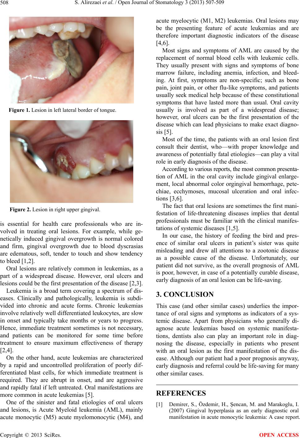
Intra-orally, bilateral palatal and lingual gingiva ap-
peared to be mildly swollen, glazed, devoid of stippling
and spongy in consistency. The color of the marginal and
papillary gingiva was dark red. Several ulcers were ob-
served on gingival and buccal mucosa. Ulcers were cov-
ered by a necrotic slough and surrounded by an erythe-
matous margin. A necrotic ulcer was also seen on left
lateral border of tongue (Figures 1 and 2).
As part of departmental routine, first we started inves-
tigating the disease with a systemic approach, and lab
tests were ordered as a part of this approach.
Haematology results revealed anemia (Hgb = 10.2
g/dL), thrombocytopenia (Plt = 49,000/mm3), and mark-
ed leukocytosis (WBC = 133,000/mm3). ESR was also
grossly elevated. Examination of the blood smear re-
vealed large cells with large nucleus with distinct nucle-
oli. Bone marrow biopsy revealed the diagnosis of acute
myelomonocytic leukemia (AML-M4).
The patient was referred to the hematology department.
Chemotherapy started, which was not successful. She
went to coma after the second cycle of chemotherapy and
passed away a couple of days later.
2. DISCUSSION
There are several etiologies for oral lesions and ulcers.
The majority of these lesions and ulcers are benign and
often self-limiting; therefore, the art of a physician is to
diagnose those sinister lesions that can be life-threaten-
ing. Lesions of different etiologies have different char-
acteristics, and proper knowledge of these characteristics
*Corresponding author.
OPEN ACCESS