W. Z. YAN, L. BAI
Copyright © 2013 SciRes. ENG
407
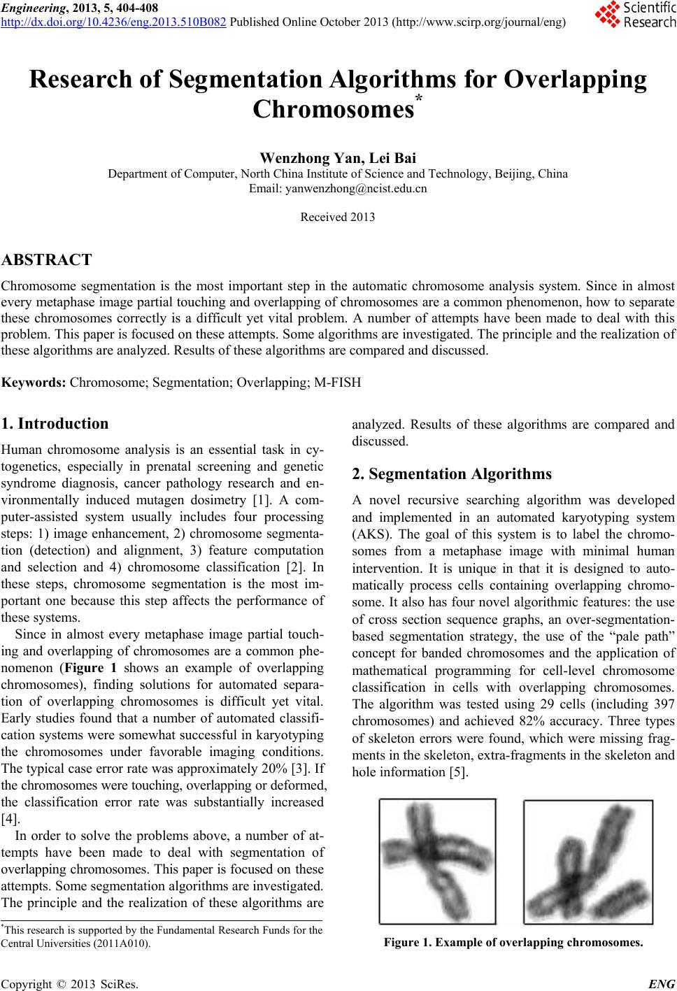
a cluster, and then evaluated based on the pixel classifi-
cation results and chromosome sizes. A hypothesis that
has a maximum-likelihood is chosen as the best decom-
position of a given cluster. About 90% of accuracy was
obtained for two or three chromosome clusters, which
consist about 95% of all clusters with two or more chro-
mosomes, and 100% accuracy was obtained for clusters
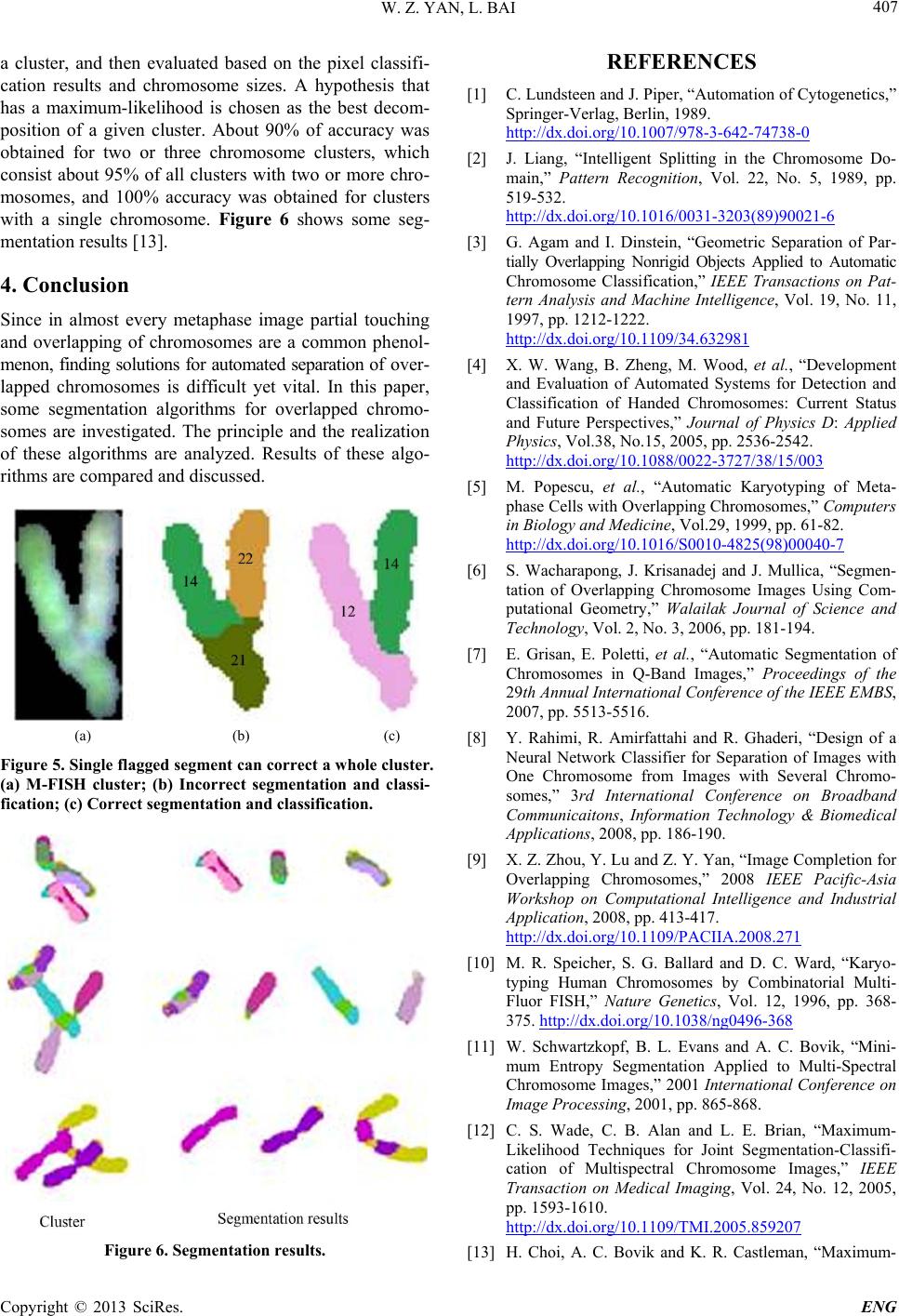
with a single chromosome. Figure 6 shows some seg-
mentation results [13].
4. Conclusion
Since in almost every metaphase image partial touching
and overlapping of chromosomes are a common phenol-
menon, finding solutions for automated separation of over-
lapped chromosomes is difficult yet vital. In this paper,
some segmentation algorithms for overlapped chromo-
somes are investigated. The principle and the realization
of these algorithms are analyzed. Results of these algo-
rithms are compare d and discus s ed.
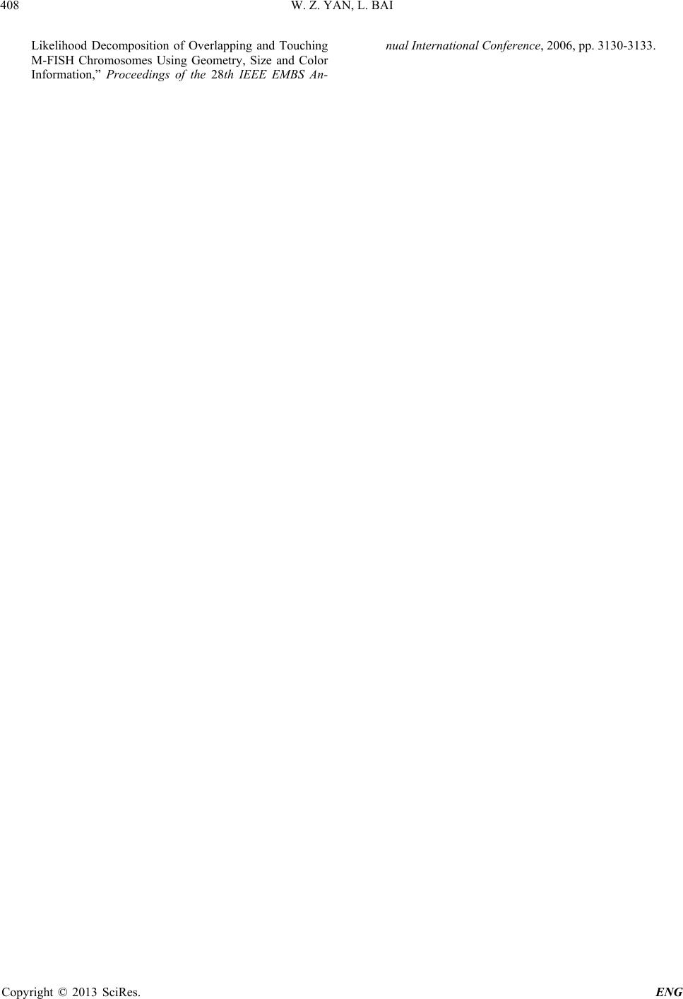
(a) (b) (c)
Figure 5. Singl e flagged segment can correct a whole cluster.
(a) M-FISH cluster; (b) Incorrect segmentation and classi-
fication; (c) Correct segmentation and classification.
Figure 6. Segmentation results.
REFERENCES
[1] C. Lundsteen and J. Piper, “Automation of Cytogenetics,”
Springer-Verlag, Berlin, 1989.
http://dx.doi.org/10.1007/978-3-642-74738-0
[2] J. Liang, “Intelligent Splitting in the Chromosome Do-
main,” Pattern Recognition, Vol. 22, No. 5, 1989, pp.
519-532.
http://dx.doi.org/10.1016/0031-3203(89)90021-6
[3] G. Agam and I. Dinstein, “Geometric Separation of Par-
tially Overlapping Nonrigid Objects Applied to Automatic
Chromosome Classification,” IEEE Transactions on Pat-
tern Analysis and Machine Intelligence, Vol. 19, No. 11,
1997, pp. 1212-1222.
http://dx.doi.org/10.1109/34.632981
[4] X. W. Wang, B. Zheng, M. Wood, et al., “Development
and Evaluation of Automated Systems for Detection and
Classification of Handed Chromosomes: Current Status
and Future Perspectives,” Journal of Physics D: Applied
Physics, Vol.38, No.15, 2005, pp. 2536-2542.
http://dx.doi.org/10.1088/0022-3727/38/15/003
[5] M. Popescu, et al., “Automatic Karyotyping of Meta-
phase Cells with Overlapping Chromosomes,” Computers
in Biology and Medicine, Vol.29, 1999, pp. 61-82.
http://dx.doi.org/10.1016/S0010-4825(98)00040-7
[6] S. Wacharapong, J. Krisanadej and J. Mullica, “Segmen-
tation of Overlapping Chromosome Images Using Com-
putational Geometry,” Walailak Journal of Science and
Technology, Vol. 2, No. 3, 2006, pp. 181-194.
[7] E. Grisan, E. Poletti, et al., “Automatic Segmentation of
Chromosomes in Q-Band Images,” Proceedings of the
29th Annual International Conference of the IEEE EMBS,
2007, pp. 5513-5516.
[8] Y. Rahimi, R. Amirfattahi and R. Ghaderi, “Design of a
Neural Network Classifier for Separation of Images with
One Chromosome from Images with Several Chromo-
somes,” 3rd International Conference on Broadband
Communicaitons, Information Technology & Biomedical
Applications, 2008, pp. 186-190.
[9] X. Z. Zhou, Y. Lu and Z. Y. Yan, “Image Completion for
Overlapping Chromosomes,” 2008 IEEE Pacific-Asia
Workshop on Computational Intelligence and Industrial
Application, 2008, pp. 413-417.
http://dx.doi.org/10.1109/PACIIA.2008.271
[10] M. R. Speicher, S. G. Ballard and D. C. Ward, “Karyo-
typing Human Chromosomes by Combinatorial Multi-
Fluor FISH,” Nature Genetics, Vol. 12, 1996, pp. 368-
375. http://dx.doi.org/10.1038/ng0496-368
[11] W. Schwartzkopf, B. L. Evans and A. C. Bovik, “Mini-
mum Entropy Segmentation Applied to Multi-Spectral
Chromosome Images,” 2001 International Conference on
Image Processing, 2001, pp. 865-868.
[12] C. S. Wade, C. B. Alan and L. E. Brian, “Maximum-
Likelihood Techniques for Joint Segmentation-Classifi-
cation of Multispectral Chromosome Images,” IEEE
Transaction on Medical Imaging, Vol. 24, No. 12, 2005,
pp. 1593-1610.
http://dx.doi.org/10.1109/TMI.2005.859207
[13] H. Choi, A. C. Bovik and K. R. Castleman, “Maximum-