 Engineering, 2013, 5, 371-375 http://dx.doi.org/10.4236/eng.2013.510B075 Published Online October 2013 (http://www.scirp.org/journal/eng) Copyright © 2013 SciRes. ENG The Expressions of P53, MDM2 in Trophoblasts of Spontaneous Abortion Mouse Model and the Relevant Researches Chunlan Zhou, Qian Wang, Menghuan Zeng, Liping Cheng, Xiaoyun Xie* Jiaxing Univer sity , Jia xing City, China Email: 863940465@qq.com, 87354674@qq.com, *99436362@qq.com Received 2013 ABSTRACT Objective: To explore the mRNA expression of the related genes of p53, MDM2, vascular endothelial growth factor (VEGF) and hypoxia inducible transcription factors-1 a (HIF-la) in villous samples of spontaneous abortion mouse models and normal pregnancy models, and to discuss the effect of p53, MDM2 on the growth of villous trophoblast cells. Methods: The abortion-prone CBAXDBA/2 matings were established as the model of spontaneous abortion and the non-abortion-prone CBAXBALB/c matings as the model of normal pregnancy. Applied q Real-time PCR method to detect the mRNA expression levels of p53, MDM2, VEGF and HIF-la in villous samples of spontaneous abortion mouse models and normal pregnancy models. Results: The relationship of the mRNA expression level of p53, MDM2, VEGF and HIF-la in vinous samples of spontaneous abortion mouse models: in the villous samples of spontaneous abortion mouse models, the expression of p53 was po sitively correlated with the expression of MD M2, HIF-la (r = 0.35; r = 0.63), and the relationship was significant (P = 0.01; P < 0.001); but negatively correlated to the expression of VEGF (r = −0.30), and the relationship was significant (P = 0.03). The expression of MDM2 was positiv ely correlated with the expr ession of HIF-la (r = 0.28), and the relationship was significant (P = 0. 04); and negatively correlated with the expression of VEGF (r = −0.08), but the relationship was not significant (P = 0.57). The expression of HIF-la was negatively correlated with the expression of VEGF (r = −0.37), and the relationship was significant (P = 0.007). The relationship of the mRNA expression level of p53, MDM2, VEGF and HIF-1 a in vinous samples of normal pregnancy models: in the vinous samples of normal pregnancy models, the expression of p53 was positively correlated with the expression of MDM2, VEGF and HIF-la (r = 0. 31; r = 0. 48; r = 0. 67), and the relationship was significant (P = 0.03; P = 0.003; P < 0.001). The expression of MDM2 was positively correlated with the expression of VEGF (r = 0. 23), but the relationship was not significant (P = 0.11); and negatively correlated with the expression of HIF-la (r = −0. 03), but the relationship was not significant (P = 0.84). The expression of HIF-la was positively correlated with the expression of VEGF ( r = 0. 35), and the relationship was significant (P = 0.01). Conclusion: angiogenesis reduces in villous sam- ples of spontaneous abortion mouse model, P53 and MDM2 involve in angiogenesis in villous samples, unlikely p53, and MDM2 have effects on normal early pregnancy villous angiogenesis and when the cell DNA damages or hypoxia exacerbates, it can induce high expression of p53, MDM2, inhibit angiogenesis in villous samples in early pregnancy. P53, MDM2 generegulate villous trophoblast cell growth by adjusting expression of HIF-1a and VEGF gene, finally influences pregnancy. Keywords: p53; MDM2; VEGF; HIF-la; Spontaneous Abortion 1. Introduction Spontaneous abortion is one of the common obstetric pathological pregnancies, its etiology is complex. In ad- dition to anatomy, endocrine, infection, chromosome, ge- netic factors, of which 40% - 80% unexplained sponta- neous abortion may be associated with immune factors. Although studies have confirmed the occurrence of spon- taneous abortion may be associated with excessive cell apoptosis, at present its specific regulation mechanism lack systematic and thorough research. The related genes of p53, MDM2 play an important role in regulating apop- tosis, occurrence mechanism of spontaneous abortion. The spontaneous abortion mouse models were chosen for the study. We applied q Real-time PCR method to detect genes expression levels in villous samples of spontane- ous abortion mouse models and normal pregnancy mod- els, discussed the effect of p53, MDM2 on pregnancy, studied the mechanism of spontaneous abortion. *Corresponding a uthor. 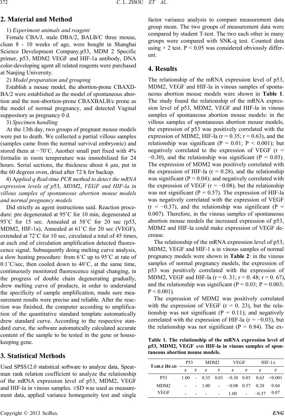 C. L. ZHOU ET AL. Copyright © 2013 SciRes. ENG 2. Material and Method 1) Experim ent animals and reagent Female CBA/J, male DBA/2, BALB/C three mouse, clean 8 - 10 weeks of age, were bought in Shanghai Science Development Company.p53, MDM 2 Specific primer, p53, MDM2 VEGF and HIF-1a antibody, DNA color-developing agent all related reagents were purchased at Nanjing University. 2) Model preparation and grouping Establish a mouse model, the abortion-prone CBAXD- BA/2 were established as the model of spontaneous abor- tion and the non-abortion-prone CBAXBALB/c prone as the model of normal pregnancy, and detected Vaginal suppository as pregnanc y 0 d. 3) Specimen handling At the 13th day, two groups of pregnant mouse models were put to death. We collected a partial villous samples (samples came from the normal survival embryonic) and stored them at −70˚C. Another small part fixed with 4% formalin in room temperature was immobilized for 24 hours. Serial sections, the thickness about 4 μm, put in the 60 degre e s oven, dried after 72 h for backup. 4) Applied q Real-time PCR method to detect the mRNA expression levels of p53, MDM2, VEGF and HIF-la in villous samples of spontaneous abortion mouse models and normal pregnancy mode ls Did strictly as agent instructions said. Reaction proce- dure: pre degenerated at 95˚C for 10 min, degenerated at 95˚C for 15 sec. Annealed at 58˚C for 20 sec (p53, MDM2, HIF-1a), Annealed at 61˚C for 20 sec (VEGF), extended at 72˚C for 10 sec, circulated a total of 45 times, at each end of circulation amplification detected fluores- cence signal. Subsequently doing melting curve analysis, a slow heating pro cedure: from 6˚C up to 95 ˚C at rate of 0.1˚C/sec, then cooled down to 40˚C, at the same time, continuously monitored fluorescence signal changing, in the progress of double chain degenerating gradually, drew melting curve of products, in order to understand the specificity of sample amplification, made sure mea- surement results were precise and reliable. After the reac- tion was finished, the computer according to amplifica- tion of the quantitative standard template automatically drew standard curve. According to the respective stan- dard curve, the software automatically calculated accurate content of the sample to be tested in the gene or house- keeping gene. 3. Statistical Methods Used SPSS12.0 statistical software to analyze data, Sp ea r- man rank relation coefficient to analyze the relationship of the mRNA expression level of p53, MDM2, VEGF and HIF-la in vinous samples. ±SD was used as measure- ment data, applied variance homogeneity test and single factor variance analysis to compare measurement data group mean. The two groups of measurement data were compared by student T-test. The two each other in many groups were compared with SNK-q test. Counted data using × 2 test. P < 0.05 was considered obviously differ- ent. 4. Results The relationship of the mRNA expression level of p53, MDM2, VEGF and HIF-la in vinous samples of sponta- neous abortion mouse models were shown in Table 1. The study found the relationship of the mRNA expres- sion level of p53, MDM2, VEGF and HIF-la in vinous samples of spontaneous abortion mouse models: in the villous samples of spontaneous abortion mouse models, the expression of p53 was positively correlated with the expression of MDM2, H IF-la (r = 0.35; r = 0.63), and th e relationship was significant (P = 0.01; P < 0.001); but negatively correlated to the expression of VEGF (r = −0.30), and the relationship was significant (P = 0.03). The expression of MDM2 was positively correlated with the expression of HIF-la (r = 0.28), and the relationship was significant (P = 0.04); and negatively corr elated with the expression of VEGF (r = −0.08), but the relationship was not significant (P = 0.57). The expression of HIF-la was negatively correlated with the expression of VEGF (r = −0.37), and the relationship was significant (P = 0.007). Therefore, in the vinous samples of spontaneous abortion mouse models the increased expression of p53, MDM2 and HIF-la could make expression of VEGF de- crease. The relationship of the mRNA exp ression lev el o f p53, MDM2, VEGF and HIF-1 a in vinous samples of normal pregnancy models were shown in Table 2: in the vinous samples of normal pregnancy models, the expression of p53 was positively correlated with the expression of MDM2, VEGF and HIF-la (r = 0. 31; r = 0. 48; r = 0. 67), and the relationship was significan t (P = 0.03; P = 0.003; P < 0.001). The expression of MDM2 was positively correlated with the expression of VEGF (r = 0. 23), but the rela- tionship was not significant (P = 0.11); and negatively correlated with the expression of HIF-la (r = −0.03), but the relationship was not significant (P = 0.84). The ex- Table 1. The relationship of the mRNA expression level of p53, MDM2, VEGF AND HIF-la in vinous samples of spon- taneous abortion mouse models. TABLE HEAD P53 MDM2 VEGF HIF-1A r P r P r P r P P53 1.00 - 0.35 0.01 −0.30 0.03 0.63 <0.001 MDM2 - - 1.00 - −0.08 0.57 0.28 0.04 - - - - 1.00 - −0.37 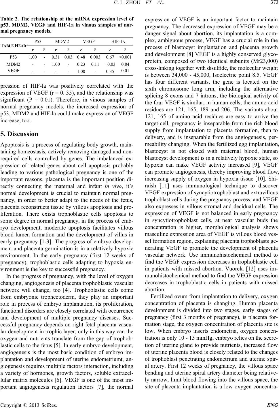 C. L. ZHOU ET AL. Copyright © 2013 SciRes. ENG 373 Table 2. The relationship of the mRNA expression level of p53, MDM2, VEGF and HIF-1a in vinous samples of nor- mal pregnancy models. TABLE HEAD P53 MDM2 VEGF HIF-1A r P r P r P r P P53 1.00 - 0.31 0.03 0.48 0.003 0.67 <0.001 MDM2 - - 1.00 - 0.23 0.11 −0.03 0.84 - - - - 1.00 - 0.35 pression of HIF-la was positively correlated with the expression of VEGF (r = 0. 35), and the relationship was significant (P = 0.01). Therefore, in vinous samples of normal pregnancy models, the increased expression of p53, MDM2 and HIF-la could make expression of VEGF increase, too. 5. Discussion Apoptosis is a process of regulating body growth, main- taining homeostasis, actively removing damaged and non- required cells controlled by genes. The imbalanced ex- pression of related genes about cell apoptosis probably leading to various pathological pregnancy is one of the important reasons, placenta is the important position di- rectly connecting the maternal and infant in vivo, it’s normal development is crucial to maintain normal preg- nancy, in order to better adapt to the needs of the fetus, placenta reconstructs tissue by villou s apoptosis and pro- liferation. There exists trophoblastic cells apoptosis to some degree in normal pregnancy, in the process of emb- ryo development, moderate apoptosis facilitates villous blood lumen formation and the development of villus in early pregnancy [1-3]. The progress of embryo develop- ment and placenta germination is in a relatively hypoxic environment. In the early pregnancy (first 12 weeks of pregnancy), trophoblastic cells adapting to hypoxia en- vironment is the key to successful pregnancy. In the progress of pregnancy, with the level of oxygen changing, angiogenesis of placenta trophoblastic vascular network will change, too [4]. Trophoblastic cells come from embryonic trophectoderm, they play an important role in process of embryo implantation, its proliferation, functional diso rders are closely cor related with o ccurrence and development of multiple pregnancy diseases. Suc- cessful pregnancy depends on right fetal placenta vascu- lar development in trophic layer, only in this way can the oxygen and nutrients translate from the gap of trophob- lastic cells to the fetus [5]. In early embryo development, angiogenesis is the most basic condition of embryo im- plantation and development of uterine endometriumt, an- giogenesis requires multiple factors inter action, inc luding a variety of hormones, growth factors, soluble extracel- lular matrix molecules [6]. VEGF is one of the most im- portant angiogenesis regulation factors [7], the normal expression of VEGF is an important factor to maintain pregnancy. The decreased expression of VEGF may be a danger signal about abortion, its implantation is a com- plex, ambiguous process, VEGF has a crucial role in the process of blastocyst implantation and placenta growth and development [8] VEGF is a highly conserved glyco- protein, composed of two identical subunits (Mr23,000) cross-linking to gether with d isu lfide, the molecu lar weight is between 34,000 - 45,000, Isoelectric point 8.5. VEGF has four different variants, the gene is located on the sixth chromosome long arm, including the alternative splicing 8 exons and 7 introns, the biological activity of the four VEGF is similar, in human cells, the amino acid residues are 121, 165, 189 and 206. The variants about 121, 165 of amino acid residues are easy to arrive the target cell, pregnancy is inseparable from the rich blood supply from implantation to placenta formation, then to delivery, and is inseparable from the angiogenesis, per- meability changing. When the fertilized egg implantation, blastocyst is not closed with maternal blood, human blastocyst development is in a relatively hypoxic state, so hypoxia can make VEGF activity increased [9], VEGF can promote angiogenesis, thereby improving blood flow, increasing supply of oxygen in hypoxia tissue [10]. Shi- raish [11] uses immunological technique to discover VEGF expression of syncytiotrophoblast and extravillous trophoblast cells during the pregnancy process, and VEGF also expresses in villous stromal and decidual cells. The expression of VEGF is not balanced in early pregnancy in syncytiotrophoblast cells, at near vascular buds the concentration is higher, morphological analysis shows masculine expression area of VEGF is villous blood ves- sel formation region, explaining placenta trophoblasts ge- nerating VEGF to promote the development of placenta vascular network. Use immunohistochemical method to find the VEGF expression decreases in trophoblastic cell in patients with missed abortion. Vuorela [12] uses im- munohistochemical method to find the VEGF expression decreases in trophoblastic cells in patients with missed abortion. Fertilized ovum from implantation to delivery, oxygen concentration of placenta is changing. Human placenta development is divided into two stages, early stages of pregnancy (first 3 months of pregnancy), is placenta for- mation stage, the oxygen concentration of placenta site is low. When embryo inserts endometria, oxygen concen- tration is only 10 - 15 mmHg, embryo relies on the secre- tion of uterine gland to provide nutrients, increased flow of uterine placenta blood is closely related to the changes of trophoblast penetrating endometrium and uterine spir- al artery. First 12 weeks of pregnancy, the villous space bending and uterine spira l artery diameter being relative- ly narrow, limit blood flowing into the villous space, the site of placenta implantation is a low oxygen concentra-  C. L. ZHOU ET AL. Copyright © 2013 SciRes. ENG tion. After 12 weeks of pregnan cy, trophoblastic cell im- merse into uterine spiral artery, remodel the vascular lay - er, make narrow artery expanded, placenta blood flow increased, corresponding placenta site oxygen concentra- tions also increased [13]. Therefore, early trophoblast de- velopment is in hypoxia environment, hypoxia has effect on trophoblast immersion. At present people have reached an agreement, placenta trophoblast angiogenesis is strictly regulated by the oxy- gen concentration. Hypoxia may lead to hypoxic cell stressed, performing for the role of hypoxia in cells has effect on oxygen sensors, initiates intracellular signal transduction pathways, activate as a transcription HIF-1 a, increased HIF-I a expression [14]. HIF-1 is a major hypoxia-inducible transcription factor, with one a subunit and one B subunit, HIF-1a is an ad- justable function unit, regulated by the oxygen concen- tration, in recent years, studies have shown that HIF-1a under certain condition has effects on apoptosis. Under hypoxia state, HI F-1 makes hig her express ion of P53 a nd inducts p21 synthesis. Suppress activity of cell cycle de- pendent kinase, makes cells stay in the G1 period, pro- motes cell apoptosis, HIF-1 as the organism regulation factor under hypoxic conditions, mediates hypoxia-in- duced apoptosis, hypoxia can increase the wild-type em- bryonic stem cell (ES, HIF-1a) apoptosis, but can not promote embryonic stem cells (ES, HIF-1β) of gene de- letion apoptosis, they incubated tumor derived from em- bryonic stem cell, also showed that HIF-1a genotype tumor apoptosis less than HIF-1β genotype hypoxic in- ducing apoptosis is mediated by HIF-1. Moritz also found early cell apoptosis is related to high expression of HIF- 1 when pancreas transplantation is for type I diabetes. VEGF is the major transcription factor of HIF-1a. Under anoxic state, the high expression of HIF-la can induce higher expression of VEGF, but the too high ex- pression of HIF-la could inhibit angiogenesis [14]. HIF- la induces apoptosis in hypoxia lying in p53 protein through combining the oxygen degradation domain, ODD and HIF-1 a, compound formation, enhance the stability of p53. The p53 protein is a short-lived protein, the intra- cellular concentration is strictly regulated by the negative accommodation factor mouse double minute gene 2 (MDM2) gene [15-17]. MDM2 is an important intracel- lular regulating p53 con centration and its activity, as p53 important activity molecules to involved in cell growth inhibition, apoptosis, regulation of cell cycle process. The expression of wild type P53 protein can stimulate high expression of MDM2 protein. Based on the p53 and MDM2 negative feedback loop, study found increased MDM2 protein can inhibit p53 protein function, thus in- creasing the negative feedback loop function. Factors that can affect the p53, MDM2 or P53-MDM2 interac- tion have an important effect on for cell fate (apoptosis, cell cycle arrest, etc.) [18]. The changing of MDM2 gene promoter sequence change can alter the expression of MDM2 protein. The expression of MDM2 protein im- proves, make p53 reduce stability, therefore change the apoptotic reaction of p53 mediating on DNA damage. [17,19]. If MDM2 identifies target p53, meanwhile combines with HIF-1a subunit, it can mediate HIF-1 a degradation, thereby affecting the expression of VEGF. Our study found, the expression of p53, MDM2 and HIF-1 are higher in villous samples of spontaneous abortion mouse models than normal pregnancy models, however the ex- pression of VEGF is lower in villous samples of sponta- neous abortion mouse models than normal pregnancy models. We hypothesized that in the progress of normal early villous samples development, Chorionic villus in is the hypoxic microenvironment, hypoxia makes the ex- pression of HIF-la gene higher, leading to the lower ex- pression of VEGF, thus prompting villus angiogenesis, ensuring the success of early pregnancy. However in the progress of spontaneous abortion, Chorionic villus cells suffer a more severe hypoxia and DNA damage, make higher expression of p53, make higher expression of MDM2 by the positive feedback regulation, and hypoxia can cause expression of HIF-la increased further, but MDM2 distinguishes target p53, meanwhile combines HIF-1 a subunit, instead mediates HIF-1 a degradation, making the expression of VEGF lower and angiogenesis decreased, at the same time making cell cycle arrested, apoptosis increased, eventually leading to the occurrence of spontaneous abortion. Therefore, based on the study of chorionic, we hypothesized that the p53, MDM2 gene plays a role in regulating during early pregnancy villous samples development, finally influencing the pregnancy. REFERENCES [1] M. Jerzak and P. Bischof, “Apoptosis in the First Trimes- ter Human Placenta: The Role in Maintaining Privilege at the Matemal-Foetal Interface and the Trophoblast Remo- deling,” The European Journal of Obstetrics & Gynecol- ogy and Reproductive Biology, Vol. 100, 2002, pp. 138- 142. http://dx.doi.org/10.1016/S0301-2115(01)00431-6 [2] D. Michael and M. Oren, “The p53-Mdm2 Module and the Ubiquitin System,” Cancer Biology, Vol. 13, 2003, pp. 49-58. http://dx.doi.org/10.1016/S1044-579X(02)00099-8 [3] G. Beckman, R. Birgander, A. Sjalander, et al., “Is p53 Polymorphism Maintained by Natural Selection?” Human Heredity, Vol. 44, 1994, pp. 266-270. http://dx.doi.org/10.1159/000154228 [4] L. V. J. Schaffer, C. Breymann, M. Gassmann and H. H. Marti, “Preserved Placental Oxygenation and Develop- ment during Severe Systemic Hypoxia,” The American Journal of Physiology Regulatory, Integrative and Com- parative Physiology, Vol. 290, No. 3, 2006, pp. 844-851. http://dx.doi.org/10.1152/ajpregu.00237.2005  C. L. ZHOU ET AL. Copyright © 2013 SciRes. ENG 375 [5] A. C. K. Ajakuma r, “Expression, Ontogeny, and Regula- tion of Hypoxia-Inducible Transcription Factors in the Human Placenta,” Biology of Reproduction, Vol. 63, No. 2, 2000, pp. 559-569. http://dx.doi.org/10.1095/biolreprod63.2.559 [6] D. E. F. J. Ingber, “Mechanochemical Switching between Growth and Differentiation during Fibroblast Growth Factor-Stimulated Angiogenesis in Vitro: Role of E xtr a- cellular Matrix,” The Journal of Cell Biology, Vol. 109, No. 1, 1989, pp. 317-330. [7] M. D. L. D. Mueller, E. Garrett and R. N. T ay lor, “Neu- trophils Infiltrating the Endometrium Express Vascular Endothelial Growth Factor: Potential Role in Endometrial Angiogenesis,” Fertility and Sterility, Vol. 74, No. 1, 2000, pp. 107-112. [8] S. SK, “Angiogenesis and Implantation,” Human Repro- duction, Vol. 15, No. S6, 2000, pp. 59-66. [9] L. O. A. Hefler, P. Husslein, C. Kainz and C. Tempfer, “Vascular Endothelial Growth Factor Serum Levels in Pregnancy and Preeclampsia,” Acta Obstetricia et Gyne- cologica Scandinavica, 2000, pp. 77-78. [10] H. H. R. W. Mart i, “Angiogenesis in Ischemic Disease,” Vol. 82, 1999, pp. 44-52. [11] S. N. K. aishi, N. Kinukawa, H. Nakano and K. Sueishi, “Immunohistochemical Localization of Vascular Endo- thelial Growth Factor in the Human Placenta,” Placenta, Vol. 17, No. 2-3, 1996, pp. 111-121. [12] P. C. O. Vuorela, M. Tulppala and E. Halmesmaki, “VEGF, Its Receptors and the Tie Receptors in Recurrent Miscarriage,” Molecular Human Reproduction, Vol. 6, No. 3, 2000, pp. 276-282. [13] G. J. H. J. Burton and A. L. Wets on, “Maternal Arterial Connections to the Placectal Intervil Lous Space during the First Trimester of Human Pregnancy,” American Jour- nal of Obstetrics & Gynecology, Vol. 181, 1999, p. 71. [14] H. D. Z. J. Skinner, J. Fang, F. Agani and B. H. Jiang, “Vascular Endothelial Growth Factor Transcriptional Ac- tivation Is Mediated by Hypoxia-Inducible Factor Lalpha, HDM2, and p74S6K1 in Response to Phosphatidylinosi- tol 3-Kinase/AKT Signaling,” The Journal of Biological Chemistry, Vol. 279, No. 44, 2004, pp. 45643-51. [15] M. Thomas, A. Kalita, S. Labrecque, et al., “Two Poly- morphic Variants of Wild-Type p53 Differ Biochemically and Biologically,” Molecular and Cellular Biology, 1999, pp. 1092-1100. [16] P. Dumont, J. I. Leu, A. Cr. Della Pietra, et al., “The Co- don 72 Polymorphic Variants of p53 Have Ma rkedly Dif- ferent Apoptotic Potential,” Nature Genetics, Vol. 33, 2003, pp. 357-365. [17] G. L. Bond, W. Hu, E. E. Bond, et al., “A Single Nucleo- tide Polymorphism in the MDM2 Promoter Attenustes the p53 Tumor Suppressor Pathway and Accelerates Tumor Formation in Humans,” Ce ll, Vol. 119, 2004, pp. 591- 602. [18] D. Michael and M. Oren, “The p53-Mdm2 Module and the Ubiquitin System,” Cancer Biology, Vol. 13, 2003, pp. 49-58. [19] Y. Hong, X. Zhang, et al., “The Role of p53 and MDM2 Polymorphisms in the Risk of Esophagel Squamaous Cell Carcinoma,” Cancer Research, Vol. 65, 2005, pp. 9582- 9587.
|