 Neuroscience & Medicine, 2013, 4, 271-276 Published Online December 2013 (http://www.scirp.org/journal/nm) http://dx.doi.org/10.4236/nm.2013.44039 Open Access NM 271 Biochemical and Neurobiological Study of Moroccan Autists Hakim Harchaoui1, Fatima-Zahra Azzaoui1, Ahmed Ahami1, Samira Bou lbar o u d 2, Mohamed El Hioui1 1Equip of Clinic and Cognitive Neurosciences and Health, Laboratory of Biology and Health, Department of Biology, Faculty of Science, IBN TOFAIL University, Kenitra, Morocco; 2Equip of Neuroendocrine Physiology, Faculty of Science, IBN TOFAIL Uni- versity, Kenitra, Morocco. Email: hakim.harchaoui@gmail.com Received August 7th, 2013; revised September 5th, 2013; accepted October 3rd, 2013 Copyright © 2013 Hakim Harchaoui et al. This is an open access article distributed under the Creative Commons Attribution License, which permits unrestricted use, distribution, and reproduction in any medium, provided the original work is properly cited. ABSTRACT Autism spectrum disorder (ASD) is a range of complex neurodevelopment disorders, characterized by social impair- ments, communication difficulties, and restricted, repetitive, and stereotyped patterns of behavior. Its diagnosis is very difficult and consists of only clinical criteria. The aim of this study is to confirm autism disorder, among three children (2 girls and 1 boy), recruited in a social association of children in a precarious situation, located in Rabat, Morocco, and to evaluate some biological and neurobiological parameters among these children. The confirmation of autistic status is realized using Questionnaire (E2) and the evaluation of biological parameters (organic acids, urinary peptides, toxic metals and essential elements) is done in urine and hair of these children. Obtained children’s scores of E2 Ques- tionnaire, is found in the favor of autism. Moreover, abnormal levels of urinary organic acids, urinary peptides, toxic and essential metals are observed. Keywords: Autism; Neurobiology; Organic Acids; Toxic Metals; Essential Elements 1. Introduction Autism, as defined by “The Diagnostic and Statistical Manual of Mental Disorders”, 4th edition (DSM IV) (2000), is a disorder of neural development characterized by impaired social interaction and verbal and non-verbal communication, and by restricted, repetitive or stereo- typed behavior. The diagnostic criteria require that sym- ptoms become apparent before a child is three years old. In Morocco, very few studies are realized about autism [1] which cannot provide the prevalence in the Kingdom. However, the prevalence of this neurodevelopmental disorder is increasing; in the USA, from 1998 to 2007, the number of 6- to 21-year-old children receiving ser- vices for an ASD (autism spectrum disorders) in public special education programs increased from 54,064 to 258,305 [2]. In the recent CDC report, it is found that the overall estimated prevalence of ASD is one in 88 chil- dren during [3]. The etiology of autism is still discussed and some stu- dies report that autism affects approximately three times as many males as females. Taken together with studies looking at families and twins, this suggests that autism has a genetic component. It is likely that a number of genes are involved. However, the exact mechanism by which genes are implicated in autism is unclear [4]. Other findings suggest that some neuroanatomical im- pairments in brain areas and/or brain chemistry changing could be involved in this neuropathology [5,6]. Moreover, some studies have reported many nutria- tional deficiencies in autism [7], and accumulations of organic acids in urine can indicate metabolic dysfunction, nutrient insufficiencies, or even microbial overgrowth [8]. The urinary organic acids test measures selected metabo- lites which serve as important diagnostic indicators of abnormal metabolism in the case of autistic children. The measurement of organic acids in urine evaluates four critical areas of metabolism: gastrointestinal function, cellular energy and mitochondrial metabolism, neuro- transmitter metabolism, and amino acid/organic acid bal- ance as influenced by vitamin/mineral cofactors [9]. In the urine of many autistic children, abnormal metabolites such as opioid peptides and [10] abnormal levels of different organic acids [11,12] were observed. Some researches  Biochemical and Neurobiological Study of Moroccan Autists 272 about this pathology implicated insufficient enzymatic activity and increased gastrointestinal permeability, and the absorption of toxic byproducts of incompletely digest- ed proteins from dairy (casein) and cereals (gluten), [13,14]. Furthermore, environmental pollution could be a pos- sible factor contributing to this disease [15]; many metals could be implicated in autism since they typically disrupt enzyme functions and cell signaling processes, generate oxygen free radicals (ROS) and elicit autoimmune reac- tions [16]. Metals involved in physiopathological proc- esses include essential elements such as zinc (Zn), copper (Cu), iron (Fe), calcium (Ca), manganese (Mn), nickel (Ni) and silicon (Si), or toxic metals such as cadmium (Cd), cobalt (Co), aluminium (Al), lead (Pb), mercury (Hg) and arsenic (As) [17,18]. The laboratory analysis of different biological matrices (urine, blood, hair) is very useful in identifying this deficiency or toxicity and allow for health professionals establishing possible strategies of treatment. The aim of this study, in one hand is to con- tribute to this field, and still virgin in Morocco, and in the other hand, to confirm autistic diagnosis of studying children and to measure some biological and neurobio- logical parameters in hair and urine of these children. 2. Patients and Methods 2.1. Ethical Clearance and Informed Consent This study was approved by the Internal Ethics Commit- tee of Faculty of Science, Kenitra, Morocco and by the GDRI of Neurosciences France-Morocco Ethics Com- mittee. The recruitment of patients is based on the fol- lowing criteria: - Written consent of the children’s parents, - Children physically healthy and not previously under- gone chelation therapy, free of any medication, with- out any casein-free and gluten-free diet and any medi- cal issues. 2.2. Patients Three children (2 girls and 1 boy) are involved in the present study. They are aged between 15 to 18 years old. The children are diagnosed for autism and they are in- serted into an associative structure in Rabat, Morocco, that cares for children suffering from neurobehavioral impairments. Patient (A): Aya, female child aged 4 years is diag- nosed with autism. Aya is the second of three sisters and she is physically normal. She had frequent infections breathing during the first months of life. She learned to walk alone between 24 and 36 months. Between 2cd and 4th year, she had an irresistible urge to eat something. At the age of 3 or 4 years, she is “going into a shell”; she became so distant and lost in thought. Moreover, she was indifferent to any affection and seems to be happier when we do not take care of her. Before 5 years, she was able to speak but not to answer. Her abnormal behavior has been discovered between 7 and 12 months. Patient (B): Salwa, female child of 18 years is diag- nosed with autism. Salwa is the younger child of her family, she is physically normal. Salwa had a difficulty of sucking and diarrhea. She learned to walk alone be- tween 8 and 12 months. Between 2cd and 4th year, she suffers from PICA syndrome. At the age of 3 or 4 years, she is “going into a shell”; she became so distant and lost in thought. Moreover, she was indifferent to any affect- tion and seems to be happier when we do not take care of her. Before 5 years, she was able to speak but not to an- swer. Her abnormal behavior has been discovered be- tween 13 and 24 months. Patient (C): Ismail 17 years old, male diagnosed with autism. Ismail is the only child in his family, he is physi- cally normal with an excellent health. He learned to walk alone between 8 and 12 months. Between 2 and 4 years, he sucks often metallic objects. A child at the age of 3 or 4 years was “locked in his shell” or so distant and lost in thought. He is indifferent to any brand of affection and seems being happier when you do not take care of him. Before 5 years, he was able to speak but not to answer. The abnormal behavior of the child has been discovered between 2 and 3 years. 2.3. Methods An E2 Questionnaire, including 79 amnestic items for the diagnosis of autism, completed by parents, is used. It covers the period from birth to age of 5 years. All items are summed to obtain a total score. If an item is applica- ble to the assessed individual, it is noted (+1), if not, it is noted (−1). The total score is the difference between the presence of autism (+) and its absence (−). A total score of “20” is in the favor of autism [19]. The urine and hair samples were collected from the patients for the following biological tests: Tests of uri- nary organic acids including neurotransmitters, fatty ac- ids, amino acids, bacterium and fungal metabolites. Test of urinary peptides including casamorphin and gluten- morphin. Test of toxic metals (Ur, Al, As, Pb, Hg, Ag) and essential elements (Ca, Mg, Na, k, Cu, Cr, Mo, Se, Be, Ru, Zn, Va, Li) in the hair samples. All the analysis is realized by Great plain laboratory, Inc (USA). The urinary organic acids and urinary peptides results are expressed as ratios to the urinary creatinin concentra- tion in mmol/mmolcreatinine. Certified hair standards and in-house standards were used as part of the labora- tory quality control and for the validation of results. Re- sults are expressed as micrograms per gram, equivalent to parts per million. Open Access NM 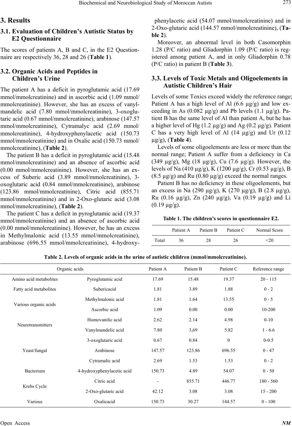 Biochemical and Neurobiological Study of Moroccan Autists Open Access NM 273 3. Results 3.1. Evaluation of Children’s Autistic Status by E2 Questionnaire The scores of patients A, B and C, in the E2 Question- naire are respectively 36, 28 and 26 (Table 1). 3.2. Organic Acids and Peptides in Children’s Urine The patient A has a deficit in pyroglutamic acid (17.69 mmol/mmolcreatinine) and in ascorbic acid (1.09 mmol/ mmolcreatinine). However, she has an excess of vanyl- mandelic acid (7.80 mmol/mmolcreatinine), 3-oxoglu- taric acid (0.67 mmol/mmolcreatinine), arabinose (147.57 mmol/mmolcreatinine), Cytramalyc acid (2.69 mmol/ mmolcreatinine), 4-hydroxyphenylacetic acid (150.73 mmol/mmolcreatinine) and in Oxalic acid (150.73 mmol/ mmolcreatinine), (Table 2). The patient B has a deficit in pyroglutamic acid (15.48 mmol/mmolcreatinine) and an absence of ascorbic acid (0.00 mmol/mmolcreatinine). However, she has an ex- cess of Suberic acid (3.89 mmol/mmolcreatinine), 3- oxoglutaric acid (0.84 mmol/mmolcreatinine), arabinose (123.86 mmol/mmolcreatinine), Citric acid (855.71 mmol/mmolcreatinine) and in 2-Oxo-glutaric acid (3.08 mmol/mmolcreatinine), (Table 2). The patient C has a deficit in pyroglutamic acid (19.37 mmol/mmolcreatinine) and an absence of ascorbic acid (0.00 mmol/mmolcreatinine). However, he has an excess in Methylmalonic acid (13.55 mmol/mmolcreatinine), arabinose (696.55 mmol/mmolcreatinine), 4-hydroxy- phenylacetic acid (54.07 mmol/mmolcreatinine) and in 2-Oxo-glutaric acid (144.57 mmol/mmolcreatinine), (Ta- ble 2). Moreover, an abnormal level in both Casomorphin 1.28 (P/C ratio) and Gliadorphin 1.09 (P/C ratio) is reg- istered among patient A, and in only Gliadorphin 0.78 (P/C ratio) in patient B (Table 3). 3.3. Levels of Toxic Metals and Oligoelements in Autistic Children’s Hair Levels of some Toxics exceed widely the reference range; Patient A has a high level of Al (6.6 µg/g) and low ex- ceeding in As (0.082 µg/g) and Pb levels (1.1 µg/g). Pa- tient B has the same level of Al than patient A, but he has a higher level of Hg (1.2 µg/g) and Ag (0.2 µg/g). Patient C has a very high level of Al (14 µg/g) and Ur (0.12 µg/g), (Table 4). Levels of some oligoelements are less or more than the normal range; Patient A suffer from a deficiency in Ca (349 µg/g), Mg (18 µg/g), Cu (7.6 µg/g). However, the levels of Na (410 µg/g), K (1200 µg/g), Cr (0.53 µg/g), B (8.5 µg/g) and Ru (0.80 µg/g) exceed the normal ranges. Patient B has no deficiency in these oligoelements, but an excess in Na (290 µg/g), K (270 µg/g), B (2.8 µg/g), Ru (0.16 µg/g), Zn (240 µg/g), Va (0.19 µg/g) and Li (0.19 µg/g). Table 1. The children’s scores in questionnaire E2. Patient APatient BPatient C Normal Score Total36 28 26 <20 Table 2. Levels of organic acids in the urine of autistic children (mmol/mmolcreatinine). Organic acids Patient A Patient B Patient C Reference range Amino acid metabolites Pyroglutamic acid 17.69 15.48 19.37 20 - 115 Fatty acid metabolites Subericacid 1.81 3.89 1.88 0 - 2 Methylmalonic acid 1.81 1.64 13.55 0 - 5 Various organic acids Ascorbic acid 1.09 0.00 0.00 10-200 Homovanilic acid 2.62 2.14 4.98 0-10 Neurotransmitters Vanylmandelic acid 7.80 3,69 5.82 1 - 6.6 3-oxoglutaric acid 0.67 0.84 0 0-0.5 Arabinose 147.57 123.86 696.55 0 - 47 Yeast/fungal Cytramalic acid 2.69 1.53 1.53 0 - 2 Bacterium 4-hydroxyphenylacetic acid 150.73 4.89 54.07 0 - 50 Citric acid - 855.71 446.77 180 - 560 Krebs Cycle 2-Oxo-glutaric acid 42.12 3.08 3.08 15 - 200 Various Oxalicacid 150.73 30.27 144.57 0 - 100 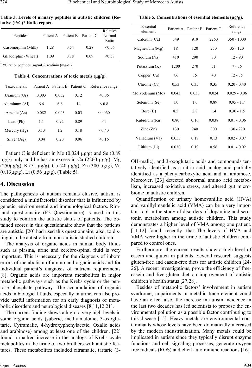 Biochemical and Neurobiological Study of Moroccan Autists 274 Table 3. Levels of urinary peptides in autistic children (Re- lative (P/C)* Ratio report. Peptides Patient A Patient B Patient C Relative Normal Ratio Casomorphin (Milk) 1.28 0.54 0.28 <0.56 Gliadorphin (Wheat) 1.09 0.78 0.09 <0.58 *P/C ratio: peptides (ng/ml)/Creatinin (mg/dl). Table 4. Concentrations of toxic metals (µg/g). Toxic metals Patient A Patient B Patient C Reference range Uranium (Ur) 0.003 0.052 0.12 <0.06 Aluminum (Al) 6.6 6.6 14 < 0.8 Arsenic (As) 0.082 0.043 0.03 <0.060 Lead (Pb) 1.1 0.92 0.89 <1 Mercury (Hg) 0.13 1.2 0.18 <0.40 Silver (Ag) 0.04 0.20 0.06 <0.16 Patient C is deficient in Mo (0.024 µg/g) and Se (0.89 µg/g) only and he has an excess in Ca (2260 µg/g), Mg (250µg/g), K (51 µg/g), Cu (40 µg/g), Zn (300 µg/g), Va (0.13µg/g), Li (0.56 µg/g), (Table 5). 4. Discussion The pathogenesis of autism remains elusive, autism is considered a multifactorial disorder that is influenced by genetic, environmental and immunological factors. Rim- land questionnaire (E2 Questionnaire) is used in this study to confirm the autistic status of patients. The ob- tained scores in this questionnaire show that the patients are autistic. [20] had used this questionnaire, also, to dis- tinguish the autistic from non-autistic studied children. The analysis of organic acids in human body fluids such as plasma, urine and cerebro-spinal fluid is very important. This is necessary for the diagnosis of inborn errors of metabolism of amino and organic acids and for individual patient’s diagnosis of nutrient requirements [8]. Organic acids are important metabolites in major metabolic pathways such as the Krebs cycle or the pen- tose phosphate pathway. The accumulation of organic acids in biological fluids, especially in urine, can also pro- vide useful information for an early diagnosis of meta- bolic disorders and neurological diseases [8,11,12,21]. The current finding shows a high to very high levels in some organic acids (suberic, methylmalonic, 3-oxoglu- taric, Cytramalic, 4-hydroxyphenylacetic, Oxalic acids and arabinose) among at least one of the children. [22] found a marked increase in the analogs of Krebs cycle metabolites in the urine of two brothers with autistic fea- tures. These metabolites included citramalic, tartaric (3- Table 5. Concentrations of essential elements (µg/g). Essential elements Patient APatient B Patient C Reference range Calcium (Ca) 349 919 2260 350 - 1000 Magnesium (Mg)18 120 250 35 - 120 Sodium (Na) 410 290 70 12 - 90 Potassium (K) 1200 270 51 7 - 36 Copper (Cu) 7.6 15 40 12 - 35 Chrome (Cr) 0.53 0.35 0.35 0.20 - 0.40 Molybdenum (Mo)0.043 0.033 0.024 0.029 - 0.06 Selenium (Se) 1.0 1.0 0.89 0.95 - 1.7 Bore (B) 8.5 2.8 1.4 0.30 - 1.5 Rubidium (Ru) 0.80 0.16 0.038 0.01 - 0.06 Zinc (Zn) 130 240 300 130 - 220 Vanadium (Va) 0.053 0.19 0.13 0.02 - 0.07 Lithium (Li) 0.030 0.19 0.56 0.01 - 0.02 OH-malic), and 3-oxoglutaric acids and compounds ten- tatively identified as a citric acid analog and partially identified as a phenylcarboxylic acid and in arabinose. Moreover, [23] detected abnormal amino acid metabo- lism, increased oxidative stress, and altered gut micro- biome in autistic children. Quantification of urinary homovanillic acid (HVA) and vanillylmandelic acid (VMA) can be a very impor- tant tool in the study of disorders of dopamine and sero- tonin metabolism among autistic children. This study demonstrates a higher level of VMA among one patient. [11,12] found, recently, that The levels of HVA and VMA were higher in the urine of autistic children com- pared to control ones. Furthermore, the current results show a high level of casein and gluten in patients. Several research suggests gluten-free and casein-free diets for autistic children [24- 26]. A recent investigations, prove the efficiency of free- casein and free-gluten diet on improvement of autistic children’s health status [27,28]. Besides of metabolic factors’ involvement in autism syndrome, impairments in metallic trace element could have an effect also; the increase in autism incidence in the last two decades has led scientists to propose the en- vironmental pollution as a possible factor contributing to this disease [15]. Heavy metals are environmental con- taminants whose levels have been dramatically increased by the modern industrialization. Many metals could be implicated in autism since they typically disrupt enzyme functions and cell signaling processes, generate oxygen free radicals (ROS) and elicit autoimmune reactions [16]. Open Access NM  Biochemical and Neurobiological Study of Moroccan Autists 275 A number of studies have implicated exposure to various metals in the development of autism [17,18,29] but, to date, the mechanisms by which metals exposure may cause autism have been difficult to ascertain. All these findings are consistent with our results that find very high levels of Ur, Al, As, Pb, Hg and Ag are registered in at least one of the patients. In the present study, impairment in the level of the es- sential element is observed; high levels of Na, K, Cr, B, Ru, Zn,Va, Li and low level of Mo and Se are found in at least of two patients. However, concerning Ca and Cu, the results show high levels in some patients and low among others. Previous studies found higher levels of Zn in both hair and teeth [18] of autistic subjects. Others [30] found a significant elevation in the concentration of Cu, Pb, and Hg and significant decrease in the concentration of Mg and Se in the hair and nail samples of autistic sub- jects. These variations could be well correlated with de- gree of autism severity. 5. Conclusions The studied children are autistic and suffer from impair- ments in organic acids, oligoelements and high levels of toxic metals. Rimland questionnaire and biological study, using analysis of biological matrix, can be effective for con- firming autistic status of patients and detecting abnormal metabolic and nutritional conditions in autistic children. In future investigations, these analyses could help to es- tablish a medical protocol for treatment or at least im- provement of health status of these children. REFERENCES [1] D. Saloua, L. R. Desviat, P. Belén, L. Fátima, U. Mag- dalena and C. Layachi, “Mutation Analysis of Phenylke- tonuria Patients from Morocco: High Prevalence of Muta- tion G352fsdelG and Detection of a Novel Mutation p.K85X,” Clinical Biochemistry, Vol. 43, No. 1-2, 2010, pp. 76-81. [2] Centers for Disease Control and Prevention (CDC), “Pre- valence of Autism Spectrum Disorders—Autism and De- velopmental Disabilities Monitoring Network, United States, 2006,” MMWR Surveillance Summary, Vol. 58, No. SS10, 2009, pp. 1-20. [3] Morbidity and Mortality Weekly Report (MMWR), “Pre- valence of Autism Spectrum Disorders—Autism and De- velopmental Disabilities Monitoring Network, 14 Sites, United States, 2008,” Surveillance Summaries, Vol. 61, No. SS03, 2012, pp. 1-19. http://www.cdc.gov/mmwr/preview/mmwrhtml/ss6103a1. htm [4] MRC, “Review of Autism Research: Epidemiology and Causes,” Medical Research Council, 2001, p. 94. [5] E. J. Carter, D. L. Williams, N. J. Minshew and J. F. Leh- man, “Is He Being Bad? Social and Language Brain Net- works during Social Judgment in Children with Autism,” PLoS One, Vol. 7, No. 10, 2012, Article ID: e47241. http://dx.doi.org/10.1371/journal.pone.0047241 [6] M. J. Kane, M. Angoa-Peréz, D. I. Briggs, C. E. Sykes, D. M. Francescutti, D. R. Rosenberg and D. M. Kuhn, “Mice Genetically Depleted of Brain Serotonin Display Social Impairments, Communication Deficits and Repetitive Behaviors: Possible Relevance to Autism,” PLoS One, Vol. 7, No. 11, 2012, Article ID: e48975. http://dx.doi.org/10.1371/journal.pone.0048975 [7] L. T. Curtis and K. Patel, “Nutritional and Environmental Approaches to Preventing and Treating Autism and At- tention Deficit Hyperactivity Disorder (ADHD): A Re- view,” The Journal of Alternative and Complementary Medicine, Vol. 14, No. 1, 2008, pp. 79-85. http://dx.doi.org/10.1089/acm.2007.0610 [8] A. Kumps, P. Duez and Y. Mardens, “Metabolic, Nutri- tional, Iatrogenic, and Artifactual Sources of Urinary Or- ganic Acids: A Comprehensive Table,” Clinical Chemis- try, Vol. 48, No. 5, 2002, pp. 708-717. [9] C. K. Burdette, “How to Assess Patient Biochemical and Nutritional Individuality through Organic and Testing Townsend Letter for Doctors and Patients,” New York, 2006. [10] P. Shattock and P. Whiteley, “Biochemical Aspects in Autism Spectrum Disorders: Updating the Opioid-Excess Theory and Presenting New Opportunities for Biomedical Intervention,” Expert Opinion on Therapeutic Targets, Vol. 6, No. 2, 2002, pp. 175-183. http://dx.doi.org/10.1517/14728222.6.2.175 [11] J. Kałużna-Czaplińska, M. Michalska and J. Rynkowski, “Determination of Tryptophan in Urine of Autistic and Healthy Children by Gas Chromatography/Mass Spectro- metry,” Medical Science Monitor, Vol. 16, No. 10, 2010, pp. 488-492. [12] J. Kałużna-Czaplińska, E. Socha and J. Rynkowski, “De- termination of Homovanillic Acid and Vanilylmandelic Acid in Urine of Autistic Children by Gas Chromatogra- phy/Mass Spectrometry,” Medical Science Monitor, Vol. 16, No. 9, 2010, pp. 445-450. [13] P. Whiteley, J. Rodgers, D. Savery and P. Shattock, “A Gluten-Free Diet as an Intervention for Autism and Asso- ciated Spectrum Disorders: Preliminary Findings,” Au- tism, Vol. 3, No. 1, 1999, pp. 45-66. http://dx.doi.org/10.1177/1362361399003001005 [14] K. Patel and L. Curtis, “A Comprehensive Approach to Treating Autism and Attention-Deficit Hyperactivity Dis- order: A Pre-Pilot Study,” The Journal of Alternative and Complementary Medicine, Vol. 13, No. 10, 2007, pp. 1091-1097. http://dx.doi.org/10.1089/acm.2007.0611 [15] E. Fombonne, “The Epidemiology of Autism: A Re- view,” Psychological Medicine, Vol. 29, No. 4, 1999, pp. 769-786. http://dx.doi.org/10.1017/S0033291799008508 [16] T. O. Kleine, “How to Diagnose Humanbrain Damage and Inflammation,” Brain Research Bulletin, Vol. 61, No. 3, 2003, pp. 227-228. http://dx.doi.org/10.1016/S0361-9230(03)00084-4 [17] A. Fido and S. Al-Saad, “Toxic Trace Elements in the Open Access NM  Biochemical and Neurobiological Study of Moroccan Autists Open Access NM 276 Hair of Children with Autism,” Autism, Vol. 9, No. 3, 2005, pp. 290-298. [18] J. B. Adams, J. Romdalvik, V. M. Ramanujam and M. S. Legator, “Mercury, Lead, and Zinc in Baby Teeth of Children with Autism versus Controls,” Journal of Toxi- cology and Environmental Health A, Vol. 70, No. 12, 2007, pp. 1046-1051. http://dx.doi.org/10.1080/15287390601172080 [19] B. Rimland, “The Differentiation of Childhood Psychoses: An Analysis of Checklists for 2218 Psychotic Children,” Journal of Autism and Childhood Schizophrenia, Vol. 1, No. 2, 1971, pp. 161-174. http://dx.doi.org/10.1007/BF01537955 [20] I. Leddet, C. Larmande, C. Barthelemy, F. Chalons, D. Sauvage and G. Lelord, “Comparison of Clinical Diagno- ses and Rimland E2 Scores in Severely Disturbed Chil- dren,” Journal of Autism and Developmental Disorders, Vol. 16, No. 2, 1986, pp. 215-225. http://dx.doi.org/10.1007/BF01531731 [21] S. A. Rogers, “Using Organicacids to Diagnose and Man- age Recalcitrant Patients,” Alternative Therapies in Health and Medicine, Vol. 12, No. 4, 2006, pp. 44-51. [22] W. Shaw, E. Kassen and E. Chaves, “Increased Urinary Excretion of Analogs of Krebs Cycle Metabolites and Arabinose in Two Brothers with Autistic Features,” Cli- nical Chemistry, Vol. 41, No. 8, 1995, pp. 1094-1104. [23] X. Ming, T. P. Stein, V. Barnes, N. Rhodes and L. Guo, “Metabolic Perturbance in Autism Spectrum Disorders: A Metabolomics Study,” Journal of Proteome Research, Vol. 11, No. 12, 2012, pp. 5856-5862. [24] B. Rimland and M. Baker, “Brief Report: Alternative Approaches to Development of Effective Treatments for Autism,” Journal of Autism and Developmental Disor- ders, Vol. 26, No. 2, 1996, pp. 237-241. http://dx.doi.org/10.1007/BF02172019 [25] J. H. Elder, M. Shankar, J. Shuster, D. Theriaque, S. Burns and L Sherrill, “The Gluten-Free, Casein-Free Diet in Autism: Results of a Preliminary Double Blind Clinical Trial,” Journal of Autism and Developmental Disorders, Vol. 36, No. 3, 2006, pp. 413-420. http://dx.doi.org/10.1007/s10803-006-0079-0 [26] C. Evans, H. R. Dunstan, T. Rothkirch, T. K. Roberts, K. L. Reichelt, R. Cosford, G. Deed, L. B. Ellis and D. L. Sparkes, “Altered Amino Acid Excretion in Children with Autism,” Nutritional Neuroscience, Vol. 11, No. 1, 2008, pp. 9-17. http://dx.doi.org/10.1179/147683008X301360 [27] C. M. Pennesi and L. C. Klein, “Effectiveness of the Glu- ten-Free, Casein-Free Diet for Children Diagnosed with Autism Spectrum Disorder: Based on Parental Report,” Nutritional Neuroscience, Vol. 15, No. 2, 2012, pp. 85- 91. [28] C. Harris and B. Card, “A Pilot Study to Evaluate Nutri- tional Influences on Gastrointestinal Symptoms and Be- havior Patterns in Children with Autism Spectrum Disor- der,” Complementary Therapies in Medicine, Vol. 20, No. 6, 2012, pp. 437-440. http://dx.doi.org/10.1016/j.ctim.2012.08.004 [29] J. K. Kern, D. A. Geier, T. Audhya, P. G. King, L. K. Sykes and M. R. Geier, “Evidence of Parallels between Mercury Intoxication and the Brain Pathology in Autism,” Acta Neurobiologiae Experimentalis, Vol. 72, 2012, pp. 113-153. [30] M. D. Lakshmi Priya and A. Geetha, “Level of Trace Ele- ments (Copper, Zinc, Magnesium and Selenium) and Toxic Elements (Lead and Mercury) in the Hair and Nail of Children with Autism,” Biological Trace Element Re- search, Vol. 142, No. 2, 2012, pp. 148-158. http://dx.doi.org/10.1007/s12011-010-8766-2
|