Neuroscience & Medicine, 2013, 4, 267-270
Published Online December 2013 (http://www.scirp.org/journal/nm)
http://dx.doi.org/10.4236/nm.2013.44038
Open Access NM
267
Benign Fibrous Histiocytoma of the Neurocranium
Chrisovalantis A. Tsimiklis*, Tom Morris
Neurosurgical Department, Royal Adelaide Hospital (RAH), North Terrace, Adelaide, Australian
Email: *ctsimiklis29@gmail.com, dr.t ommorris@gmail.com
Received October 8th, 2013; revised November 5th, 2013; accepted December 3rd, 2013
Copyright © 2013 Chrisovalantis A. Tsimiklis, Tom Morris. This is an open access article distributed under the Creative Commons
Attribution License, which permits unrestricted use, distribution, and reproduction in any medium, provided the original work is
properly cited.
ABSTRACT
Presented is a case of benign fibrous histiocytoma (BFH) involving the calvarium of a 25 years old lady who noticed a
depression in her occiput associated with localised pain. Imaging revealed a tumour eroding through the inn er and outer
skull tables, closely associated with major underlying dural sinuses. She underwent complete macroscopic resection of
the tumour and reconstru ction of a titanium mesh craniop lasty. Histology favoured a benign process with a diagnosis of
BFH of the calvarium given. At 1 year follow-up, the patient is asymptomatic and has not developed recurrence of the
tumour.
Keywords: Benign Fibrous Histiocytoma; Skull Tumour; Neurocranium
1. Introduction
BFH is most often encountered as a soft-tissue neoplasm
involving the skin, and although less common, BFH in-
volving other sites including the bony skeleton is seen.
Involvement of the skull is particularly rare with only
one other case of BFH involving the neurocranium re-
ported in the literature [1]. Here we report a case of a
symptomatic BFH involving the occipital bone and dis-
cuss the role for minimising the degree of resection in
these benign ca ses.
2. Case Report
Presentation and examination. A bump was first noted
by this 25 years old lady on her occiput 18 months prior
to presentation. It was steadily increasing over this period
and she had associated pain at the site but was otherwise
neurologically intact. There were no overlying changes
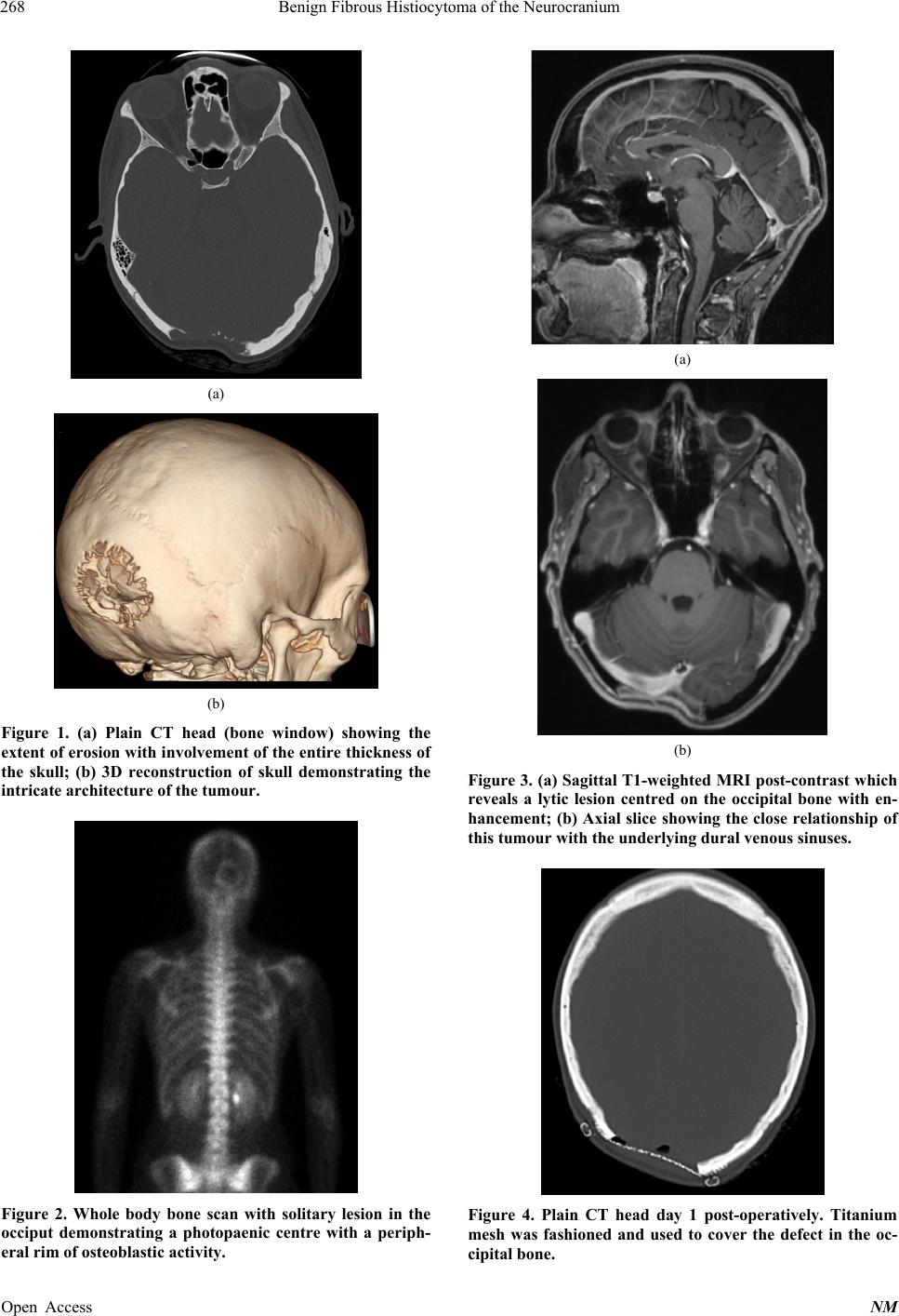
to the scalp tissue itself. A CT head revealed an expansile
lytic process to the right of the midline invo lving the oc-
cipital bone, measuring 40 × 45 mm, and resulting in loss
of both the inner and outer tables of the skull (Figures
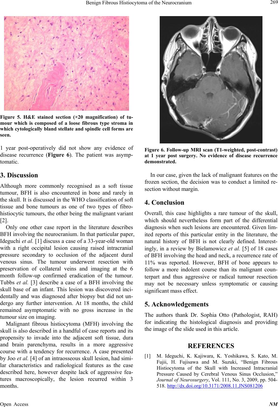
1(a) and (b)). A bone scan showed a solitary photo-
paenic occipital bone lesion with a peripheral rim of os-
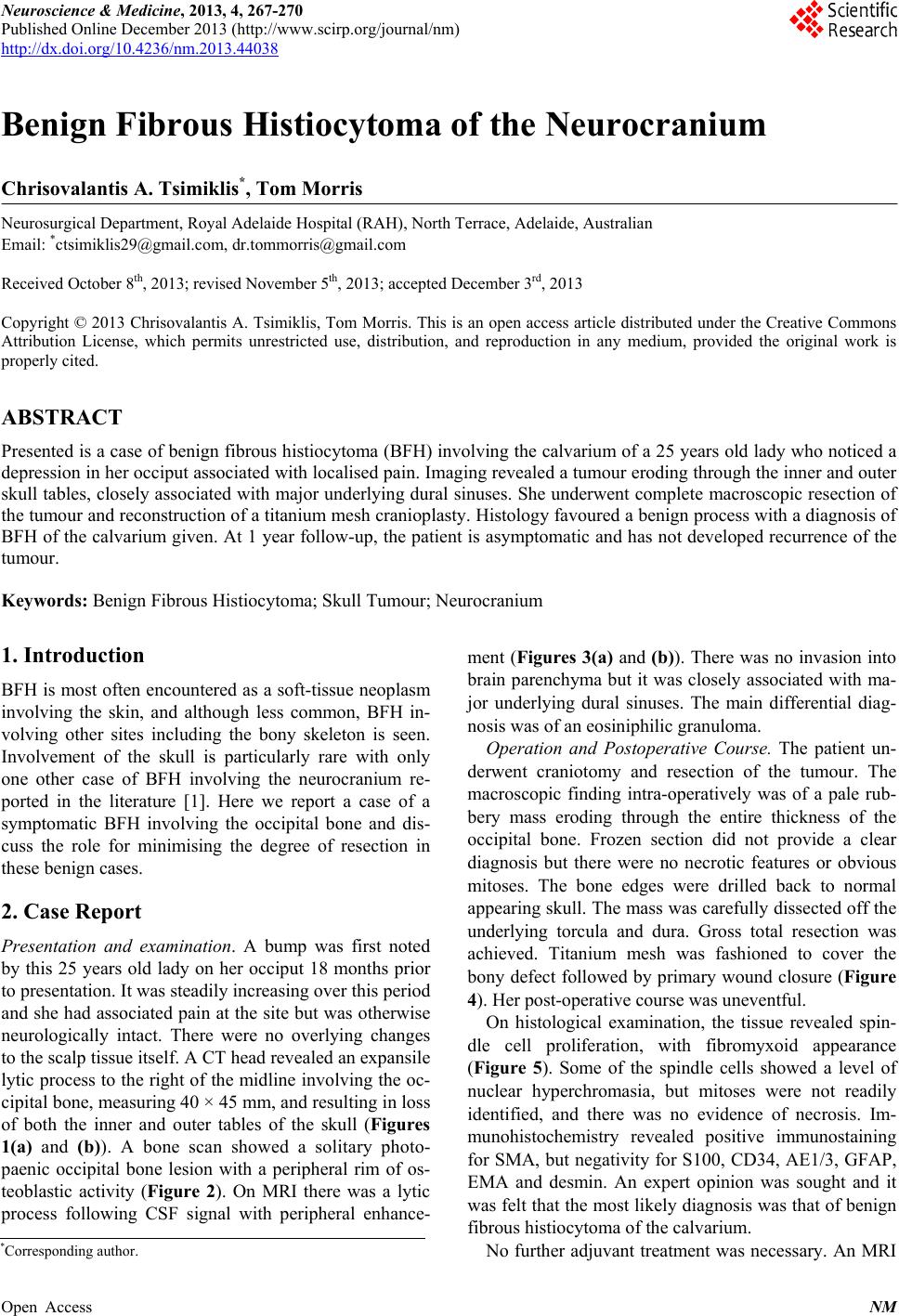
teoblastic activity (Figure 2). On MRI there was a lytic
process following CSF signal with peripheral enhance-
ment (Figures 3(a) and (b)). There was no invasion into
brain parenchyma but it was closely associated with ma-
jor underlying dural sinuses. The main differential diag-
nosis was of an eosiniphilic granuloma.
Operation and Postoperative Course. The patient un-
derwent craniotomy and resection of the tumour. The
macroscopic finding intra-operatively was of a pale rub-
bery mass eroding through the entire thickness of the
occipital bone. Frozen section did not provide a clear
diagnosis but there were no necrotic features or obvious
mitoses. The bone edges were drilled back to normal
appearing skull. The mass was carefully dissected off the
underlying torcula and dura. Gross total resection was
achieved. Titanium mesh was fashioned to cover the
bony defect followed by primary wound closure (Figure
4). Her post-operative course was uneventful.
On histological examination, the tissue revealed spin-
dle cell proliferation, with fibromyxoid appearance
(Figure 5). Some of the spindle cells showed a level of
nuclear hyperchromasia, but mitoses were not readily
identified, and there was no evidence of necrosis. Im-
munohistochemistry revealed positive immunostaining
for SMA, but negativity for S100, CD34, AE1/3, GFAP,
EMA and desmin. An expert opinion was sought and it
was felt that the most likely diagnosis was that of b enign
fibrous histiocytoma of the calv arium.
No further adjuvant treatment was necessary. An MRI
*Corresponding a uthor.
Benign Fibrous Histiocytoma of the Neurocranium
268
(a)
(b)
Figure 1. (a) Plain CT head (bone window) showing the
extent of erosion with involvement of the entire thickness of
the skull; (b) 3D reconstruction of skull demonstrating the
intricate architecture of the tumour.
Figure 2. Whole body bone scan with solitary lesion in the
occiput demonstrating a photopaenic centre with a periph-
eral rim of osteoblastic activity.
(a)
(b)
Figure 3. (a) Sagittal T1-weighted MRI post-contrast which
reveals a lytic lesion centred on the occipital bone with en-
hancement; (b) Axial slice showing the close relationship of
this tumour with the underly ing dural venous sinuses.
Figure 4. Plain CT head day 1 post-operatively. Titanium
mesh was fashioned and used to cover the defect in the oc-
cipital bone.
Open Access NM
Benign Fibrous Histiocytoma of the Neurocranium 269
Figure 5. H&E stained section (×20 magnification) of tu-
mour which is composed of a loose fibrous type stroma in
which cytologically bland stellate and spindle cell forms are
seen.
1 year post-operatively did not show any evidence of
disease recurrence (Figure 6). The patient was asymp-
tomatic.
3. Discussion
Although more commonly recognised as a soft tissue
tumour, BFH is also encountered in bone and rarely in
the skull. It is discussed in the WHO classification of soft
tissue and bone tumours as one of two types of fibro-
histiocytic tumours, the other being the malignant variant
[2].
Only one other case report in the literature describes
BFH involving the neurocranium. In that particular paper,
Ideguchi et al. [1] discuss a case of a 33-year-old woman
with a right occipital lesion causing raised intracranial
pressure secondary to occlusion of the adjacent dural
venous sinus. The tumour underwent resection with
preservation of collateral veins and imaging at the 6
month follow-up confirmed eradication of the tumour.
Tubbs et al. [3] describe a case of a BFH involving the
skull base of an infant. This lesion was discovered inci-
dentally and was diagnosed after biopsy but did not un-
dergo any further intervention. At 18 months, the child
remained asymptomatic with no gross increase in the
tumour size on imaging.
Malignant fibrous histiocytoma (MFH) involving the
skull is also described in a handful of case reports and its
propensity to invade into the adjacent soft tissue, dura
and brain parenchyma, results in a more aggressive
course with a tendency for recurrence. A case presented
by Joo et al. [4] of an intraosseous skull lesion, had simi-
lar characteristics and radiological features as the case
described here, however despite lack of aggressive fea-
tures macroscopically, the lesion recurred within 3
months.
Figure 6. Follow-up MRI scan (T1-weighted, post-contrast)
at 1 year post surgery. No evidence of disease recurrence
demonstrated.
In our case, given the lack of malignant features on the
frozen section, the decision was to conduct a limited re-
section without margin.
4. Conclusion
Overall, this case highlights a rare tumour of the skull,
which should nevertheless form part of the differential
diagnosis when such lesions are encountered. Given lim-
ited reports of this particular entity in the literature, the
natural history of BFH is not clearly defined. Interest-
ingly, in a review by Bielamowicz et al. [5] of 18 cases
of BFH involving the head and neck, a recurrence rate of
11% was reported. However, BFH of bone appears to
follow a more indolent course than its malignant coun-
terpart and thus aggressive or radical tumour resection
may not be necessary unless symptomatic or causing
significant mass effect.
5. Acknowledgements
The authors thank Dr. Sophia Otto (Pathologist, RAH)
for indicating the histological diagnosis and providing
the image of the slide used in this article.
REFERENCES
[1] M. Ideguchi, K. Kajiwara, K. Yoshikawa, S. Kato, M.
Fujii, H. Fujisawa and M. Suzuki, “Benign Fibrous
Histiocytoma of the Skull with Increased Intracranial
Pressure Caused by Cerebral Venous Sinus Occlusion,”
Journal of Neurosurgery, Vol. 111, No. 3, 2009, pp. 504-
518. http://dx.doi.org/10.3171/2008.11.JNS081206
Open Access NM
Benign Fibrous Histiocytoma of the Neurocranium
Open Access NM
270
[2] C. D. M. Fletcher, J. A. Bridge, P. Hogendoorn and F.
Mertens, “WHO Classification of Tumours of Soft Tissue
and Bone,” 4th Edition, Vol. 5, WHO Press, Geneve,
2013.
[3] R. S. Tubbs, D. R. Kelly, J. L. Pugh, M. Loukas and W. J.
Oakes, “Benign Fibrous Histiocytoma of the Skull Base,”
Journal of Neurosurgery, Vol. 106, 2007, pp. 65-67.
[4] M. Joo, G. J. Lee, Y.-C. Koh, O.-K. Kwon and Y.-K.
Park, “Primary Intraosseous Malignant Fibrous Histiocy-
toma of the Skull: A Case Report,” Journal of Korean
Medical Science, Vol. 18, 2003, pp. 609-613.
[5] S. Bieclamowi cz, M. S. Dauer, B. Chang and M. C. Zim-
merman, “Non-Cutaneous Benign Fibrous Histiocytoma
of the Head and Neck,” Otolaryngology Head and Neck
Surgery, Vol. 113, No. 1 1995, pp. 140-146.
http://dx.doi.org/10.1016/S0194-5998(95)70159-1