Journal of Cancer Therapy, 2013, 4, 1403-1410
Published Online November 2013 (http://www.scirp.org/journal/jct)
http://dx.doi.org/10.4236/jct.2013.49167
Open Access JCT
1403
Plasma Levels of Angiotensin-Converting Enzymes 1 and 2
and AGTR2 (T1247G and A5235G) Gene Polymorphisms
Are Associated to Breast Cancer Progression*
Maria Del Carmen Garcia Molina Wolgien1, Ismael Dale Cotrim Guerreiro da Silva1,
Afonso Celso Pinto Nazário1, Clovis Riyuchi Nakaie2, Silvana Aparecida Alves Corrêa de Noronha3,
Samuel Marcos Ribeiro de Noronha3, Gil Facina1
1Gynecology Department, Escola Paulista de Medicina/Universidade Federal de São Paulo (EPM-UNIFESP), São Paulo, Brazil;
2Biophisics Department, Escola Paulista de Medicina da Universidade Federal de São Paulo (EPM-UNIFESP), São Paulo, Brazil;
3Surgery Department, Escola Paulista de Medicina/Universidade Federal de São Paulo (EPM-UNIFESP), São Paulo, Brazil.
Email: cmolinawolgien@uol.com.br, ismael.dale@gmail.com, nazarioafonso@hotmail.com, cnakaie@unifesp.br,
silaac@globo.com, labgineco@globo.com, gilfacina@hotmail.com
Received October 4th, 2013; revised November 1st, 2013; accepted November 9th, 2013
Copyright © 2013 Maria Del Carmen Garcia Molina Wolgien et al. This is an open access article distributed under the Creative
Commons Attribution License, which permits unrestricted use, distribution, and reproduction in any medium, provided the original
work is properly cited.
ABSTRACT
Background: Breast cancer is the most common type of cancer among women. Diagnosed and treated timely, patients
may have good prognostics. In Brazil, in 2012, the estimate of new cases was 52,680 and the number of registered
deaths in 2012 was 12,852. The Renin-Angiotensin System (RAS) is known for its role in arterial hypertension and in
other cardiovascular diseases. Angiotensin-Converting Enzyme 2 (ACE2) is the key to Ang-(1-7) formation, and coun-
terbalances the ACE1/AngII/AGTR1 axis actions. RAS components have complex interactions with different tissues
and their actions are not restricted to the cardiovascular system. Recently, the RAS has been associated with different
types of cancers and in particular with gynecological cancers. Objectives: Our aim is to investigate possible associa-
tions between allelic distribution of two genetic polymorphisms in the AGTR2 receptor with ACEs 1 and 2 plasma lev-
els among women with breast cancer. Patients and Methods: Patients with breast cancer were genotyped for two poly-
morphisms of the AGTR2 (T1247G and A5235G). Genotyping assays (TaqMan) were performed with genomic DNA
extracted from blood cells. ACEs plasma level measurements were conducted in women from the breast-cancer group
(N = 53). ACEs were measured in the plasma of these patients using ELISA kits. Results: SNPs genotype distribution
is correlated with ACEs plasma levels. ACEs plasma levels are also correlated with clinical variables and ACE2 high
levels are associated with better prognostics. Conclusions: Changes in circulating levels of ECA1/AngII ECA2/
Ang-(1-7) determine the magnitude of the inflammatory response that an individual can trigger and the variation in
ACE 1 and 2 plasma level measurements in the blood of breast cancer patients suggests an association with the process of
mammary carcinogenesis. Thus, the RAS may be associated with the process of mammary carcinogenesis by both
genotypic variations of RAS components and by circulating levels of ACEs.
Keywords: Angiotensin-Converting Enzyme 1; Angiotensin-Converting Enzyme 2; Angiotensin II Type 2 Receptor;
Breast Neoplasm; ACEs Plasma Level; Genetic Polymorphisms
1. Introduction
Breast cancer can be defined as a malignant proliferation
of epithelial cells that line the ducts and lobules of the
breast [1]. Generally, cancer is considered as a disease
that originates from progressive genetic alterations in
oncogenes and in tumor suppressor genes combined to
chromosomal abnormalities [2]. Breast cancer is among
the most frequently diagnosed cancers and the second
leading cause of cancer death among U.S. women [3].
22% of new cancer cases in women around the world are
caused by malignancies of the breast. In Brazil, as in
*Funding: This work was supported by the Sao Paulo State Research
Foundation (FAPESP) by Grants numbers 2007/56480-0, 2008/50776-
7, and 2008/54383-0.
Conflict of interest statement: None declared.
Plasma Levels of Angiotensin-Converting Enzymes 1 and 2 and AGTR2 (T1247G and A5235G) Gene
Polymorphisms Are Associated to Breast Cancer Progression
1404
many other countries, the incidence of breast cancer has
been increasing. In 2012, 52,680 new cases of the disease
were estimated.
There are risk factors classically associated with breast
cancer, like sex (99% women), age (generally over 40
years), personal history for cancer (risk goes up based on
previously found gynecological cancers), family history,
menstrual history (early menarche and late menopause
increase risk), obstetric history (increased risk for nul-
liparous and primiparous over 35 years), nutritional fac-
tors (high-fat diets and intake of alcohol abuse are asso-
ciated with an increased risk) and genetic (gene muta-
tions in BRCA 1 and 2) [1].
More recently, as the number of studies on the inte-
grated operation of RAS components increases, the RAS
is no longer interpreted only as a linear proteolytic cas-
cade, but instead, as a complex humoral system in which
several agonists, besides the traditional AngII, may be
bound to multiple receptors, and offset the effects of each
other to control the (in)activation of different intracellular
pathways [4]. Angiotensin-Converting Enzyme 2 (ACE2)
seems to be the key to Ang-(1-7) formation which coun-
terbalances ACE1/AngII/AGTR1 axis effects. RAS com-
ponents have complex interactions with different tissues
and their actions are not restricted to the cardiovascular
system.
Among the recent approaches to study the RAS, it is
noteworthy to mention the two-axis concept, in which
one axis is composed of the peptidase Angiotensin-
Converting Enzyme 2 (ACE2), the hormone Ang-(1-7)
and the receptor MAS1, synergistically acting on organs
like the heart, liver and kidneys, and having opposite
actions to those assigned to the traditional ECA1/AngII/
AGTR1 axis [5]. Nowadays, Ang-(1-7) is robustly asso-
ciated with hypotensional actions and with inhibition of
calcium release from intracellular stores and with volt-
age-dependent calcium channel inactivation. This hep-
tapetide hormone also inhibits inflammatory pathways
and activates proapoptotic signals, in a tissue-specific
pattern [6,7]. Peptidase activity of ACE2 seems to be the
key to Ang-(1-7) formation, which counterbalances effects
triggered by the ACE1/AngII/AGTR1 axis. RAS compo-
nents have complex interactions with different tissues
and their actions are not restricted to the cardiovascular
system [8,9]. Studies about the role of the AGTR2 re-
ceptor in these recent approaches of the RAS are lacking.
2. Patients and Methods
2.1. Patients
Fifty-three (53) women with histologically confirmed breast
cancer were included in this study. Breast cancer patients
were admitted to and treated at the Mastology Section,
Division of Gynecology and Obstetrics, in Hospital Sao
Paulo, Sao Paulo, Brazil. Control group women were
admitted to the Menopause session/Gynecology Depart-
ment, also in Hospital Sao Paulo, Sao Paulo, Brazil. Ad-
missions occurred from December 2009 to December
2012. A structured questionnaire was applied to obtain
detailed information on demographic factors, as well as
menstrual and reproductive history. Clinical variables
like TNM category and axillary lymph node status were
also assessed in breast cancer patients. This study is ap-
proved by the Research Ethics Committee (REC) of the
Federal University of São Paulo under number 0424/09.
2.2. DNA Extraction
Blood samples were collected and immediately stored at
80˚C for posterior genomic DNA extraction. DNA ex-
traction was performed through GFX® Kit for blood cells
(GE HealthCare Life Sciences, Pittsburg, USA) accord-
ing to the manufacturer’s instructions.
2.3. AGTR2 SNPs Genotypin g
The Custom TaqMan® SNP Genotyping Assays (AGTR2)
(Applied Biosystems, Palo Alto, CA, USA) were used
for allelic discrimination, with a 40X PCR mix (Applied
Biosystems, Palo Alto, CA, US) specific to each AGTR2
SNP studied (T1247G rs5950584 and A5235G
rs1403543). This Assays-by-DesignSM (Applied Biosys-
tems, Palo Alto, CA, USA) is an assay for SNP genotype-
ing and gene expression based on sequence information
(Table 1) submitted by the customer. To obtain specific
DNA sequences amplification, we employed a 7500
Real-Time PCR system (Applied Biosystems, Palo Alto,
CA, USA) using 10 μl of TaqMan® Universal PCR Mas-
ter Mix, No AmpErase® UNG (2X), 0.5 l of specific
assay mix (T1247G and A5235G) containing the primers
and probes, 1 to 20 ng of genomic DNA and sterile water
for a total volume of 20 μl. PCR conditions were an ad-
ditional step at 50˚C for 2 minutes, and an initial step at
Table 1. Sequences that contained the mutation region for
each one of polymorphisms studied using Assays-by-Design
service, or TaqMan® SNP Genotyping Assays (AGTR2)
(Applied Biosystems).
Polymorphisms Sequences and mutations used for
TaqMan® SNP Genotyping Assays
AGTR2-T1247G
(rs5950584; Chr X)
(Pre-designed assay)
AGTR2-A5235G (rs1403543;
Chr X intron)
(Pre-designed assay)
GACAGGAGTTTACGATTATT[T/G]
GGTTGACCATTTTTTAA
TTAACACTGTATTTTGCAAAACTC
CT[A/G]AATTATTTAGCTGCTGTTT
CTCTTA
Open Access JCT
Plasma Levels of Angiotensin-Converting Enzymes 1 and 2 and AGTR2 (T1247G and A5235G) Gene
Polymorphisms Are Associated to Breast Cancer Progression
1405
95˚C for 10 minutes, followed by 40 cycles of denaturing
to 92˚C for 15 seconds and annealing/extension at 60˚C
for 1 minute. A negative control without template DNA
was used in each run. The allelic discrimination was de-
termined by analysis using SDS Software v. 1.3.1 (Ap-
plied Biosystem®s, Palo Alto, CA, USA).
2.4. Measurement of ACEs 1 and 2 in Plasma
Blood plasma samples were collected from the 53 wo-
men of the breast cancer group, the anticoagulant EDTA
was added, and these samples were centrifuged for 15
minutes at 1000 xg at 2˚C - 8˚C. The plasma removed
was stored at 80˚C until use. Plasma levels of ACEs 1
and 2 were measured with commercial ELISA kits (Uscn
Life Science Inc., Wuhan, China). ELISA standard
curves were generated with the diluent standard kit seri-
ally diluted to obtain seven different concentration points
(25 ng/ml; 12.5 ng/ml, 6.25 ng/ml, 3.12 ng/ml, 1.56
ng/ml, 0.78 ng/ml, 0.39 ng/ml; white and 0 ng/ml). Next,
plasma samples were thawed at RT and diluted 1:100 in
PBS 0.02 mol/L (pH = 7.0 to 7.2). Then, 100 mL of each
sample, the white point and the seven dilution ones were
placed in a 96-well assay plate coated with monoclonal
antibody specific for ACEs 1 and 2. These specimens
were incubated for 2 h at 37˚C. After this first incubation
period, the liquid was removed without washing and 100
mL of detection reagent A (polyclonal antibody conju-
gated to biotin specific for ACEs 1 and 2) were added.
The plate containing the sample was again incubated for
1 h at 37˚C, and then sealed. After this second incubation
period, the solution was aspirated and washed with 350
μl of the kit wash solution for 1.5 min. The plate was
dried out on paper towels. Rinsing was performed three
times. Next, 100 mL of detection reagent B (radish per-
oxidase conjugated to avidin HRP) working solution
were added and the sealed plates were incubated for 30
min at 37˚C. Plates were washed again (repeating proce-
dure described above) and 90 µl of TMB Substrate Solu-
tion were added to each well and the plate was again in-
cubated for 15 minutes at 37˚C protected from light. Fi-
nally, 50 mL of the kit stop solution (sulfuric acid) were
added and the plate was brought to a microplate reader
where spectrophotometric readings at 450 nm were made.
Each reading was taken in duplicates to obtain average
values and each standard and target sample was sub-
tracted from the average zero standard optical density.
GraphPad Prism 5.0 was used to generate a standard
curve (as recommended by the manufacturer), displaying
the ACE concentration, in the y-axis, and absorbance, in
the x-axis. The detection limits for the ACE1 assay is
ECA1 0.39 - 25 ng/mL, being 0.14 ng/mL the minimum
detectable dose. For ACE2, the detection limits are 0.78 -
50 ng/mL, being the minimum detectable dose equal to
0.27 ng/mL.
3. Results
3.1. AGTR2 SNPs Allelic Distribution and
Association with Breast Cancer
Among the two polymorphisms here studied, there could
only be found an association between allelic distributions
with case/control groups for SNP AGTR2 (A5235G).
However, both SNPs of the AGTR2 were associated with
at least one clinical variable (data not shown).
3.2. Correlation of ACE1 Plasma Levels with
AGTR2 SNPs Allelic Distribution and with
Clinical Variables
Plasma levels of ACE1 in breast cancer patients ranged
from 0.16 ng/ml to 115 ng/ml. For data analysis, these
patients were divided into three groups (0.1 - 1.0; 1.0 to
10, and >10 ng/ml) according to ACE1 plasma levels. In
each of these three groups, the following ACE1 plasma
levels were detected [mean ± SE (N)], respectively: [0.37
± 0.04 (27)], [3.9 ± 0.8 (13)], [48.6 ± 0, 04 (13)]. After
that, genotype distribution for the AGTR2 polymor-
phisms according to descriptive and/or clinical variables
(age, clinical stage, presence of lymph node metastasis
and histological grade) were correlated to the plasma le-
vels of ACE1 among these patients.
Correlation of ACE1 plasma levels with AG TR2
(A5235G) SNP genotype distribution—The SNP A5235G
genotype distribution correlated with plasma levels of
ACE1 (p < 0.0001***, chi-square), being the highest
ACE2 plasma levels (0.1 to 1.0ng/ml) the most abundant
level detected among homozygous GG individuals (GG =
54%), intermediary levels (1.0 to 10.0 ng/ml) predomi-
nated among homozygous AA individuals (38%) and
lower ACE1 levels (<0.1 ng/ml) among heterozygous
ones (52%) (Figure 1(a)).
Correlation of ACE1 plasma levels with AG TR2
(T1247G) SNP genotype distribution—The SNP A5235G
genotype distribution correlated with plasma levels of
ACE1 (p = 0.0027**, chi-square). Most of the patients are
homozygous TT carriers and, among these, intermediary
ACE1 plasma levels predominated (88%). Higher ACE1
plasma levels predominated among heterozygous patients
and, curiously, only lower ACE1 plasma levels were de-
tected among GG carriers (Figure 1(b)).
Correlation of ACE1 plasma levels with age—No sta-
tistically significant difference could be observed in the
age of the three ACE1 plasma levels groups (p = 0.69, t
test). The age of these patients in years (mean ± sd) in
each ACE2 concentration range were: ACE1 from 0.1 to
1.0 ng/ml (59 ± 12), ACE1 from 1.0 to 10.0 ng/ml (55 ±
19) and ACE1 > 10 ng/ml (57 ± 14) (Figure 2).
Open Access JCT
Plasma Levels of Angiotensin-Converting Enzymes 1 and 2 and AGTR2 (T1247G and A5235G) Gene
Polymorphisms Are Associated to Breast Cancer Progression
Open Access JCT
1406
Figure 1. Polymorphism genotype distribution of AGTR2 (A5235G or T1247G) and correlation with the concentration of
plasma levels of ACE1 or ACE 2. (a) AGTR2 (A5235G) and ACE1; (b) AGTR2 (T1247G) and ACE1; (c) AGTR2 (A5235G)
and ACE2; (d) AGTR2 (T1247G) and ACE2.
Figure 2. Age distribution of the patients according to each group separately for the concentration of ACE1 or ACE 2. (a)
Age and ACE1; (b) Age and ACE2.
Correlation of ACE1 plasma levels with breast cancer
clinical stage—Breast cancer patients’ clinical stage (I/II
and III/IV) was correlated to ACE1 plasma levels (01,
1.0, 1.0 to 10.0; >10 ng/ml) and statistically significant
differences were observed between these groups (p =
0.025*; chi-square test). High ACE1 plasma levels (>10.0
ng/ml) was the most abundant enzyme concentration
range detected among patients diagnosed with initial
clinical stages (I and II) and the least abundant among
patients in advanced clinical stages (III and IV). Patients
with more advanced clinical stages (III and IV) predo-
minated in the intermediary range (1.0 and 10 ng/ml) of
ACE1 plasma levels (Figure 3( a )).
Correlation of ACE1 plasma levels with lymph node
Plasma Levels of Angiotensin-Converting Enzymes 1 and 2 and AGTR2 (T1247G and A5235G) Gene
Polymorphisms Are Associated to Breast Cancer Progression
1407
Figure 3. Classification of clinical stage, lymph node status and histological grade for each of the patients according to the
correlation with the concentration of plasma levels of ACE1 or ACE 2. (a) Clinical stage and ACE1; (b) Lymph node status
and ACE1; (c) Histological grade and ACE1; (d) Clinical stage and ACE2; (e) Lymph node status and ACE2; (f) Histological
grade and ACE2.
status—Statistically significant differences were observed
between the groups (p = 0.0019**; chi-square test), High
ACE1 plasma levels (>10.0 ng/ml) was the most abun-
dant enzyme concentration range detected among pa-
tients with negative lymph node status and the least
abundant among patients with positive lymph node status.
Intermediary ACE1 plasma levels (1.0 to 10.0 ng/ml)
predominated among patients with positive lymph node
status (Figure 3 (b)).
Correlation of ACE1 plasma levels with histological
grade—Statistically significant differences were observed
between the groups (p = 0.0029**; chi-square test), being
the intermediary ACE1 plasma levels the most abundant
concentration among patients with histological grade G3
and the least abundant among patients with histological
grades G1 and G2 (Figure 3(c)).
3.3. Correlation of ACE2 Plasma Levels with
AGTR2 SNPs Allelic Distribution and with
Clinical Variables
Plasma levels of ACE2 in breast cancer patients ranged
from 27.5 ng/ml to 108.5 ng/ml. For data analysis, these
patients were divided into three groups (<50; 50 - 100; e
>100 ng/ml) according to ACE2 plasma levels. In each
of these three groups, the following ACE2 plasma levels
were obtained [mean ± SE (N)], respectively: [39.7 ± 1.3
(28)]; [64.5 ± 3.6 (18)]; [105.3 ± 1.00 (7)]. After that,
genotype distribution for the AGTR2 polymorphisms ac-
cording to descriptive and/or clinical variables (age, cli-
nical stage, presence of lymph node metastasis and his-
tological grade) were correlated to the plasma levels of
ACE1 among these patients.
Correlation of ACE2 plasma levels with AG TR2
(A5235G) SNP genotype distribution—The SNP A5235G
genotype distribution correlated with plasma levels of
ACE2 (p < 0.0001***, chi-square), being the highest
ACE2 plasma levels (>100 ng/ml) the most abundant
level detected among homozygous individuals (AA =
29% and GG = 58%) and intermediary levels (50 to 100
ng/ml) predominated among heterozygous individuals
(AG = 56%) (Figure 1(c)).
Correlation of ACE2 plasma levels with AG TR2
(T1247G) SNP genotype distribution—The SNP T1247G
genotype distribution correlated with plasma levels of
ACE1 (p < 0.0001**, chi-square), being the highest ACE2
plasma levels (>100 ng/ml) the most abundant level de-
tected among polymorphic homozygous individuals (GG
= 37%), lower levels (<50 ng/ml) among heterozygous
ones (TG = 17%) and intermediary levels among homo-
zygous (TT = 88%). Curiously, no GG carriers were de-
tected with intermediary ACE2 plasma levels, nor were
detected heterozygous ones with intermediary levels
(Figure 1(d)).
Correlation of ACE2 plasma levels with age—No sta-
tistically significant difference could be observed in the
Open Access JCT
Plasma Levels of Angiotensin-Converting Enzymes 1 and 2 and AGTR2 (T1247G and A5235G) Gene
Polymorphisms Are Associated to Breast Cancer Progression
1408
average age of the patients in each of the three ACE2
plasma levels ranges (p = 0.69, t test). The age of these
patients in years (mean ± sd) in each ACE2 concentration
range were: ACE2 < 50 ng/ml (58 ± 13), ACE2 from 50
to 100 ng/ml (56 ± 15) and ACE2 > 100 ng/ml (61 ± 15)
(Figure 2).
Correlation of ACE2 plasma levels with breast cancer
clinical stage—Breast cancer patients’ clinical stage (I/II
and III/IV) was correlated to ACE1 plasma levels (01,
1.0, 1.0 to 10.0; >10 ng/ml) and statistically significant
differences were observed between experimental groups
(p = 0.025*; chi-square test). High ACE2 plasma levels
(>100 ng/ml) was the most abundant enzyme concentra-
tion range detected among patients diagnosed with initial
clinical stages (I and II) and the least abundant among
patients in advanced clinical stages (III and IV). Exactly
the opposite pattern was observed for the lowest ACE2
concentration range (<50 ng/ml) (Figure 3(d)).
Correlation of ACE2 plasma levels with lymph node
status—Statistically significant differences were observed
between experimental groups (p = 0.0019**; chi-square
test). High ACE2 plasma levels (>100 ng/ml) was the
most abundant enzyme concentration range detected
among patients with negative lymph node status and the
least abundant among patients with positive lymph node
status. Exactly the opposite pattern was observed for the
lowest ACE2 concentration range (<50 ng/ml) (Figure
3(e)).
Correlation of ACE2 plasma levels with histological
grade—Statistically significant differences were observed
between experimental groups (p = 0.0029**; chi-square
test). Intermediary ACE2 plasma levels (50 to 100 ng/ml)
was the most abundant enzyme concentration range de-
tected among patients diagnosed with histological grades
G1 and G2 and the least abundant among histological
grade G3 patients (Figure 3(f)).
4. Discussion
4.1. ACE1 and ACE2 Plasma Levels and
Genotypic Distribution
This study represents the first one to report correlations
between ACEs 1 and 2 plasma levels and genotype dis-
tribution of AGTR2 polymorphisms and clinical charac-
teristics among breast cancer patients.
The balance between ACE1/AngII and ACE2/Ang-
(1-7) circulating levels seems to be key in determining
the magnitude of the inflammatory response that the
body will trigger. High circulating levels of ACE1/AngII
are associated with longer and more severe inflammatory
responses, while the opposite occurs when larger amounts
of ACE2/Ang-(1-7) are observed [9,10]. It is broadly
known that inflammatory processes are associated with
the process of carcinogenesis [10,11], therefore, it seems
licit to considered that high levels of circulating ACE1/
AngII and low ones of ACE2/Ang-(1-7) may be associ-
ated with the risk of developing breast cancer and its
prognosis, being the opposite also true.
ACEs circulating levels in Brazilian women with breast
cancer are correlated with the genotype distribution of
SNPs T1247G and A5235G AG TR2. However, in a case-
control study we observed that only the allelic distribu-
tion of SNP T5235G AGTR2 is associated with breast
cancer risk (data not shown). The number of heterozy-
gous individuals with high circulating levels of ACE1
(>10 ng/ml) is about 30%, this number drops to about 5%
for ACE2. We can also observe high levels of ACE1
among wild homozygous subjects (AA = 15%), and high
levels of ACE2 in 30% of these women. The distribution
profile of women homozygous polymorphic (GG) is very
similar between the different levels of ACEs.
Allelic distribution of SNP T1247G is not associated
with breast cancer risk (data not shown), however, high
doses of ACE2 (>10 ng/ml) are reduced in homozygous
wild (TT). Comparing ACEs 1 and 2 plasma level pro-
files, we observe that high levels of ACE2 are confined
to the homozygous genotypes (GG, or TT), while in the
case of ACE1 it is present only in the genotypes contain-
ing at least one T allele (TT or GT).
In summary, genetic predisposition to cancer is deter-
mined by a complex combination of many components
within cellular context. And certainly, within this breast
cancer cellular context, although very heterogeneous
between different subtypes, there is an important role
being played by the set of SNPs that each one carries in
its genome. Also, our results show that the level of cir-
culating peptidases ACE1 and ACE2 might affect the
process of mammary carcinogenesis through the classic
systemic RAS and not through the tissue RAS (tRAS)
[8].
4.2. ACE1 and ACE2 Plasma Concentrations
and Clinical Variables
Fortunately, no statistically significant difference could
be observed in the average age of the patients in each of
the three plasma levels ranges of ACEs 1 and 2.
In general, high levels of ACE2 are observed in sub-
jects with a better chance to have a good prognostic,
what could be explained by the recent RAS two-axis
concept. When we compare the intermediate circulating
concentrations of ACEs in patients with breast cancer for
the three clinical conditions (clinical stage, lymph node
status and histological grade), we can observe that these
patients predominate among the advanced stages of the
disease, while the opposite occurs for ACE2 intermedi-
ary levels. Therefore, this data allows us to infer that, in
Open Access JCT
Plasma Levels of Angiotensin-Converting Enzymes 1 and 2 and AGTR2 (T1247G and A5235G) Gene
Polymorphisms Are Associated to Breast Cancer Progression
1409
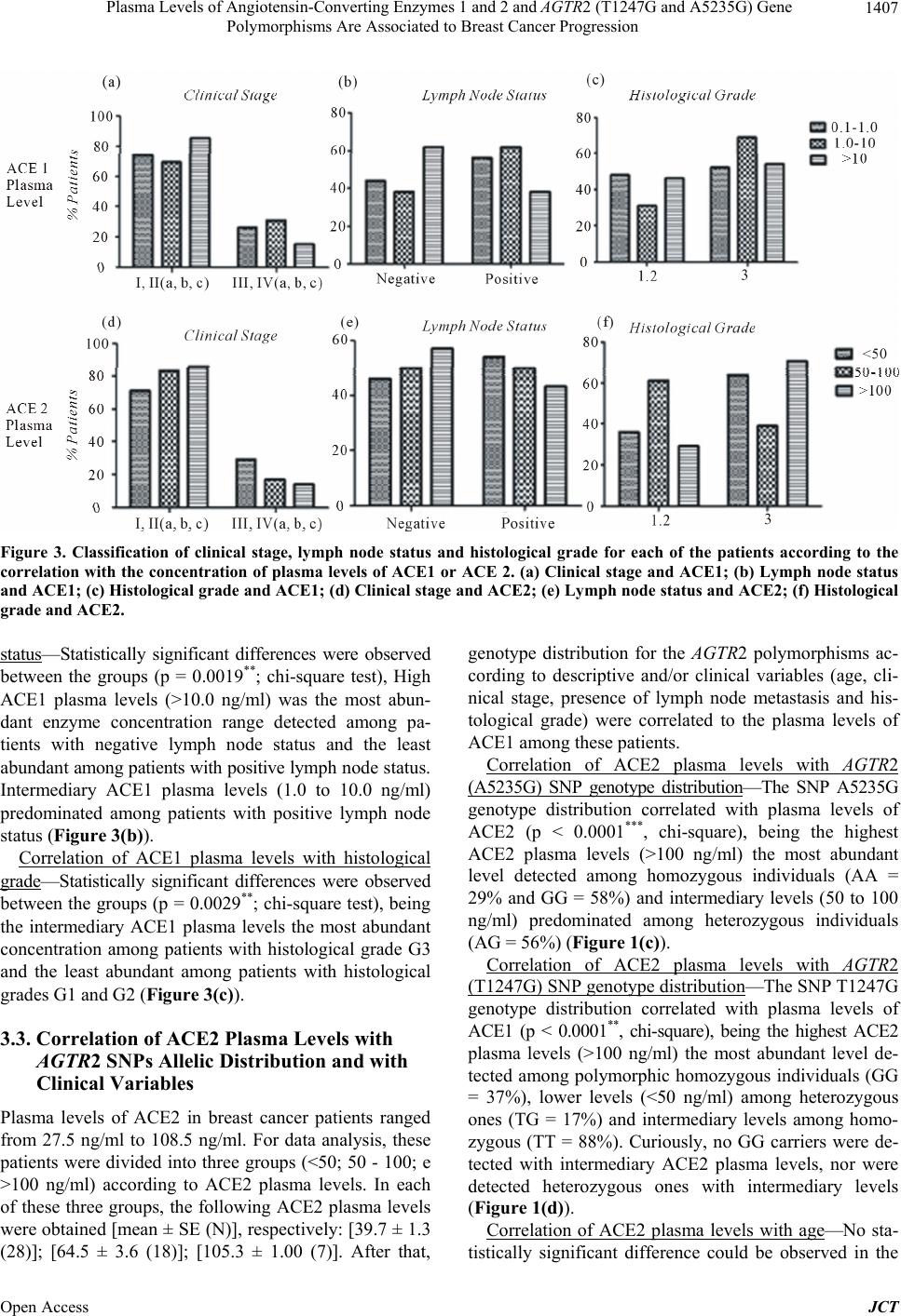
general, ECA1 seems to be associated with advanced
stages of the disease and, on the other hand, the ACE2
seems to be more associated with the early stages, giving
it a protector status against cancer. These ACEs plasma
levels data here presented, combined to other recent ob-
servations that Ang-(1-7) attenuates lung cancer metasta-
sis, has a protective effect by inhibiting cell proliferation
[11-16] and that genetic polymorphisms of the RAS com-
ponents are associated with gynecological cancer risk
and progression [17,18], give another piece of evidence
that the RAS may be associated with breast cancer.
Correlation between peptidases plasma levels and his-
tological grade has a different pattern. In the variable
histological grade, an inversion was observed, i.e., high
levels of ACE1 is associated with the early stages (grades
1 and 2) and high levels of ACE2 is associated with the
advanced stage (grade 3) of the disease. This can be un-
derstood as some kind of feedback for recovering the
patient from the pathological condition, if one has ex-
treme concentrations of circulating enzymes.
5. Conclusions
Our results show that ACE enzymes can be related to
worsening or improvement of breast cancer, as well as,
of other types of cancer.
Therefore, the use of enhancers or inhibitors of these
enzymes in cancer therapy should be considered, espe-
cially when there are little therapeutic options available
(triple-negative breast cancer, for example) or when they
are administered in an adjuvant regimen with other anti-
neoplastic compounds.
6. Acknowledgements
We thank Sao Paulo State Research Foundation (FAPESP)
for the financial support.
REFERENCES
[1] J. S. Berek, “Berek & Novak’s Gynecology,” 14th Edi-
tion, Wolters Kluwer Health/Lippincott Williams & Wil-
kins, Philadelphia, 2011, p. 1539.
[2] M. Smalley and A. Ashworth, “Stem Cells and Breast
Cancer: A Field in Transit,” Nature Reviews Cancer, Vol.
3, No. 11, 2003, pp. 832-844.
http://dx.doi.org/10.1038/nrc1212
[3] A. Jemal, R. Siegel, J. Xu and E. Ward, “CA Cancer,”
Journal of Clinical, Vol. 60, No. 5, 2010, pp. 277-300.
[4] A. Ribeiro-Oliveira Jr., A. I. Nogueira, R. M. Pereira, W.
W. Boas, R. A. Dos Santos and A. C. Simões e Silva,
“The Renin-Angiotensin System and Diabetes: An Up-
date,” Journal of Vascular Health and Risk Management,
Vol. 4, No. 4, 2008, pp. 787-803.
[5] T. P. Wong, K. Y. Ho, E. K. Ng, E. S. Debnam and P. S.
Leung, “Upregulation of ACE2-ANG-(1-7)-Mas Axis in
Jejunal Enterocytes of Type 1 Diabetic Rats: Implications
for Glucose Transport,” American Journal of Physiology,
Endocrinology and Metabolism, Vol. 303, No. 5, 2012,
pp. E669-E681.
http://dx.doi.org/10.1152/ajpendo.00562.2011
[6] G. Li, N. Xi and D. H. Wang, “Investigation of Angio-
tensin II Type 1 Receptor by Atomic Force Microscopy
with Functionalized Tip,” Nanomedicine, Vol. 1, No. 4,
2005, pp. 306-312.
http://dx.doi.org/10.1016/j.nano.2005.10.004
[7] S. Arumugam, R. A. Thandavarayan, S. S. Palaniyandi, et
al., “Candesartan Cilexetil Protects from Cardiac Myosin
Induced Cardiotoxicity via Reduction of Endoplasmic Re-
ticulum Stress and Apoptosis in Rats: Involvement of
ACE2-Ang(1-7)-Mas Axis,” Toxicology, Vol. 291, No. 1-
3, 2012, pp. 139-145.
http://dx.doi.org/10.1016/j.tox.2011.11.008
[8] M. Tahmasebi, J. R. Puddefoot, E. R. Inwang and G. P.
Vinson, “The Tissue Reninangiotensin System in Human
Pancreas,” Journal of Endocrinology, Vol. 161, No. 2,
1999, pp. 317-322.
[9] D. G. Passos-Silva, T. Verano-Braga and R. A. Santos,
“Angiotensin-(1-7): Beyond the Cardio-Renal Actions,”
Clinical Science, Vol. 124, No. 7, 2013, pp. 443-456.
[10] U. N. Das, “Angiotensin-II Behaves as an Endogenous
Pro-Inflammatory Molecule,” Journal of Association of
Physicians of India, Vol. 53, 2005, pp. 472-476.
[11] Y. R. Qian, Y. Guo, H. Y. Wan, L. Fan, Y. Feng, L. Ni, Y.
Xiang and Q. Y. Li, “Angiotensin-Converting Enzyme 2
Attenuates the Metastasis of Non-Small Cell Lung Cancer
through Inhibition of Epithelial-Mesenchymal Transi-
tion,” Oncology Reports, Vol. 29, No. 6, 2003, pp. 2408-
2414.
[12] J. R. Puddefoot, U. D. K. Udeozo, S. Barker and G. P.
Vinson, “The Role of Angiotensin II in the Regulation of
Breast Cancer Cell Adhesion and Invasion,” Endocrine-
Related Cancer, Vol. 13, No. 3, 2006, pp. 895-903.
http://dx.doi.org/10.1677/erc.1.01136
[13] P. E. Gallagher, K. Cook, D. Soto-Pantoja, J. Menon and
E. A. Tallant, “Angiotensin Peptides and Lung Cancer,”
Current Cancer Drug Targets, Vol. 11, No. 4, 2011, 2011,
pp. 394-404.
[14] Y. Feng, H. Wan, J. Liu, et al., “The Angiotensin-Con-
verting Enzyme 2 in Tumor Growth and Tumor-Associat-
ed Angiogenesis in Non-Small Cell Lung Cancer,” On-
cology Reports, Vol. 23, No. 4, 2010, pp. 941-948.
[15] S. A. A. C. Noronha, W. Bernardo, A. J. Barros, C. R.
Nakaie, S. I. Shimuta, I. D. C. G. Silva and S. M. R. No-
ronha, “Effects on Cell Viability and on Apoptosis in
Tumoral (MCF-7) and in Normal (MCF10A) Epithelial
Breast Cells after Human Chorionic Gonadotropin and
Derivated-Angiotensin Peptides Treatments,” Journal of
Cancer Therapy, Vol. 4, No. 7, 2013, pp. 65-69.
[16] I. Binda Neto, S. M. R. Noronha, S. A. A. C. Noronha, M.
D. C. M. Wolgien, A. J. Barros, C. R. Nakaie, S. I. Shi-
muta, G. Facina and I. D. C. G. Silva, “Angiotensin-(1-7)
and Human Chorionic Gonadotropin (hCG) Modulate the
Open Access JCT
Plasma Levels of Angiotensin-Converting Enzymes 1 and 2 and AGTR2 (T1247G and A5235G) Gene
Polymorphisms Are Associated to Breast Cancer Progression
Open Access JCT
1410
Nuclear Transcription Factors or Nuclear Receptors Genes
in the Tumorigenic Undifferentiated Breast Cancer Cell
Line SKBR3,” Journal of Cancer Therapy, Vol. 4, No.
7A, 2013, pp. 70-74.
http://dx.doi.org/10.4236/jct.2013.47A011
[17] S. A. A. Corrêa, S. M. R. Noronha, N. C. Nogueira-de-
Souza, C. V. V. Carvalho, A. M. M. Costa, J. J. Linhares,
M. T. V. Gomes and I. D. G. Silva, “Association between
the Angiotensin-Converting Enzyme (Insertion/Deletion)
and Angiotensin II Type 1 Receptor (A1166C) Polymor-
phisms and Breast Cancer among Brazilian Women,”
Journal of Renin Angiotensin Aldosterone System, Vol.
10, No. 1, 2009, pp. 51-58.
http://dx.doi.org/10.1177/1470320309102317
[18] S. A. Correa-Noronha, S. A. Ribeiro de Noronha, C. Ale-
crim, A. D. Mesquita, G. S. Brito, M. G. Junqueira, D. B.
Leite, C. V. Carvalho and I. D. Silva, “Association of An-
giotensin-Converting Enzyme I Gene I/D Polymorphism
with Endometrial but Not with Ovarian Cancer,” Gyne-
cology Endocrinology, Vol. 28, No. 11, 2012, pp. 889-
891.