International Journal of Otolaryngology and Head & Neck Surgery, 2013, 2, 248-252
Published Online November 2013 (http://www.scirp.org/journal/ijohns)
http://dx.doi.org/10.4236/ijohns.2013.26052
Open Access IJOHNS
A Comparative Study of Closure of Tympanic Membrane
Perforation between Chemical Cauterization and Fat Plug
Myringoplasty
Manabendra Debnath, Swagata Khanna
Department of ENT, Gauhati Medical College & Hospital, Guwahati, India
Email: swagatakhanna@sify.com
Received July 13, 2013; revised August 15, 2013; accepted September 2, 2013
Copyright © 2013 Manabendra Debnath, Swagata Khanna. This is an open access article distributed under the Creative Commons
Attribution License, which permits unrestricted use, distribution, and reproduction in any medium, provided the original work is
properly cited.
ABSTRACT
The purpose of closure of small dry tympanic membrane perforation s is to restore the continuity of the tympanic mem-
brane in order to improve hearing and decrease the incidence of middle ear infections. Small and cost effective proce-
dures like Chemical Cauter ization and Fat Plug Myringoplasty have been found to be effective in healing small central
perforations with significant hearing improvement. A study was carried out in 69 patients with central perforations in
the Department of ENT, Gauhati Medical College & Hospital for the duration of 1 year from Aug ust 11 to July 12 . Out
of 69 patients, 36 underwent Chemical Cauterization and 33 Fat Plug Myringoplasty. Pre- and post-operative hearing
assessments of each patient were done and statistically analyzed. The success rate was found to be 83.33% and 90.9%
respectively. In both groups, there was statistically significant hearing improvement with a P value 0.01. From our study,
it was found that the two procedures are simple, reliable and also lessened the morbidity of the patient. Thus we rec-
ommend the use of these two official procedures for the treatment of central perforations of tympanic membrane less
than 5 mm.
Keywords: Fat Plug Myringoplasty; Chemical Cauterization; Paper Patching; Central Perforations
1. Introduction
Tympanic membrane perforations result mainly from
infectious and traumatic etiologies. Perforations result
from acute otitis media and trauma heal spontaneously in
the majority of cases. But if there is repeated infections
or if the infection is persisten t, there is less possibility of
spontaneous healing of these perforations. Long-standing
tympanic membrane perforations may cause conductive
hearing loss and middle ear infection even if they are
small. The purpose of closure of chronic dry tympanic
membrane perforations is to restore the continuity of the
tympanic membrane in order to improve hearing and
decrease the incidence of middle ear infections.
Myringoplasty is the procedure of closure of the per-
forations of the pars tensa of the tympanic membrane.
The first attempt at myringoplasty was recorded by Mar-
cus Banzer in 1640 [1]. The first surgical closure of
tympanic membrane perforations using an autograft (a
full thickness free skin g raft) was performed by Berthold
in 1878 and he introduced the term “Myringoplasty” [1].
Although Tympanoplasty (myringoplasty + ossicular
chain reconstruction) is the ideal treatment, simple and
cost effective procedures like chemical cauterization and
fat plug myringoplasty h ave been found to be effectiv e in
healing small central perforations with significant hear-
ing improvement.
Fat Plug Myringoplasty has many advantages com-
pared with conventional myringoplasty using temporalis
fascia, as it is short, simple, cost-effective and minimally
invasive. Fat from ear lobule has been in use for last 15 -
20 years. Microscopic study of fat from ear lobule
showed that the fat cells are more compact and contain
more fibrous tissue. It provides scaffolding for the
growth of tympanic membrane epithelium and mucus
membrane by bridging the gap. In 1962, Ringenberg
successfully used adipose tissue (Fat) to repair a small
tympanic membrane pe rforation [2].
M. DEBNATH, S. KHANNA 249
2. Aims and Objectives
1) To study the efficacy of chemical cauterization in
small central perforations of tympanic membrane.
2) To study the efficacy of Fat Plug Myringoplasty in
small central perforations of tympanic membrane.
3) To evaluate the outcome of these two procedures
and compare them.
3. Materials and Methods
The study was carried out in the Department of ENT,
Gauhati Medical College and Hospital, Guwahati for
duration of 1 year from August 2011 to July 2012. The
study group included patients above 15 years of age with
central perforations of tympanic membrane up to 5 mm
in size. The locations of the perforations in th e pars tensa
were variable from patient to patient as demonstrated in
Figure 1. This study consisted of a total of 69 patients
out of which 36 underwent Chemical Cauterization and
33 underwent Fat Plug Myringoplasty.
Criteria for patient selection:
1) Inclusion criteria:
a) Patients with central perforation of tympanic mem-
brane following Chronic Suppurative Otitis Media or
traumatic perforations of tympanic membrane.
b) At least 2 months old dry perforations were in-
cluded.
c) Central perforations not more than 5 mm (35%) by
visual assessment were included in the study.
2) Exclusion criteria:
a) History of previous ear surgery;
b) Attico-antral disease;
c) Active discharge;
d) Nasopharyngeal pathology;
22
11
16
5
2
6
4
3
P ostero-inferior
Postero-
superior
Anter-inferior
Antero-superior
Post-
inferio r+Po st. s
up.
P ost-inf+Ant.-
inf
Ant.
Inf+ant.sup
Ant. Sup.+Post.
Sup
Figure 1. Pie diagram showing location of perforations in
different quadrants of pars tensa.
e) Eustachian tube dysfunction and;
f) Active rhinosinusitis.
Hearing assessment both before and after the proce-
dures was done by Tuning fork tests and Pure Tone
Audiometry.
Nasal Endoscopy was done to rule out any naso-
pharyngeal pathology, Eustachian tube dysfunction and
rhinosinusitis etc.
3.1. Chemical Cauterization (Paper Patching)
1) Patients with central perforations less than 5 mm
selected according to the section criteria mentioned
above.
2) Patients were properly explained about the proce-
dure and possible outcome and written and informed
consent taken.
3) Cotton ball soaked in 4% xylocaine was applied in
the external auditory cana l.
4) Under microscope, the edges of the tympanic mem-
brane perforations were cauterized by using cotton tipped
applicator dipped in trichloroacetic acid.
5) After the procedure, a thin paper preferably tissue
paper was placed over the perforation followed by appli-
cation of 1 - 2 drops of steroid containing antibiotic ear
drop.
6) The perforation is inspected after 2 weeks and if
required patient is called for subsequent sitting. A maxi-
mum of four applications were made.
7) After complete healing of the perforation the patient
is followed up at 6 weeks and Pure Tone Audiometry
was done to assess the hearing improvement.
3.2. Fat Plug Myringoplasty
1) After proper written and informed consent the con-
cerned ear was prepared by betadine solution and then
properly draped.
2) 2% xylocaine + adrenaline was infiltrated in the
posterior aspect of the pinna and also in the external
auditory canal.
3) Now by giving an incision in the posterior aspect of
the ear lobule fat graft was harvested and the incision site
was closed by 4 - 0 Et hilon.
4) Now under an operating microscope the edges of
the tympanic membrane perforation were de-epithelial-
ised (Figure 2).
5) The harvested fat plug is then wedged like a dumb-
bell one part of which is in the middle ear and other lat-
eral to tympanic membrane then it is supported by a few
pieces of g el foam from outside.
6) A course of antibiotic and anihistaminic drugs was
given in the post-operativ e period
7) The patient was then followed up after 2 weeks, 6
weeks and then at 3 months and Pure Tone Audiometry
was done to assess the hearing improvement (Figure 3).
Open Access IJOHNS
M. DEBNATH, S. KHANNA
250
Figure 2. Central perforation (right tympanic membrane)
of pars tensa.
Figure 3. Healed right tympanic membrane perforation.
3.3. Statistical Analysis
The hearing gain after the two procedures was analyzed
with Student’s t-test, and the Pearson correlation coeffi-
cient (r) was determined by using Statistical Package
for Social Science (SPSS) version 10.0. The other cal-
culations performed for the present study was done by
spreadsheets developed by Microsoft Excel (2007). A p
value of 0.05 or less was considered significant for the
study.
4. Results and Observations
The present study consisted of 69 patients out of which
40 were male and 29 female. The average age of the
study group was 35 years. Out of 69 patients 30 patients
had bilateral central perforations and 39 had unilateral
perforations. 55 patients had perforations due to infective
etiology and 14 had due to trauma. Out of 69 patients 36
underwent chemical cauterization and 33 underwent Fat
Plug Myringoplasty.
In the group that underwent Chemical Cauterization
the mean preoperative hearing threshold (dB) was 24.66
± 3.69 and after the procedure the mean hearing thresh-
old (dB) was 16 ± 2.03 and there was a mean gain of
about 8.66 ± 3.69 dB (Figure 4) which was found to be
statistically significant(r = 0.849, p = 0.01). Out of 36
patients who underwent Chemical Cauterization 6 pa-
tients had residual perforations. The success rate was
found to be 83.33%.
In the group that underwent Fat Plug Myringoplasty
the mean preoperative hearing threshold (dB) was 33.83
± 4.41 and after the procedure the mean hearing thresh-
old (dB) was 17.83 ± 2.84 and there was a mean gain of
about 16 ± 4.02 dB (Figure 5) which was found to be
statistically significant (r = 0.782, p = 0.01). Out of 33
patients who underwent Fat Plug Myringoplasty 3 pa-
tients had residual perforations. The success rate was
found to be 90.9%.
Therefore, in our study we have found that Fat Plug
Myringoplasty is a better option (success rate being
90.9%) for the closure of Tympanic Membrane perfora-
tion less than 5 mm with only one surgical sitting.
Whereas in Chemical Cauterization single application
may not be sufficient in all cases.
In both the groups mentioned in the study, the number
of patients having residual perforations (i.e. 6 out of 36
in Chemical Cauterization and 3 out of 33 in Fat Plug
Myringoplasty) was not found to be statistically signifi-
cant.
0
5
10
15
20
25
30
35
147 10131619222528
Pre oper ative
he aring
threshold(dB)
Postope rative
he aring
threshold(dB)
‐‐‐‐No.ofPatients‐‐‐‐>
HearingTHreshold(dB)>
Figure 4. Line diagram showing comparison between pre
and post operative hearing threshold (dB) of patients un-
dergoing chemical cauterization.
0
5
10
15
20
25
30
35
40
45
147 10131619222528
‐‐‐‐Hearingthr eshold(dB)‐‐‐>
‐‐‐‐‐‐‐‐‐No.ofpatients‐‐‐‐‐‐‐‐‐>
Pre
operative
he aring
th resho ld( d
B)
Post
operative
he aring
th resho ld( d
B)
Figure 5. Line diagram showing comparison between pre
and post-operative hearing threshold (dB) of patients un-
dergoing fat plug myringoplasty.
Open Access IJOHNS
M. DEBNATH, S. KHANNA 251
5. Discussion
The first recorded use of silver nitrate to stimulate clo-
sure of tympanic membrane perforations is by William
Wilde in 1848 [3]. The office chemical myringoplasty
was introduced by Roosa in 1876 and was popularized by
Derlacki in the 1950s who reported good results and the
procedure came to be known as the Derlacki method [4].
In his method he used trichloroacetic acid to cauterize
perforation followed by antibiotic sufflation and covering
with sterile cotton pledgets and prescribing Euthymol ear
drops. Two decades later, Derlacki reported that he and
his colleagues (Shambaugh, Harrison and Clemis) at
Otologic Professional Associates at Chicago and cumula-
tively treated 1277 pars tensa perforations between 1953
and 1972 and had successfully healed at least 1027 of
them (80.4%) [5].
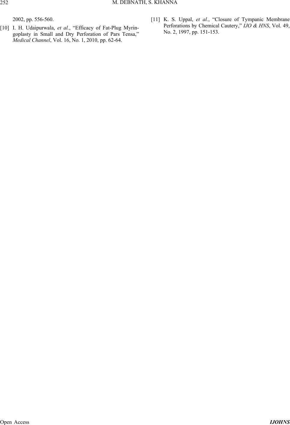
From our study it was found that the two procedures
showed significant improvement in post operative hear-
ing threshold. The overall success rate of 90.9% in pa-
tients undergoing Fat Plug Myringoplasty and 83.33 % in
patients undergoing Chemical Cauterization observed in
the present study is in agreement with the rates of 80% -
92% reported in other studies. No significant difference
in the tympanic membrane perforation closure rate was
observed according to the perforation sites in different
quadrants of the tympanic membrane.
Dursun E. et al. found out in 2008 that there were no
statistically significant differences in tympanic mem-
brane closure rates between paper patch, fat and peri-
chondrium myringoplasty which is also found in our
study [6].
Landsberg R. performed fat plug myringoplasty on a
population comprising 27 adults and 11 children. The
speech reception threshold improved significantly in his
study. This is also seen in this study [7].
Nelson C. Goldman performed chemical cauterization
in 11 patients and found 82.7% success rate [8]. Similar
outcome is also seen in a study by Louis F. Scaramella et
al. where success rate with chemical cauterization was
84.2% which holds true in our study also [9].
Jadia S. et al. in their study found 92% and 88.8% out-
come with fat plg myringoplasty and chemical cauteriza-
tion which is also seen in this study [4].
Iqbal Hussain Udaipurwala et al. in 2008 in their stud y
achieved 95.2% success rate with Fat Plug Myrin-
goplasty for central perforations of tympanic membrane
which is consistent with this study [10].
Kuljit S. Uppal et al. in 1997 achieved 78% success
rate with chemical cautery of tympanic membran e perfo-
rations which is consistent with this study [11].
6. Conclusion
A reliable tympanic membrane closure rate in small cen-
tral perforations was achieved with Fat Plug Myrin-
goplasty and Chemical Cauterization without general
anaesthesia in an official setting with a good amount of
success. These two procedures are easy to perform, reli-
able with a satisfactory outcome, virtually simple and
rapid risk free and also lessen the financial burden and
morbidity on the part of the patient. Although both
methods can be considered as convenient official proce-
dures, the success rate is higher in Fat Plug Myrin-
goplasty (90.9%), and this procedure can be recom-
mended as a better alternative/preferable choice for first
line treatment of central perforations of the tympanic
membrane in pars tensa less than 5 mm in size.
7. Acknowledgements
We are highly grateful to The Principal and the Superin-
tendant of Gauhati Medical College and Hospital, Gu-
wahati for allowing us to carry out the study in this In-
stitution.
REFERENCES
[1] U. Chalishazar, “Fat Plug Myringoplasty,” Indian Journal
of Otolaryngology and Head Neck Surgery, Vol. 57, No.
1, 2005, pp. 43-44.
[2] J. C. Ringerberg, “Closure of Tympanic Membrane Per-
forations by the Use of Fat,” Laryngoscope, Vol. 88, No.
6, 1978, pp. 982-993.
http://dx.doi.org/10.1288/00005537-197806000-00010
[3] W. R. Wilde and A. Hewson, “Practical Observations on
Aural Surgery and the Nature and Treatment of Diseases
of the Ear,” Blanchard & Lea, Philadelphia, 1853, pp.
292-293.
[4] S. Jadia, et al., “Office Procedure for the Management of
Central Perforation: A Comparative Study,” Indian Jour-
nal of Otology, Vol. 16, 2010, pp. 5-9.
[5] E. L. Derlacki, “Office Closure of Central Tympanic
Membrane Perforation: A Quarter Century Experience,”
Transactions of the American Academy of Ophthalmology,
Vol. 77, 1973, pp. 53-56.
[6] E. Dursun, et al., “Comparison of Paper Patch, Fat and
Perichondrium Myringoplasty in Repair of Small Tym-
panic Perforations,” Otolaryngology Head Neck Surgery,
Vol. 138, No. 3, 2008, pp. 353-356.
http://dx.doi.org/10.1016/j.otohns.2008.01.003
[7] R. Landsberg, G. Fishman, A. DeRowe, E. Berco and G.
Berger, “Fat Graft Myringoplasty: Results of a Long-
Term Follow-Up,” Journal of Otolaryngology, Vol. 35,
No. 1, 2006, pp. 44-47.
http://dx.doi.org/10.2310/7070.2005.4124
[8] N. C. Goldman, “Chemical Closure of Chronic Tympanic
Membrane Perforations,” ANZ Journal of Surgery, Vol.
77, No. 10, 2007, pp. 850-851.
http://dx.doi.org/10.1111/j.1445-2197.2007.04256.x
[9] L. F. Scaramella, et al., “Effectiveness in Non-Surgical
Office Closure of Tympanic Membrane Pars Tensa Per-
forations,” Ear, Nose and Throat Journal, Vol. 81, No. 8,
Open Access IJOHNS
M. DEBNATH, S. KHANNA
Open Access IJOHNS
252
2002, pp. 556-560.
[10] I. H. Udaipurwala, et al., “Efficacy of Fat-Plug Myrin-
goplasty in Small and Dry Perforation of Pars Tensa,”
Medical Channel, Vol. 16, No. 1, 2010, pp. 62-64.
[11] K. S. Uppal, et al., “Closure of Tympanic Membrane
Perforations by Chemical Cautery,” IJO & HNS, Vol. 49,
No. 2, 1997, pp. 151-153.