Jour nal of Biosci enc e s an d M e dic ine s, 2013, 1, 10-13 JBM
http://dx.doi.org/10.4236/jbm.2013.12003 Published Online October 2013 (http://www.scirp.org/journal/jbm/)
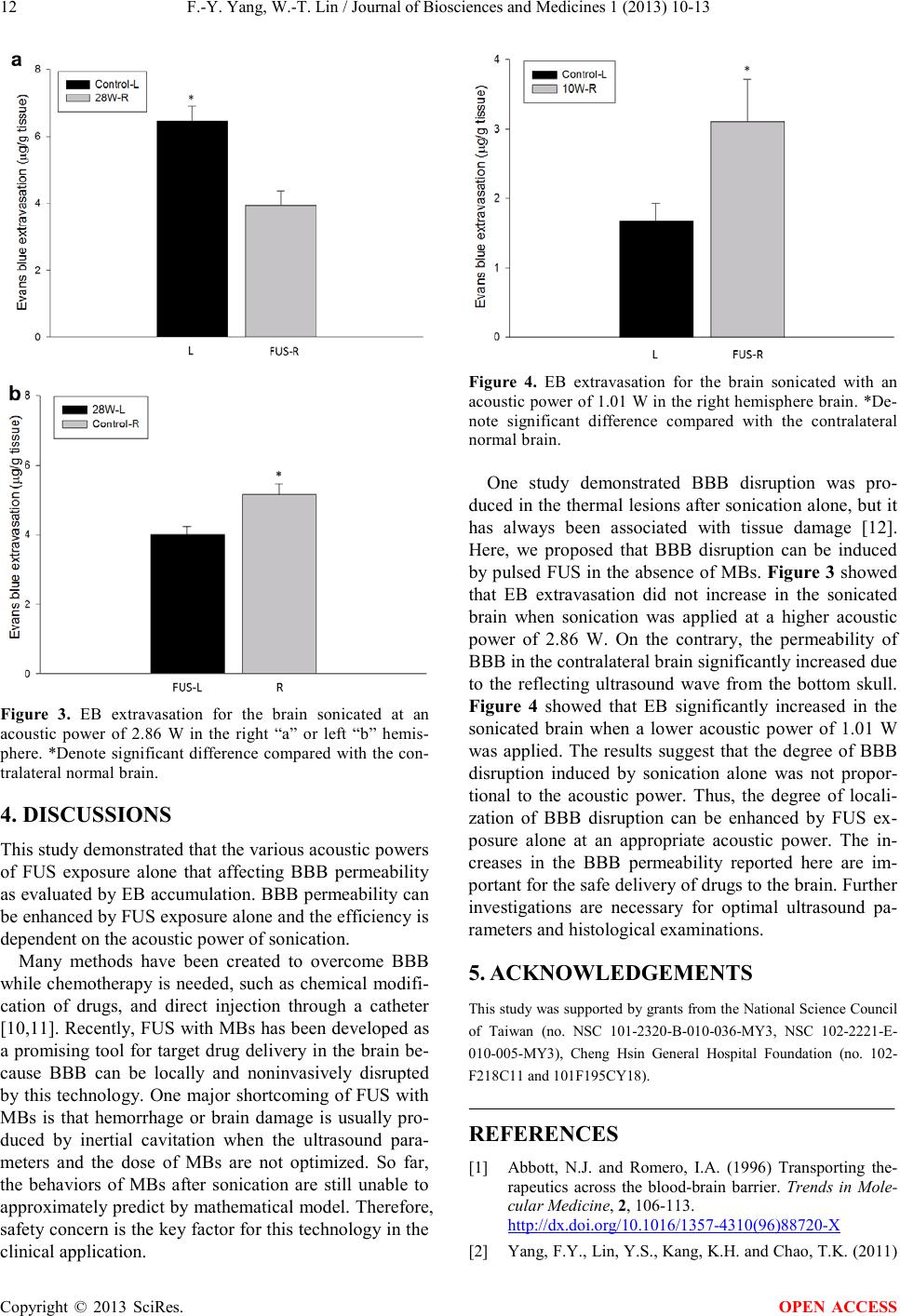
OPEN ACCESS
The effects of ultrasound on blood-brain barrier
Feng-Yi Yang, Wei-Ting Lin
Department of Biomedical Imaging and R adi ological Sciences , National Yang-Ming University, Taipei, Taiwan
Email: fyyang@ym.edu.tw, lin_79_03_26@yahoo.com.tw
Received August 2013
ABSTRACT
The brain is protected from the entry of foreign sub-
stances by blood-brain barrier (BBB), but becomes a
barrier while chemotherapy is needed for the brain
diseases. Ultrasound with microbubbles (MBs) has
been shown to noninvasively increase the permeabili-
ty of the BBB in the normal tissue and brain tumor.
The real mechanism for disruption is still unknown.
Hemorrhage was usually found in the sonicated re-
gion of the brain. Thus, treatment safety is the pri-
mary concern when considering clinical application
of BBB disruption induced by ultrasound in the pre-
sence of MBs. Here we investigate the effects of ul-
trasound on the permeability of BBB whether the
MBs were administered. The data reveals that Evans
blue (EB) a ccu mulation w as highest in t he brain after
sonication with MBs. However, the permeability of
BBB also can be significantly increased by ultrasound
alone. These results demonstrated that noninvasive
disruption of BBB by ultrasound alone with no dam-
age i s p ossible.
Keywords: Ultrasound; Blood-Brain Barrier;
Permeability; Drug Delivery; Brain Disease
1. INTRODUCTION
Most brain diseases are hard to treat with chemotherapeu-
tics due to the inability of molecules to pass the bloo d-brain
barrier (BBB). The endothelial cells of the brain are
tightl y fused to each othe r known as tight j unctions [1].
Several methods have been developed to disrupt the
BBB to facilitate drug delivery. Recently, it has been
shown that BBB can be locally and noninvasively dis-
rupted by focused ultrasound (FUS) in the presence of
microbubbles (MBs) [2-4]. However, small areas of
erythrocyte extravasation were found in the sonicated
site [5,6]. Interactions between the ultrasound and
MBs—which include oscillatory forces, acoustic cavi-
tation, and shear stress related to streaming of fluid
around the bubbles—are likely to trigger various physi-
ological responses [7]. The side effect of hemorrhage
may b e induced by the widening of the tight junc tion or
vessel damage after FUS sonication. In this study, we
investigate if BBB can be disrupted noninvasively by
FUS without MBs administration in order to avoid the
brain damage from cavitation effects.
2. METHODS
2.1. Animal Prepara tion
A total of t welv e male Spr ague-Dawley rats weighing
from 280 to 350 g were used in these experiments. All
the procedures of the animal experiment adhered to the
Guidelines for Care and Use of Experimental Animals
by our institutional animal committee.
2.2. Ultrasound System
FUS was produced by a 1 MHz single-element focused
transducer (A392S, Panametrics, Waltham, MA, USA)
with a diameter of 38 mm and a radius of curvature of
63.5 mm. The half-maximum of the pressure amplitude
of the focal zone had a diamete r and length o f 3 and 26
mm, respectively. The transducer was mounted on a
removable cone filled with deionized and degassed wa-
ter whose tip was capped by a polyurethane memb r ane ,
and the center of the focal spot was at approximately
5.7 mm below the cone tip. FUS beam was precisely
targeted using a stereotaxic apparatus (Stoelting, Wood
Dale, IL, USA). A function generator (33220A, Agilent
Inc., Palo Alto, USA) was connected to a power am-
plifier (500-009, Advanced Surgical Systems, Tucson,
AZ) to drive the FUS transducer and a power meter/
sensor module (Bird 4421, Ohio, USA) was used to
measure the input electrical power. The animal posi-
tioning for the sonication arrangement was the same as
our previous works [8,9]. The rat was laid prone be-
neath the cone tip and ultrasound transmission gel
(Pharmaceutical Innovations, Newark, NJ, USA) was
used to maximize the transmission of ultrasound be-
tween the transducer and the rat’s b r ai n.
2.3. Soncation
Ultrasound contrast agent (UCA, SonoVue, Bracco In-