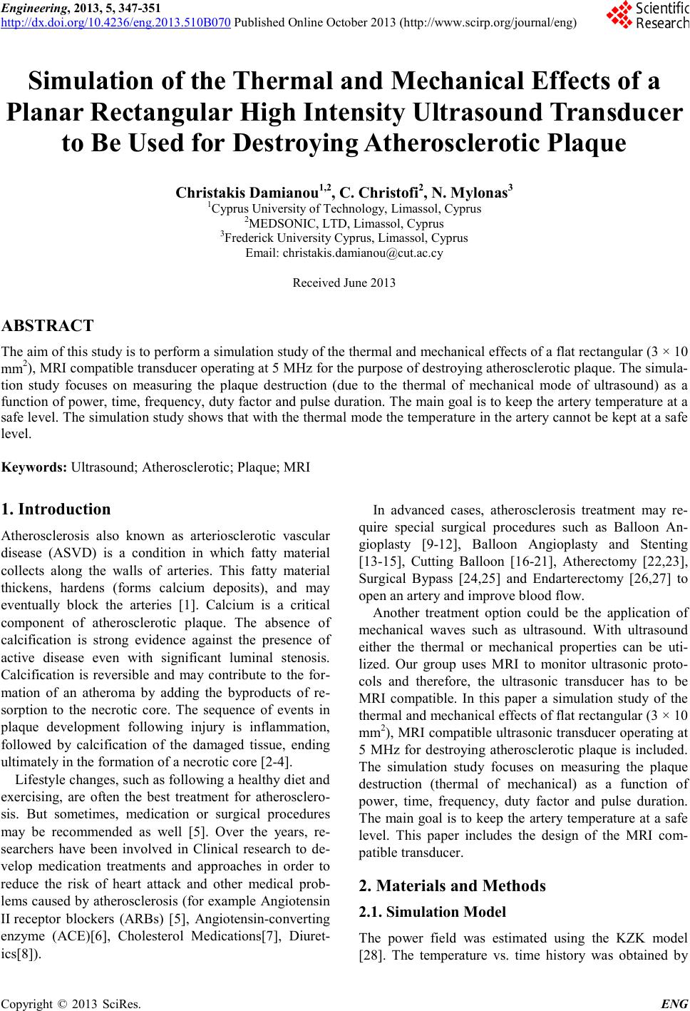
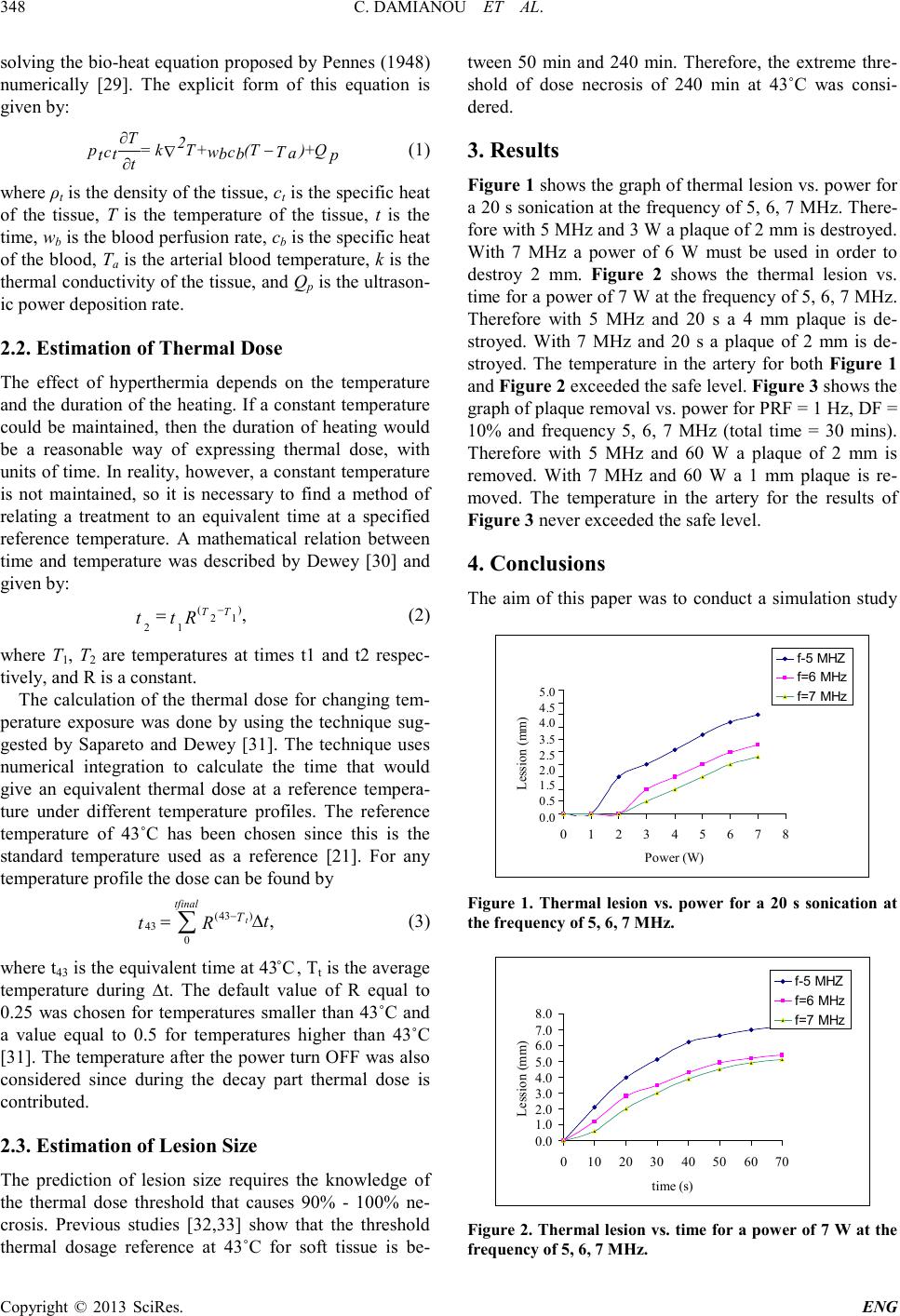
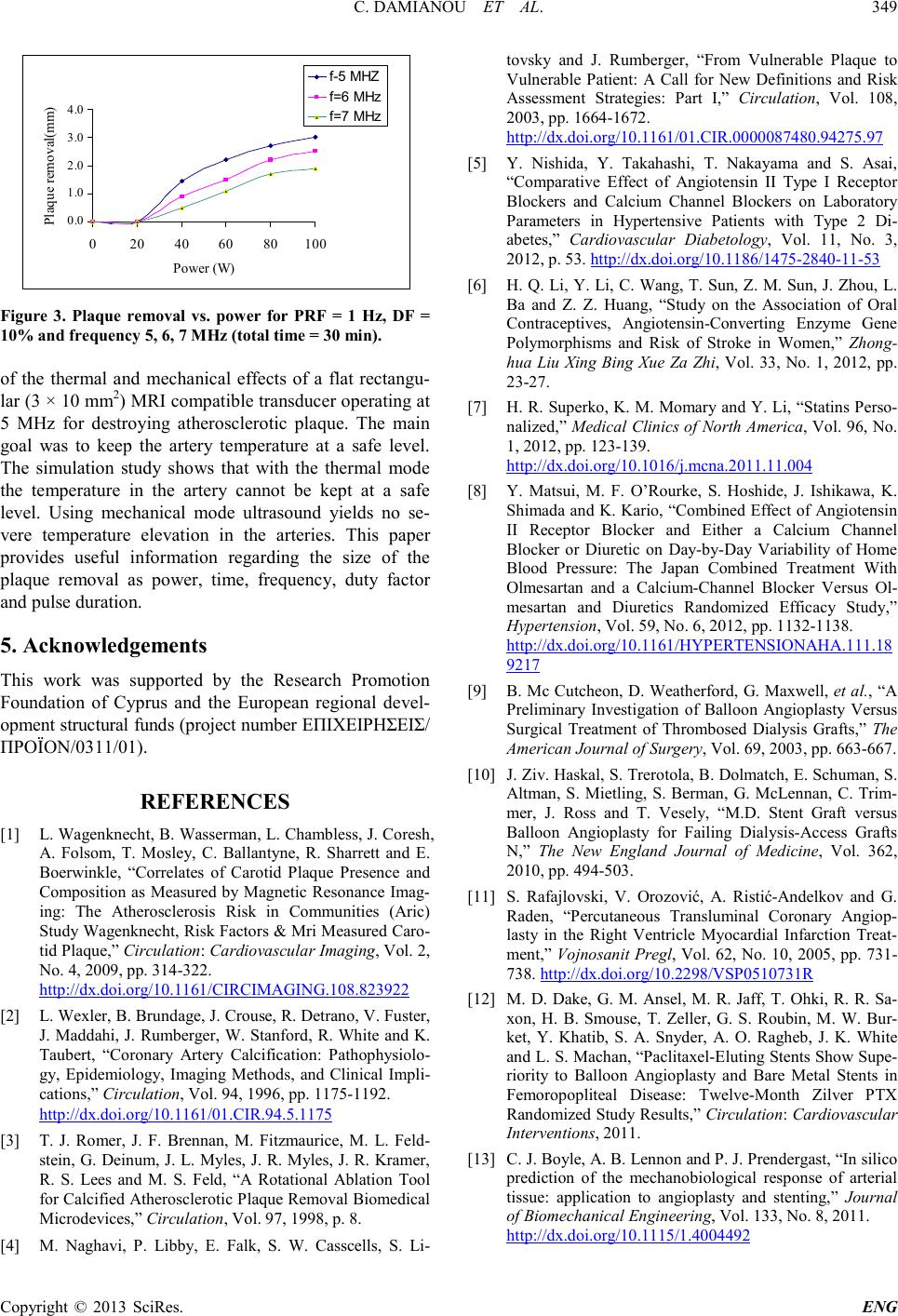
Engineering, 2013, 5, 347-351
http://dx.doi.org/10.4236/eng.2013.510B070 Published Online Octob er 2013 (http://www.scirp.org/journal/eng)
Copyright © 2013 SciRes. ENG
Simulation of the Thermal and Mechanical Effects of a
Planar Rectangular High Intensity Ultrasound Transducer
to Be Used for Destroying Atherosclerotic Plaque
Christakis Damianou1,2, C. Christofi2, N. Mylonas3
1Cyprus University of Technology, Limassol, Cyprus
2MEDSONIC, LTD, Limassol , C ypru s
3Frederick University Cyprus, Limassol, Cyprus
Email: christakis.damianou@cut.ac.cy
Received June 2013
ABSTRACT
The aim of this study is to pe rform a simulation study of the thermal and mechanical effects of a flat rectangular (3 × 10
mm2), MRI compatible transducer operating at 5 MHz for the purpose of destroying atherosclerotic plaque. The simula-
tion study focuses on measuring the plaque destruction (due to the thermal of mechanical mode of ultrasound) as a
function of power, time, frequency, duty factor and pulse duration. The main goal is to keep the artery temperature at a
safe level. T he simulation st udy shows that with the therma l mode the te mperature in the artery cannot be kept at a safe
level.
Keywords: Ultrasound; Atheroscle r o tic ; Plaque; MRI
1. Introduction
Atherosclerosis also known as arteriosclerotic vascular
disea se (ASVD) is a condition in which fatty material
collects along the walls of arteries. This fatty material
thickens, hardens (forms calcium deposits), and may
eventually block the arteries [1]. Calcium is a critical
component of atherosclerotic plaque. The absence of
calcification is strong evidence against the presence of
active disease even with significant luminal stenosis.
Calcification is reversible and may contribute to the for-
mation of an atheroma by adding the byproducts of re-
sorption to the necrotic core. The sequence of events in
plaque development following injury is inflammation,
followed by calcification of the damaged tissue, ending
ultimately in the formation of a necrotic core [2-4].
Lifestyle changes, such as follo wing a healthy diet and
exercising, are often the best treatment for atherosclero-
sis. But sometimes, medication or surgical procedures
may be recommended as well [5]. Over the years, re-
searchers have been involved in Clinical research to de-
velop medication treatments and approaches in order to
reduce the risk of heart attack and other medical prob-
lems caused by atherosclerosis (for example Angiotensin
II receptor blockers (ARBs) [5], Angiotensin-converting
enzyme (ACE)[6], Cholesterol Medications[7], Diuret-
ics[8]).
In advanced cases, atherosclerosis treatment may re-
quire special surgical procedures such as Balloon An-
gioplasty [9-12], Balloon Angioplasty and Stenting
[13-15], Cutting Balloon [16 -21], Atherectomy [22,23],
Surgical Bypass [24,25] and Endarterectomy [26,27] to
open an artery and improve blood flow.
Another treatment option could be the application of
mechanical waves such as ultrasound. With ultrasound
either the thermal or mechanical properties can be uti-
lized. Our group uses MRI to monitor ultrasonic proto-
cols and therefore, the ultrasonic transducer has to be
MRI compatible. In this paper a simulation study of the
thermal and mechanical effects of flat rectangular (3 × 10
mm2), MRI compatible ultrasonic transducer operating at
5 MHz for destroying atherosclerotic plaque is included.
The simulation study focuses on measuring the plaque
destruction (thermal of mechanical) as a function of
power, time, frequency, duty factor and pulse duration.
The main goal is to keep the artery temperature at a safe
level. This paper includes the design of the MRI com-
patible transducer.
2. Materials and Methods
2.1. Simulation Model
The power field was estimated using the KZK model
[28]. The temperature vs. time history was obtained by
C. DAMIANOU ET AL.
Copyright © 2013 SciRes. ENG
348
solvi ng t he bio-heat equation proposed by Pennes (1948)
numerically [29]. The explicit form of this equation is
given by:
T2
= kT+(T)+
pQ
cwc Ta
t bb
tp
t
(1)
where ρt i s t he de nsit y of the ti ssue, ct is the specific heat
of the tissue, T is the temperature of the tissue, t is the
time, wb is the blood perfusion rate, cb is the specific heat
of the blood, Ta is the arterial blood temperature, k is the
ther mal co nducti vity of t he tissue , and Qp is the ultrason-
ic power deposition rate.
2.2. Estimation of Thermal Dose
The effect of hyperthermia depends on the temperature
and the duration of the heating. If a constant temperature
could be maintained, then the duration of heating would
be a reasonable way of expressing thermal dose, with
units o f time. In reali ty, ho wever, a constant temperature
is not maintained, so it is necessary to find a method of
relating a treatment to an equivalent time at a specified
reference temperature. A mathematical relation between
time and temperature was described by Dewey [30] and
given by:
()
21
21
,
TT
tt
R
=
(2)
where T1, T2 are temperatures at times t1 and t2 respec-
tively, and R is a constant.
The calculation of the thermal dose for changing tem-
perature exposure was done by using the technique sug-
gested by Sapareto and Dewey [31]. The technique uses
numerical integration to calculate the time that would
give an equivalent thermal dose at a reference tempera-
ture under different temperature profiles. The reference
temperature of 43˚C has been chosen since this is the
standard temperature used as a reference [21]. For any
temperature profile the dose can be found by
(43 )
43 0
,
t
tfinal
T
t
tR
= ∆
(3)
where t43 is the eq uivale nt time at 4 3˚C, Tt is the average
temperature during t. The default value of R equal to
0.25 was chosen for temperatures smaller than 43˚C and
a value equal to 0.5 for temperatures higher than 43˚C
[31]. The temperature after the power turn OFF was also
considered since during the decay part thermal dose is
contributed.
2.3. Estimation of Lesion Size
The prediction of lesion size requires the knowledge of
the thermal dose threshold that causes 90% - 100% ne-
crosis. Previous studies [32,33] show that the threshold
thermal dosage reference at 43˚C for soft tissue is be-
tween 50 min and 240 min. Therefore, the extreme thre-
shold of dose necrosis of 240 min at 43˚C was consi-
dered.
3. Resul ts
Figure 1 sho ws the grap h of t her mal lesi on vs . power for
a 20 s sonication at the frequency of 5, 6, 7 MHz. There-
fore with 5 MHz and 3 W a plaque of 2 mm is destr o ye d.
With 7 MHz a power of 6 W must be used in order to
destroy 2 mm. Figure 2 shows the thermal lesion vs.
time for a power of 7 W at the frequency of 5, 6, 7 MHz.
Therefore with 5 MHz and 20 s a 4 mm plaque is de-
stroyed. With 7 MHz and 20 s a plaque of 2 mm is de-
st r oye d . The temperature in the artery for both Figure 1
and Figure 2 exceeded the safe level. Figure 3 sho ws the
graph of plaque removal vs. power for PRF = 1 Hz, DF =
10% and frequency 5, 6, 7 MHz (total time = 30 mins).
Therefore with 5 MHz and 60 W a plaque of 2 mm is
removed. With 7 MHz and 60 W a 1 mm plaque is re-
mo v e d . The temperature in the artery for the results of
Figure 3 never exceeded the safe level.
4. Conclusions
The aim of this paper was to conduct a simulation study
Figure 1. Thermal lesion vs. power for a 20 s sonication at
the frequency of 5, 6 , 7 MHz.
Figure 2. Thermal lesion vs. time f or a pow er of 7 W at the
frequency of 5, 6, 7 M Hz.
0 ,0
0 ,5
1 ,0
1 ,5
2 ,0
2 ,5
3 ,0
3 ,5
4 ,0
4 ,5
5 ,0
012345678
Power (W)
Lession (mm)
f-5 MHZ
f=6 MHz
f=7 MHz
0 .0
1 .0
2 .0
3 .0
4 .0
5 .0
6 .0
7 .0
8 .0
010 20 3040 50 60 70
time (s)
Lession (mm)
f-5 MHZ
f=6 MHz
f=7 MHz
C. DAMIANOU ET AL.
Copyright © 2013 SciRes. ENG
349
Figure 3. Plaque removal vs. power for PRF = 1 Hz, DF =
10% and frequency 5, 6, 7 MHz (total time = 30 min).
of the thermal and mechanical effects of a flat rectangu-
lar (3 × 10 mm2) MRI compatible transducer operating at
5 MHz for destroying atherosclerotic plaque. The main
goal was to keep the artery temperature at a safe level.
The simulation study shows that with the thermal mode
the temperature in the artery cannot be kept at a safe
level. Using mechanical mode ultrasound yields no se-
vere temperature elevation in the arteries. This paper
provides useful information regarding the size of the
plaque removal as power, time, frequency, duty factor
and pulse duration.
5. Acknowledgemen ts
This work was supported by the Research Promotion
Foundation of Cyprus and the European regional devel-
opment structural funds (project number ΕΠΙΧΕΙΡΗΣΕΙΣ/
ΠΡΟΪΟΝ/0311/01).
REFERENCES
[1] L. Wagenknecht, B. Wasserman, L. Chambless, J. Coresh,
A. Folsom, T. Mosley, C. Ballantyne , R. Sh arrett and E.
Boerwinkl e, “Correlates of Carotid Plaque Presence and
Composition as Measured by Magnetic Resonance Imag-
ing: The Atherosclerosis Risk in Communities (Aric)
Stud y Wagenknecht , Risk Fact or s & Mri Measu red Car o-
tid Plaqu e,” Circulation: Cardiovascular Imaging, Vol. 2,
No. 4, 2009, pp. 314-322.
http://dx.doi.org/10.1161/CIRCIMAGING.108.823922
[2] L. Wexler, B. Brundage, J. Crouse, R. Detrano, V. Fuster,
J. Maddahi, J. Rumberger, W. Stanford, R. White and K.
Taubert, “Coronary Artery Calcification: Pathophysiolo-
gy, Epidemiology, Imaging Methods, and Clinical Impli-
cations,” Circulation, V ol. 94, 1996, pp. 1175-1192.
http://dx.doi.org/10.1161/01.CIR.94.5.1175
[3] T. J. Romer, J. F. Brennan, M. Fitzmaurice, M. L. Feld-
stein, G. Deinu m, J. L. Myles, J. R. Myles, J. R. Kramer,
R. S. Lees and M. S. Feld, “A Rotational Ablation Tool
for Calci fied Athero sclerot ic P laque Removal Biomedical
Micro devices,” Circulation, Vol. 97, 1998, p. 8.
[4] M. Naghavi, P. Libby, E. Falk, S. W. Casscells, S. Li-
tovsky and J. Rumberger, “From Vulnerable Plaque to
Vulnerable Patient: A Call for New Definitions and Risk
Assessment Strategies: Part I,” Circulation, Vol. 108,
2003, pp. 1664-1672.
http://dx.doi.org/10.1161/01.CIR.0000087480.94275.97
[5] Y. Nishida, Y. Takahashi, T. Nakayama and S. Asai,
“Comparative Effect of Angiotensin II Type I Receptor
Blockers and Calcium Channel Blockers on Laboratory
Parameters in Hypertensive Patients with Type 2 Di-
abetes,” Cardiovascular Diabetology, Vol. 11, No. 3,
2012, p. 53. http://dx.doi.org/10.1186/1475-2840-11-53
[6] H. Q. Li, Y. Li, C. Wang, T. Sun, Z. M. Sun, J. Zhou, L.
Ba and Z. Z. Huang, “Study on the Association of Oral
Contraceptives, Angiotensin-Converting Enzyme Gene
Polymorphisms and Risk of Stroke in Women,” Zhong-
hua Liu Xing Bing Xue Za Zhi, Vol. 33, No. 1, 2012, pp.
23-27.
[7] H. R. Superko, K. M. M o mary and Y. Li, “Statins Perso-
nalized,” Medical Clinics of North America, Vol. 96, No.
1, 2012, pp. 123-139.
http://dx.doi.org/10.1016/j.mcna.2011.11.004
[8] Y. Matsui, M. F. O’Rourke, S. Hoshide, J. Ishikawa, K.
Shimada and K. Kario, “Combined Effect of Angiotensin
II Receptor Blocker and Either a Calcium Channel
Blocker or Diuretic on Day-by-Day Variability of Home
Blood Pressure: The Japan Combined Treatment With
Olmesartan and a Calcium-Channel Blocker Versus Ol-
mesartan and Diuretics Randomized Efficacy Study,”
Hypertension, Vol. 59, N o. 6, 2012, pp. 1132-1138.
http://dx.doi.org/10.1161/HYPERTENSIONAHA.111.18
9217
[9] B. Mc Cu tcheon, D. Weatherford, G. Maxwell, et al., “A
Preliminary Investigation of Balloon Angioplasty Versus
Surgical Treatment of Thrombosed Dialysis Grafts,” The
American Journal of Surgery, Vol. 69, 2003, pp. 663-667.
[10] J. Ziv. Haskal, S. Treroto la, B. Dolmatch, E. Schuman, S.
Alt man, S. Mietling, S. Berman, G. McLennan, C. Tri m-
mer, J. Ross and T. Vesely, M.D. Stent Graft versus
Balloon Angioplasty for Failing Dialysis-Access Grafts
N,” The New England Journal of Medicine, Vol. 362,
2010, pp. 494-503.
[11] S. Rafajlovski, V. Orozović, A. Ristić-Andelkov and G.
Raden, “Percutaneous Transluminal Coronary Angiop-
lasty in the Right Ventricle Myocardial Infarction Treat-
ment,” Vojnosanit Pregl, Vol. 62, No. 10, 2005, pp. 731-
738. http://dx.doi.org/10.2298/VSP0510731R
[12] M. D. Dake, G. M. Ansel, M. R. Jaff, T. Ohki, R. R. Sa-
xon, H. B. Smouse, T. Zeller, G. S. Roubin, M. W. Bur-
ket, Y. Khatib, S. A. Snyder, A. O. Ragheb, J. K. White
and L. S. Machan, “Paclitaxel -Elu ting Stents Show Supe-
riority to Balloon Angioplasty and Bare Metal Stents in
Femoropopliteal Disease: Twelve-Month Zilver PTX
Randomized Study Results,” Circulation: Cardiovascular
Interventions, 2011.
[13] C. J. Boyle, A. B. Lennon and P. J. Prendergast, “In silico
prediction of the mechanobiological response of arterial
tissue: application to angioplasty and stenting,” Journal
of Biomechanical Engineering, Vol. 133, No. 8, 2011.
http://dx.doi.org/10.1115/1.4004492
0 ,0
1 ,0
2 ,0
3 ,0
4 ,0
020 4060 80100
Power (W)
Plaque removal(mm)
f-5 MHZ
f=6 MHz
f=7 MHz
4.0
3.0
2.0
1.0
0.0
C. DAMIANOU ET AL.
Copyright © 2013 SciRes. ENG
350
[14] T. Abu-Tair, C. Martin and C. Kamp ma n n , “Acute Aortic
Dissection after Balloon Angioplasty of a Recoarctation
and Treatment by Stenting and Distal Membrane Fene-
stration in a Child ,” Heart, Vol. 97, No. 20, 2011, pp.
1699-1700.
http://dx.doi.org/10.1136/heartjnl-2011-300709
[15] H. Takebayashi, S. Haruta, H. Kohno, et al., “Immediate
and 3-Month Follow-Up Outcome after Cutting balLoon
Angioplasty for Bifurcation Lesions,” Journal of Inter-
ventional Cardiology, Vol. 17, 2004, pp. 1-7.
http://dx.doi.org/10.1111/j.1540-8183.2004.00246.x
[16] L. J. Bergers en, S. B. Perry and J. E. Lock, “Effect of
Cutting Balloon Angioplasty on Resistant Pulmonary Ar-
te ry S tenosis,” Ameri can Journal of Cardiology, Vol. 15,
2003, pp. 185-189.
http://dx.doi.org/10.1016/S0002-9149(02)03107-7
[17] J. F. Rhodes, G. K. Lane and C. I. Mesia, “Cutting Bal-
loon Angioplasty for Children with Small-Vessel Pulmo-
nary Artery Stenoses,” Circulation: Cardiovascular In-
terventions, Vol. 55, 2002, pp. 73-77.
http://dx.doi.org/10.1002/ccd.10031
[18] G. M. Ansel, N. S. Sample, C. F. Bo tti III Jr, et al ., “Cut-
ting Balloon Angioplasty of the Popliteal and Infrapopli-
teal Vessels for Symptomatic Limb Isc h e mi a ,” Circula-
tion: Cardiovascular Interventions, Vol. 61, 2004, pp. 1-4.
http://dx.doi.org/10.1002/ccd.10731
[19] C. Engelke, C. Sandhu, R. A. Morgan, et al., “Using
6-mm Cutting Balloon Angioplasty in Patients with Re-
sistant Peripheral Artery Stenosis: Preliminary Results,”
AJR, Vol. 179, 2002, pp. 619-623.
http://dx.doi.org/10.2214/ajr.179.3.1790619
[20] K. Kasirajan and P. A. Schneider, “Early Outcome of
“Cutting” Balloon Angioplasty For Infrainguinal Vein
Graft Stenosis,” Journal of Vascular Surgery, Vol. 39,
2004, pp. 702-708.
http://dx.doi.org/10.1016/j.jvs.2003.10.046
[21] R. D. Safian, C. L. Gri n e s, M. A. M ay, A. Licht enber g, N.
Juran, T. L. Schreiber, G. Pavlides, T. B. Meany, V. Sa-
vas and W. W. O’Neill, “Clinical and Angiographic Re-
sults of Transluminal Extraction Coronary Atherectomy
in Saphenous Vein Bypass Grafts,” Circulation, Vol. 89,
1994, pp . 302-312.
http://dx.doi.org/10.1161/01.CIR.89.1.302
[22] F. Mangiacapra, G. R. Heyndrickx, E. Puymirat, A. J.
Peace, W. Wijns, B. De Bruyne and E. Bar bato, “Com-
parison of drug-Eluting Versus Bare-Metal Stents after
Rotational Atherectomy for the Treatment of Calcified
Coronary Lesions,” International Journal of Cardiology,
Vol. 153, No. 3, 2012, pp. 373-376.
http://dx.doi.org/10.1016/j.ijcard.2011.11.048
[23] S. K. Forouzannia, M. H. Abdollahi, S. J. Mirhosseini, H.
Hosseini, S. H. Moshtaghion, A. Golzar, N. Naserzadeh,
S. M. Ghoraishian and T. Emami Meybodi, “Clinical
Outcome and Cost in Patients with Off-Pump vs. On-
Pump Coronary Artery Bypass Surgery,” Acta Medica
Iranica, Vol. 49, No. 7, 2011, pp. 414-419.
[24] E. J. Lee, K. H. Choi, J. S. Ryu, S. B. Jeon, S. W. Lee, S.
W. Park, S. J. Park, J. W. Lee, S. J. Choo, C. H. Chung, S.
H. Jung, D. W. Kang, J. S. Kim and S. U. Kwon, “Stroke
Risk after Coronary Artery Bypass Graft Surgery and
Extent of Cerebral Artery Atherosclerosis,” American
College of Cardiology Foundation, Vol. 57, No. 18, 2011,
pp. 1811-1818.
http://dx.doi.org/10.1016/j.jacc.2010.12.026
[25] J. T. McGinn Jr, M. A. Shariff, T. M. Bhat, B. Azab, W. J.
Molloy, E. Quat trocchi, M. Farid, A. M. Eichorn, Y. D.
Dlugacz and R. A. Silverman , Prevalence of Dysglyce-
mia among Coronary Artery Bypass Surgery Patients
with No Previous Diabetic History,” Journal of Cardio-
thoraci c Surgery, Vol. 6, 2011, p. 104.
http://dx.doi.org/10.1186/1749-8090-6-104
[26] A. Redzek, B. Mihajlović, P. Kovacević, N. C. Adić, K.
Pavlović, L. Vel icki, “Patency of internal thoracic artery
and vein grafts according to revascularized coronary ar-
tery properties,” Med Pregl., Vol. 64, No. 3-4, 2011, pp.
137-142. http://dx.doi.org/10.2298/MPNS1104137R
[27] A. Siani, F. Accrocca, L. M. Siani, R. Gabrielli, R. Anto-
nelli, G. A. Giordano and G. Marcucci, “Prosthetic Caro-
tid Bypass Graft for In-Stent Restenosis Performed for
Post-Endarterectomy Recurrent Stenosis: Technical De-
tails,” Giornale di Chirurgia, Vol. 33, No. 3, 2012, pp.
95-97.
[28] R. J. McDonald, H. J. Cloft and D. F. Kallmes, “Intracra-
nial Hemorrhage is Much More Common after Carotid
Stenting than after Endart erectomy: Evidence from the
National Inpatient Sample,” Stroke, Vol. 42, No. 10, 2011,
pp. 2782-7278.
http://dx.doi.org/10.1161/STROKEAHA.111.618769
[29] J. E. Soneson and M. R. Myers, “Gaussian rep resentati on
of High-Intensity Focused Ultrasound Beams,” Journal of
the Acou stical So ciety of America, Vol. 122, No. 5, 2007,
pp. 2526 -2531.
[30] M. Pennes, “Analysis of Tissue and Arterial Blood Tem-
perature in the Resting Human Forearm,” Journal of Ap-
plied Physics, Vol. 1, 1948, pp. 93-122.
[31] W. Dewey, L. Hopwood, S. Sap ar eto and L. Gerwecki,
“Cellular Responses to Combinations of Hyperthermia
and Radiation,” Radiology, Vol. 123, 1977, pp. 463-474.
[32] S. Sapareto and W. Dewey, “Thermal Dose Determina-
tion in Cancer Therapy,” International Journal of Radia-
tion Oncology Biology Physics, Vol. 10, 1984, pp. 787-
800. http://dx.doi.org/10.1016/0360-3016(84)90379-1
[33] W. Jansen and J. Haverman, “Histological Changes in the
Skin and Subcutaneous Tissues of Mouse Legs after
Treatment with Hyperthermia,” Pathology: Research and
Practice, Vol. 186, 1983, pp. 247-253.
http://dx.doi.org/10.1016/S0344-0338(11)80542-X