Open Journal of Gastroenterology, 2013, 3, 303-306 OJGas
http://dx.doi.org/10.4236/ojgas.2013.36052 Published Online October 2013 (http://www.scirp.org/journal/ojgas/)
When the skin meets the bowel: About a case*
Ganzetti Giulia1#, Campanati Anna1, Di Sario Antonio2, Rubini Corrado3, Zizzi Antonio3,
Tarantino Giuseppe2, Benedetti Antonio2, Offidani Annamaria1
1Dermatological Clinic, Department of Clinical and Molecular Medicine, Polytechnic Marche University, Ancona, Italy
2Gastroenterologic Clinic, Department of Clinical and Molecular Medicine, Polytechnic Marche University, Ancona, Italy
3Anatomic Pathology Institute, Department of Neurosciences, Polytechnic University of Marche, Ancona, Italy
Email: #giulia.ganzetti@alice.it
Received 23 July 2013; revised 28 August 2013; accepted 15 September 2013
Copyright © 2013 Ganzetti Giulia et al. This is an open access article distributed under the Creative Commons Attribution License,
which permits unrestricted use, distribution, and reproduction in any medium, provided the original work is properly cited.
ABSTRACT
Ulcerative colitis (UC) is a chronic relapsing inflam-
matory disease of the colorectal region which may be
complicated by extraintestinal manifestations, as cu-
taneous involvement. Immune dysregulation resulting
in a lymphocyte mediated destructive process has
been suggested as a possible pathogenetic link be-
tween gut and skin-related manifestations. The au-
thors describe an atypical clinical aspect of lichen ru-
ber planus in a patient affected by vitiligo, alopecia
areata and ulcerative colitis.
Keywords: Ulcerative Colitis; Vitiligo; Lichen Ruber
Planus; Alopecia Areata; Autoimmunity
1. INTRODUCTION
Ulcerative colitis (UC) is a chronic relapsing inflam-
matory disease of the colorectal region still remains to be
the unknown etiology [1].
Up to 40% of IBD patients may be complicated by
extraintestinal manifestations and their prevalence ap-
pears higher in Crohn disease than ulcerative colitis.
Among the organ systems involved, the skin is one of the
most commonly affected: up to a third of IBD patients
develops cutaneous manifestations, as erythema nodo-
sum, pyoderma gangrenosum, aphthous stomatitis, pso-
riasis, vitiligo, alopecia [1,2].
Vitiligo is an acquired disease affecting 0.2% - 1% of
the worldwide population and characterized by progres-
sive, patchy, multifocal loss of skin pigmentation, over-
lying hair and mucous membranes [3].
Lichen planus (LP) is a chronic inflammatory eruption
of the skin, scalp, nails, and mucous membranes with a
reported prevalence rates of 0.5% - 2.2%. Its classic pre-
sentation is characterized by 4 p’s, purple, polygonal,
pruritic papules [4,5].
Alopecia Areata (AA) is a chronic inflammatory hair
disease resulting in a Th1-mediated non-scarring hair
loss [2,11].
The authors describe the first case of concomitant li-
chen ruber planus, vitiligo and alopecia areata in a pa-
tient affected by ulcerative colitis.
2. CASE REPORT
A 50-year-old male patient was referred to our Derma-
tological Department for the clinical evaluation of mul-
tiple non itchy patches on the extensor surface of the
right lower limb. Same erythematous lesions were de-
tected on the right elbow (Figures 1(A) and (B)).
Lesions had started one year previously and progres-
sively increased in number. Physical examination de-
monstrated erythematous, finely scalying psoriasiform
eruption, localized on a vitiligo patch.
Patient’s anamnesis evidenced vitiligo since 1987,
ulcerative colitis for eight years; no personal nor familiar
history of skin diseases and further autoimmune diseases
were reported.
Ulcerative colitis was previously treated with pred-
nisone, beclomethasone dipropionate and metronidazole
ad it was in actual clinical remission: no specific inflam-
matory signs were found at endoscopy and laboratory
data, including CRP and faecal calprotectin, were within
normal ranges.
No previous treatments were done for vitiligo and pso-
riasiform lesions.
A skin biopsy was performed on one skin patch
showing a dermal band-like chronic inflammatory in-
filtrate damaging basal membrane, lymphocytic exo-
citosis and intraepithelial cytoid bodies. Histologic fea-
tures were compatible with lichen ruber planus (Figure
2(A) and (B)). Moreover, an oral examination showed a
*Authors declare no conflicts of interest.
*Corresponding a uthor.
OPEN ACCESS
G. Giulia et al. / Open Journal of Gastroenterology 3 (2013) 303-306
304
fine whityish network on the left cheek mucosa com-
patible with oral lichen planus (Figure 3(A)).
Based on the limited skin surface involved by vitiligo
and lichen ruber planus, we opted for a topical therapy
with clobetasol foam 0.05% daily for one month then
gradually tapered for another month with completely
resolution of both skin diseases.
After six weeks, the patient returned to our attention
for multiple non-scarring hair loss patched of the beard.
The pull test was positive and a video-dermatoscopic
analysis identified hyperkeratotic plugs (“yellow dots”),
hair follicles containing cadaverized hairs (“black dots”)
and broken hairs considered as pathognomonic features
of alopecia areata (Figures 3( B ) and (C)).
For alopecia areata, we suggested one month topical
treatment with methylprednisolone aceponate 0.1% five
days per week and a chili derivative product during the
week end, with partial clinical resolution of the lesions.
Figure 1. (A) Multiple erythematous, finely scalying patches on the extensor surface of the right lower limb localized on a viti-
ligo patch; (B) Erythematous, finely scalying plaque on the right elbow.
Figure 2. (A) Dermal band-like chronic inflammatory infiltrate damaging basal membrane, lymphocytic exocitosis and in-
traepithelial cytoid bodies; (B) A detail.
Figure 3. (A) Fine whityish network on the left cheek mucosa; (B) Multiple non-scarring hair loss patched of the beard; (C)
Video-dermatoscopic findings of an alopecic patch.
Copyright © 2013 SciRes. OPEN ACCESS
G. Giulia et al. / Open Journal of Gastroenterology 3 (2013) 303-306 305
3. DISCUSSION
Many gastrointestinal tract disorders show cutaneous
manifestations and several skin diseases may have asso-
ciated to gastrointestinal lesions. Thus, the skin could
mirror an occult gut disease [1,6]. Immune dysregulation
resulting in a lymphocyte mediated destructive process
have been suggested as a possible pathogenetic link be-
tween gut and skin-related manifestations [6].
Cytokines are important mediators of immunity and an
unbalance in the cytokine network may largely determine
autoimmune disease susceptibility and severity [7].
Classically, UC has been associated with Th2 media-
ted pathways with elevated levels of IL-5 and IL-13 [7].
However, recent evidence have suggested an impor-
tant role of IL-23 and Th17 pathways both in IBD and
vitiligo and lich en ruber planus; thus it could represent a
further critical “meeting” point between the skin and the
bowel [8-10] .
In UC IL-23 may play important roles in controlling
the differential Th1/Th17 balance [10].
In vitiligo, IL-17A seems to dramatically induce IL-1β,
IL-6, and TNF-α production in keratinocytes and fibro-
blasts providing evidence of the influence of a complex
Th17 cell-related cytokine environment in local depig-
mentation in addition to CD8(+) cell-mediated melano-
cyte destruction [9].
In oral lichen planus, the proportion of local and peri-
pheral Th1 and Th17 cells appears significantly increa-
sed and they may be associated with its pathogenesis [8].
Furthermore, alopecia areata is characterized by in-
creased serum levels of IL-2, IFN-γ, IL-13, IL-17A and it
emphasizes an altered T helper (Th1, Th2 and Th17) cell
function and a reduced serum TGF-β1 levels suggested
defect in Tregs function [11].
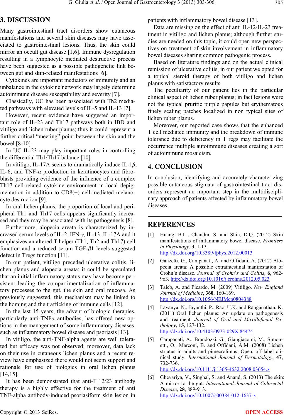
In our patient, vitiligo preceded ulcerative colitis, li-
chen planus and alopecia areata: it could be speculated
that an initial inflammatory statu s may have become per-
sistent leading the compartimentalization of inflamma-
tory processes to the gut, the skin and oral mucosa. As
previously suggested, this mechanism may be linked to
the homing and the trafficking of immune cells [12].
In the last 15 years, the advent of biologic therapies,
particularly anti-TNFα antibodies, has offered new op-
tions in the management of some inflammatory diseases,
such as inflammatory bowel disease and psoriasis [13].
In vitiligo, the anti-TNF-alpha agents are well tolera-
ted but efficacy was not observed; moreover, data lack
on their use in cutaneous lichen planus and a recent re-
view have emphasized there would not seem support and
rationale for use of biologics in oral lichen planus
[14,15].
It has been demonstrated that anti-IL12/23 antibody
therapy is a highly effective for the treatment of anti
TNF-alpha antibody-induced psoriasiform skin lesion in
patients with inflammatory bowel disease [13].
Data are missing on the effect of anti IL-12/IL-23 trea-
tment in vitiligo and lichen planus; although further stu-
dies are needed on this topic, it could open new perspec-
tives on treatment of skin involvement in inflammatory
bowel diseases sharing common pathogenic process.
Based on literature findings and on the actual clinical
remission of ulcerative colitis, in our patient we opted for
a topical steroid therapy of both vitiligo and lichen
planus with satisfactory results.
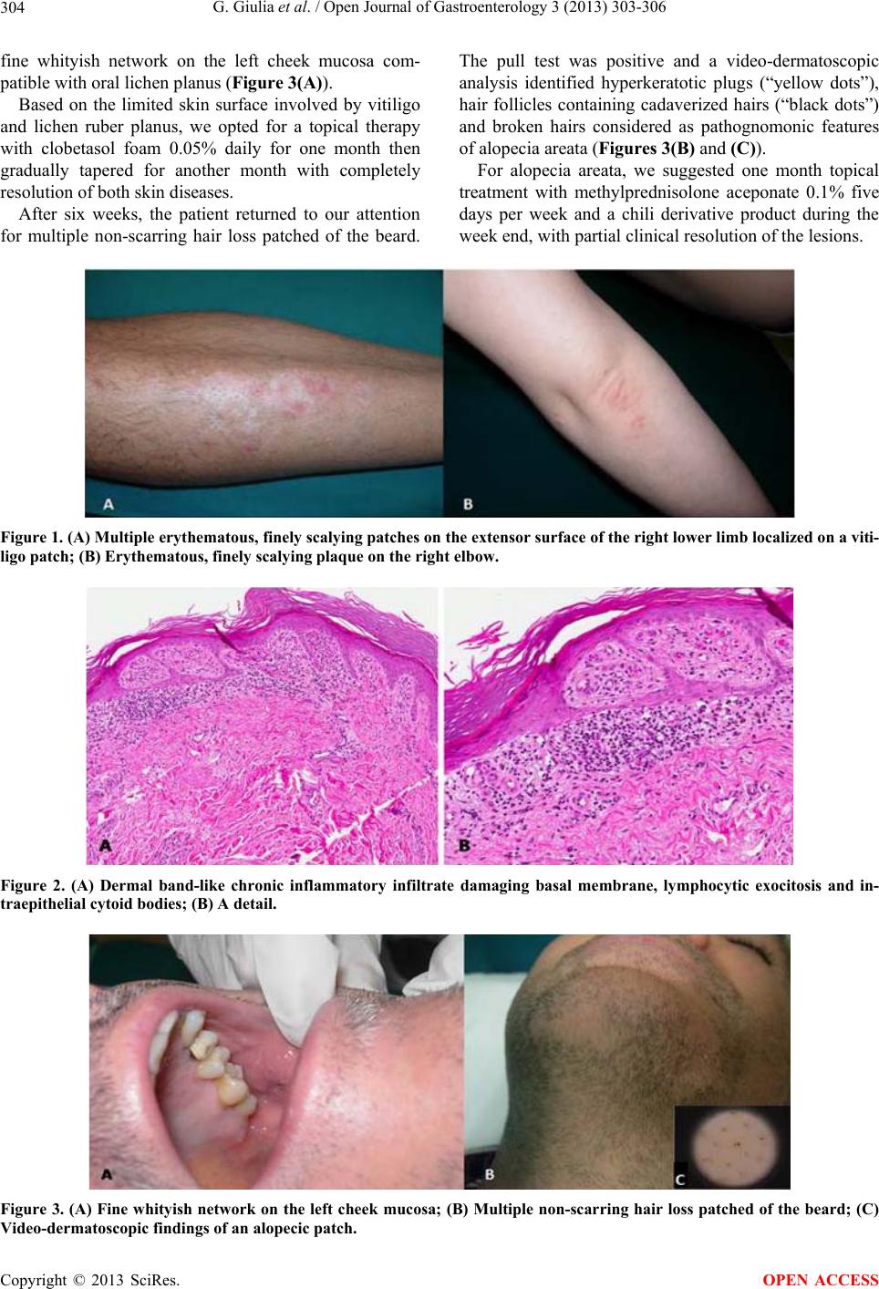
The peculiarity of our patient lies in the particular
clinical aspect of lichen ruber planus; in fact lesions were
not the typical pruritic purple papules but erythematous
finely scaling patches localized in non typical sites of
lichen ruber pl anu s .
Moreover, our reported case shows that the enhanced
T cell mediated immunity and the breakdown of immune
tolerance due to deficiency in T regs may facilitate the
occurrence multiple autoimmune diseases creating a sort
of autoimmune mosaicism.
4. CONCLUSION
In conclusion, identifying and accurately characterizing
possible cutaneous stigmata of gastrointestinal tract dis-
orders represent an important step in the multidiscipli-
nary approach of patients affected by inflammatory bowel
diseases.
REFERENCES
[1] Huang, B.L., Chandra, S. and Shih, D.Q. (2012) Skin
manifestations of inflammatory bowel disease. Frontiers
in Physiology, 3, 1-13.
http://dx.doi.org/10.3389/fphys.2012.00013
[2] Ganzetti, G., Campanati, A. and Offidani, A. (2012) Alo-
pecia areata: A possible extraintestinal manifestation of
Crohn’s disease. Journal of Crohns and Colitis, 6, 962-
963. http://dx.doi.org/10.1016/j.crohns.2012.05.023
[3] Taieb, A. and Picardo, M. (2009) Vitiligo. New England
Journal of Medicine, 360, 160-169.
http://dx.doi.org/10.1056/NEJMcp0804388
[4] Lavanya, N., Jayanthi, P., Rao, U.K. and Ranganathan, K.
(2011) Oral lichen planus: An update on pathogenesis
and treatment. Journal of Oral and Maxillofacial Pa-
thology, 15, 127-132.
http://dx.doi.org/10.4103/0973-029X.84474
[5] Campanati, A., Brandozzi, G., Giangiacomi, M., Simon-
etti, O., Marconi, B. and Offidani, A.M. (2008) Lichen
striatus in adults and pimecrolimus: Open, off-label cli-
nical study. International Journal of Dermatology, 47,
732-736.
http://dx.doi.org/10.1111/j.1365-4632.2008.03654.x
[6] Ghevariya, V., Singhal, S. and Anand, S. (2013) The skin:
A mirror to the gut. International Journal of Colorectal
Disease, 28, 889-913.
http://dx.doi.org/10.1007/s00384-012-1637-x
Copyright © 2013 SciRes. OPEN ACCESS
G. Giulia et al. / Open Journal of Gastroenterology 3 (2013) 303-306
306
[7] Van Wijk, F. and Cheroutre H. (2010) Mucosal T cells in
gut homeostasis and inflammation. Expert Review of
Clinical Immunology, 6, 559-566.
http://dx.doi.org/10.1586/eci.10.34
[8] Xie, S., Ding, L., Xiong, Z. and Zhu, S. (2012) Implica-
tions of Th1 and Th17 cells in pathogenesis of oral lichen
planus. Journal of Huazhong University of Science and
Technology-Medical Sciences, 32, 451-457.
http://dx.doi.org/10.1007/s11596-012-0078-7
[9] Kotobuki, Y., Tanemura, A., Yang, L., Itoi, S., et al.
(2012) Dysregulation of melanocyte function by Th17-
related cytokines: Significance of Th17 cell infiltration in
autoimmune vitiligo vulgaris. Pigment Cell & Melanoma
Research, 25, 219-230.
http://dx.doi.org/10.1111/j.1755-148X.2011.00945.x
[10] Kobayashi, T., Okamoto, S., Hisamatsu, T., et al. (2008)
IL23 differentially regulates the Th1/Th17 balance in ul-
cerative colitis and Crohn’s disease. Gut, 57, 1682-1689.
http://dx.doi.org/10.1136/gut.2007.135053
[11] Tembhre, M.K. and Sharma, V.K. (2013) T helper and
regulatory T cell cytokines in the peripheral blood of ac-
tive alopecia areata. British Journal of Dermatology, 169,
543-548. http://dx.doi.org /10.1111/bjd.12396
[12] Mann, E.R., McCa rthy, N.E., Peake, S.T. C., et al. (2012)
Skin- and gut-homing molecules on human circulating γδ
T cells and their dysregulation in inflammatory bowel
disease. Clinical & Experimental Immunology, 170 , 122-
130. h ttp://dx.doi.org /10.1111/j.1365-2249.2012.04649.x
[13] Tillack, C., Ehmann, L.M., Friedrich, M., et al. (2013)
Anti-TNF antibody-induced psoriasiform skin lesions in
patients with inflammatory bowel disease are character-
ised by interferon-γ-expressing Th1 cells and IL-17A/IL-
22-expressing Th17 cells and respond to anti-IL-12/IL-23
antibody treatmen t. Gut.
http://www.ncbi.nlm.nih.gov/pubmed/23468464
[14] Alghamdi, K.M., Khurrum, H., Taieb, A. and Ezzedine,
K. (2012) Treatment of generalized vitiligo with anti-
TNF-α Agents. Journal of Drugs in Dermatology, 11,
534-539.
[15] O’Neill, I.D. and Scully, C. (2013) Biologics in oral
medicine: Ulcerative disorders. Oral Diseases, 19, 37-45.
http://dx.doi.org/10.1111/j.1601-0825.2012.01931.x
Copyright © 2013 SciRes. OPEN ACCESS