Surgical Science, 2013, 4, 453-458
http://dx.doi.org/10.4236/ss.2013.410089 Published Online October 2013 (http://www.scirp.org/journal/ss)
Femoral Hernia: A Review of the Clinical Anatomy and
Surgical Treatment *
Makio Mike#, Nobuyasu Kano
Department of Surgery, Kameda Medical Center, Kamogawa, Japan
Email: #mmike@ace.ocn.ne.jp
Received September 2, 2013; revised October 1, 2013; accepted October 9, 2013
Copyright © 2013 Makio Mike, Nobuyasu Kano. This is an open access article distributed under the Creative Commons Attribution
License, which permits unrestricted use, distribution, and reproduction in any medium, provided the original work is properly cited.
ABSTRACT
Purpose: Femoral hernia is a kind of ventral hernia that surgeons commonly encounter, second in frequency only to
inguinal hernia. Femoral hernias often require emergency surgery because of incarceration or strangulation of the intes-
tine. In addition, intestinal resection may need to be considered based on intestinal viability. Definitive preoperative
diagnosis and strategic planning for surgery are thus important. The surgeon should consider the operation in the con-
text of the clinical anatomy of the abdominal cavity. Therefore the essence of the clinical anatomy and treatment of
femoral hernia is described. Methods: The medical records of 38 patients who underwent femoral hernia repair be-
tween March 2006 and November 2011 were retrospectively analyzed. Results: Femoral hernioplasty was performed
with original mesh repair or Ruggi’s repair plus iliopubic tract repair (or Bassini’s repair). The mean patient age was
76.7 years, and a female predominance was apparent. Twenty-four patients underwent emergency surgery with a diag-
nosis of in carcerated femoral hernia. Nine patien ts showed intestinal strangulation and underwent resection of the small
intestine. Four patients developed complications. One patient died due to aspiration pneumonia. No recurrences were
encountered after 6 months to 6 years of follow-up. Conclusion: Femoral hernia is an important surgical cond ition with
high rates of incarceration/strangulation and in testinal resection. Correct preoperative diagnosis of femoral hernia and a
strict operative strategy are important. The original mesh repair is effective and easy to perform.
Keywords: Femoral Hernia; Clinical Anatomy; Mesh Repair; Ruggi’s Repair
1. Introduction
Femoral hernia is a ventral hernia commonly encoun-
tered by surgeons in clinical p ractice, although less com-
mon than inguinal hernia. Femoral hernia often needs an
emergency operation because of incarceration or stran-
gulation [1]. In addition, in testinal resection may need to
be considered after taking into account the viability of
the intestine. A definitive preoperative diagnosis and
strategic plan for surgery are thus important. The choice
of operation should be considered based on the clinical
anatomy of the abdominal cavity. Since an understanding
of the anatomy of this region is comparable to an under-
standing of the basic factors behind the clinical anatomy
of the entire abdominal cavity, the essence of the clinical
anatomy and treatment of femoral hernia are described.
2. Anatomy of the Inguinal and
Femoral Region
The basic format of the peritoneal configuration of the
body walls in fetal life is shown in Figure 1. Basic inter-
pretations of the fascial composition of the bod y circum-
ference have been provided by Tobin et al. [2] and Satoh
[3]. According to these interpretations, the structure of
the body below the diaphragm can be simplified as a
straight intestine within a cylindrical body. The basis of
body composition can then be divided into the composi-
tion of the peritoneal cavity (composition in the cylinder)
and the composition of the body wall (composition of the
cylinder wall). In the former, the cranial abdomen in-
cludes the dorsal and ventral mesentery, while the caudal
abdomen includes only the dorsal mesentery involving
the intestine. The latter has a ringed composition, with
the body walls symmetrical in relation to the central po-
sition of the muscle layer (Figure 1). The trunk has typi-
cally been regarded as an onion-like, multi-layered
structure [4]. Corresponding to the subcutaneous superfi-
*Conflicts of interest: The authors declare that they have no potential
conflicts of interest, including specific financial interests, relationships
or affiliations relevant to the subject matter or materials discussed in
the manuscript.
#Corresponding author.
C
opyright © 2013 SciRes. SS
M. MIKE, N. KANO
454
’s
Meckel’s
Figure 1. Basic structures of the abdominal wall and em-
bryonic peritoneal cavity. The basis of body composition
can then be divided into the composition of the peritoneal
cavity and the composition of the body wall. The body walls
are symmetrical in relation to the central position of the
muscle layer.
cial fascia (Camper’s fascia) and subcutaneous deep fas-
cia (Scarpa’s fascia), deep subperitoneal fascia and su-
perficial subperitoneal fascia exist circumferentially
around the abdominal wa ll ( Fig u r e 1).
Corresponding to the innominate fascia as an outer
aponeurosis (investing fascia) along with the external
oblique aponeurosis, the transversalis fascia (investing
fascia) extends circumferentially around the abdominal
wall as an inner aponeurosis with the transversus ab-
dominis muscle. The transversalis fascia can th us be rec-
ognized as the endoabdominal fascia. The fascial struc-
tures present in the body wall are embryologically repre-
sented by the term superficial or deep, defined in relation
to the skin surface. The terminology used throughout the
body for anatomical terms should be used given the con-
tinuity of the abdominal cavity. It is also important to
understand the differences between the concepts of fascia
and muscle aponeurosis.
The orifice of the femoral hernia, called the femoral
ring, is bordered laterally by the femoral vein, anteriorly
by the iliopubic tract, and medially b y Cooper’s ligament
[5] (Figure 2). The iliopubic tract represents a th ickening
of the transversalis fascia, and the transversalis fascia
should extend to the femoral canal and further to Coo-
per’s ligament according to the concepts proposed by
Tobin et al. [2] and Satoh [3]. The transversalis fascia is
originally considered fragile, but this aponeurosis is
thought to become toughened by intertwining fibers from
the transversus abdominis aponeurosis and the internal
oblique apon eurosis. In the posterior wall of the inguinal
canal, three layers can reportedly be separated: the inter-
nal oblique aponeurosis; transversus abdominis aponeu-
rosis; and transversalis fascia [6].
3. Materials and Surgical Method
The majority of cases of femoral hernias involve elderly
women, but here, the more complex procedure for men is
described.
The start of surgery is the incision along the inguinal
ligament. The superficial and deep fasciae of the subcu-
taneous tissue are incised to reach the innominate fascia.
By peeling back the innominate fascia toward the oval
fossa, the underlying viscera are exposed, covering the
superior and deep subperitoneal fasciae, transversalis
fascia and peritoneum (the term sac represents all fasciae
for convenience) (Figure 3). Here, the inguinal canal is
exposed with an incision into the external oblique fascia,
and the spermatic cord is secured and taped in the region
of the pubic tubercle. An incision is made from the in-
ternal inguinal ring to the pubic tub ercle. The neck of the
sac should be seen wrapped in the superficial and deep
subperitoneal fasciae and the peritoneum (Figure 4).
Here, the sac is dissected from the iliopubic tract and
Cooper’s ligament. Lateral d issection is not performed at
this point. The transv ersalis fascia is incised between the
iliopub i c tract and Cooper’s ligament usin g Kelly forceps,
and the femoral ring takes on a triangular shape (Figure
Cooper’s
Figure 2. Anatomy around the femoral ring. The femoral
ring is structured with the iliopubic tract, Cooper’s liga-
ment and the medial border of the external iliac vein.
Figure 3. Innominate fascia and fascia lata. The innominate
fascia is dissected caudally from the groin. A hernial sac can
be confirmed covered with superficial and deep subperito-
neal fascia and transverse fascia.
Copyright © 2013 SciRes. SS
M. MIKE, N. KANO 455
Cooper’s
Figure 4. Cross-section of the cranial and caudal sides of the
(a), arrow). Opening the superficial subperitoneal fascia
o strangula-
tio
fascia. An incision is made into the posterior wall of the
inguinal canal from the internal ring to the pubic tubercle.
The neck should be seen as a sac wr apped in superficial and
deep subperitoneal fascia. The relationship between the
fascia is shown in the cranial (A) and caudal (B) portions of
the inguinal ligament.
5
introduces a space between the superficial and deep sub-
peritoneal fasciae on the medioventral side, and dissec-
tion of this space allows the neck of the sac to be taped
(Figure 5). Fatty tissue is present in the space between
the superficial and deep fasciae, and the space is rela-
tively loose to dissect, allowing easy taping of the neck
of the sac. In this taping, blun t-tipped forceps with a long,
strong curvature are convenient to use. Holding a Nela-
ton’s catheter or cotton tape through the neck of the sac,
reduction of the sac to the abdominal side can be at-
tempted. Alternatively, reduction of the sac to the ab-
dominal side can be tried by compressing the sac at the
oval fossa. If reduction of the sac to the abdominal side is
impossible, incision of the deep subperitoneal fascia and
peritoneum may be needed to open the sac at the superior
portion of the iliopubic tract and confirm the contents. If
peeling the sac at the oval fossa, five sheaths from the
ventral side can also be dissected to reach the real sac
(actual sac) as shown in Figure 4(b). A femoral hernia is
not incarcerated or strangulated at the femoral ring, but is
considered to be incarcerated and strangulated with
stenosis of the sac itself. The sac thus needs to be opened
and the constricted area eventually released.
In cases of suspected bowel necrosis due t
n of the intestine, the affected seg ment of the intestine
is not resected immediately. Replacing the segment back
into the peritoneal cavity for 10 min with support from
Figure 5. Dilatation of the femoral ring and method for
e seromuscular sutur es of the intestine near good blood
emoral hernia
is
was contaminated, Ruggi’s repair [10,11]
an
taping the neck of the sac. The femoral ring is dilated with
an incision in the transversalis fascia (A). The superior
subperitoneal fascia is opened on the medioventral side, and
dissection is performed between the superficial and deep
subperitoneal fasciae from mediodorsal to laterodorsal. The
neck of the sac can then be taped by performing the same
procedure from ventral to lateral.
th
flow is preferable, and improvements can be obtained by
paying attention to th e color of the bowel.
The basic concept of surgical repair for f
to cover the femoral ring and groin area with mesh [6].
Thus, a 7.6-cm × 15-cm lightweight mesh (Prolene
Soft®; ETHICON, Inc., Somerville, NJ, USA) is used.
First the mesh is sutured to Cooper’s ligament and folded
to overlap the pubic tubercle by about 1.5 cm and to en-
sure sufficient coverage of the supravesical hernia [7]
(Figure 6(a)). The return line of the mesh is then sutured
to the iliopubic tract or to the shelving portion of the in-
guinal ligament (Figure 6(b)). After folding the mesh,
the posterior wall of the inguinal canal is covered as in
Lichtenstein’s repair, providing posterior support (Fig-
ure 6(c)) [8,9].
If the surgery
d anterior iliopubic tract repair [12] are performed. In
other words, 1-0 nylon sutures are added between Coo-
per’s ligament and the ilio pubic tract or the shelv ing por-
tion of the inguinal ligament from the pubic tubercle to
close the femoral ring (Figure 7(a)), and then anterior
iliopubic tract repair is performed as posterior reinforce-
ment of the inguinal canal (Figure 7(b) ). However, since
excessive femoral ring closure has been reported to lead
to the obstruction of the external iliac vein, care is re-
quired.
Copyright © 2013 SciRes. SS
M. MIKE, N. KANO
456
Cooper’s
Figure 6. Femoral hernia repair in clean operation. (a) The
narrow side of the mesh is sutured to Cooper’s ligament; (b)
The mesh is sutured to the iliopubic tract or shelving por-
tion of the inguinal ligament; (c) The posterior wall of the
inguinal canal is reinforced, as in Lichtenstein’s repair.
Cooper’s
Cooper’s
Figure 7. Femoral hernia repair in dirty operation. (a)
tract repair.
ch 2006 and November 2011, a total of 38
ssion
thought to represent about 2% - 4% of
ad support against pressure based on Pascal’s
Ruggi’s repair: sutures between Cooper’s ligament and the
shelving portion of the inguinal ligament or the iliopubic
tract from the inside; (b) After the above anterior iliopubic
4. Results
Between Mar
patients with femoral hernia underwent surgery using an
inguinal approach in our institution. These patients in-
cluded 29 women (76.3%) and 9 men (23.7%), with a
mean age of 76.7 years (range, 53 - 99 years). Primary
hernias accounted for 97 .4% of cases (37 of 38), whereas
only 2.6% of cases (1 of 38) was con sidered to represent
recurrent femoral hernia after a femoral approach.
Twenty-four patients (63.2%) underwent emergency sur-
gery with a diagnosis of incarceration or strangulation of
femoral hernia. Nine patients showed strangulation and
underwent resection of the small intestine. All resections
were performed through the inguinal incision. First in-
traoperative inspection showed strangulated intestines
with color change evident in the wall, changing to a vi-
able coloration in 4 patients after abo ut 10 - 15 min in the
abdominal cavity. All patients underwent definitive sur-
gery. Mesh repair was used in 30 patients (78.9%), while
8 patients (21.1%) were treated using Ruggi’s repair with
iliopubi c tract re pai r or with Bassi ni ’s repair.
Four patients developed complications (3 patients as
emergencies). Two cases of complications were related
to wound infection, and one involved peritonitis due to
delayed perforation of the intestine that required re-op-
eration with intestinal resection. Another had massive
bloody ascitic fluid because of an impending rupture of
an abdominal aortic aneurysm at the hernia operation and
died due to aspiration pneumonia 1 month 5 days after
operation. No recurrences were detected after 6 months
to 6 years follow-up (average follow-up period, 9.7
months).
5. Discu
Femoral hernia is
inguino-femoral hernias [1,13-16], but it is often over-
looked during inguinal hernia repair [15]. Many reports
have described intraoperative identification of femoral
hernia after preoperative misdiagnosis [17]. Accurate
preoperative diagnosis of the hernia is important for
achieving optimal surgical results.
The anatomy of femoral hernia is often stated incor-
rectly, and the terminology of the surrounding area for
these hernias has not been strictly defined. An under-
standing restricted to the local anatomy of the inguinal
and femoral regions is thus a limitation. Understanding
the basic fascial composition of the body circumference
as described by Tobin et al. [2] and Sato [3] clarifies th e
inguinal and femoral anatomy. As a matter of course, the
pathologies involving this area also become more under-
standable.
For surgical procedures, hernia repair uses the princi-
ple of “bro
Copyright © 2013 SciRes. SS
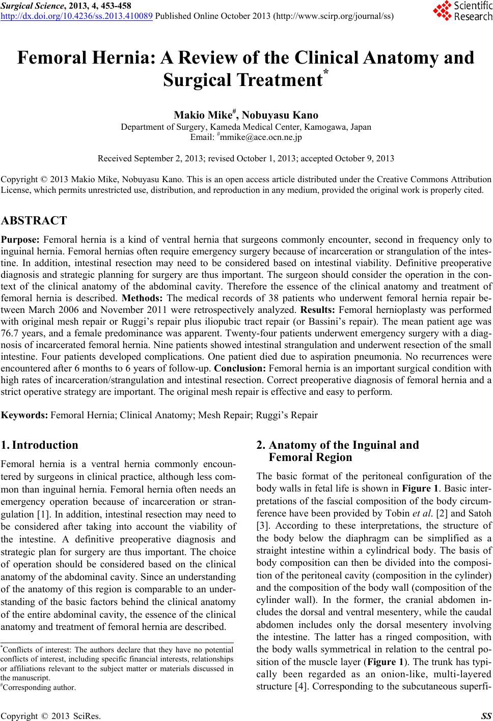
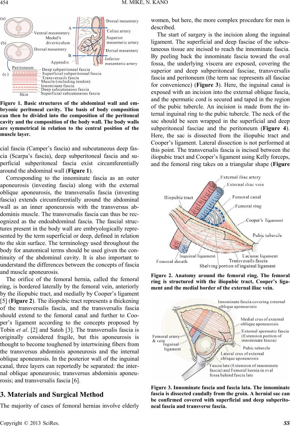
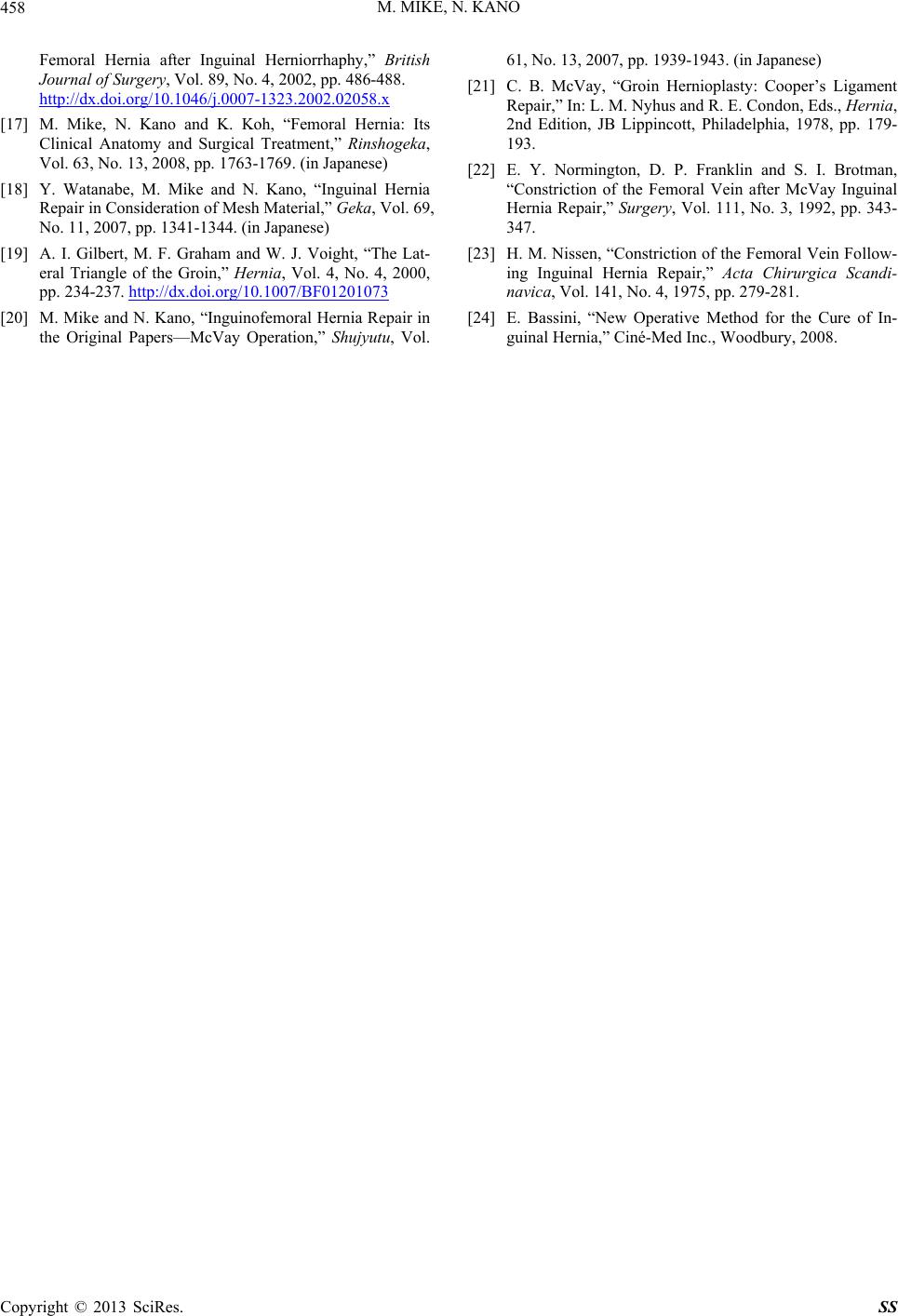
M. MIKE, N. KANO 457
la
com-
m
e preoperative diagnosis
an
an important surgical pathology with
rceration/strangulation and intestin
[1] U. Dahlstrand. Sandblom and U.
Gunnarsson, Hernia Repair. A
w and minimal biological response”. We therefore used
a lightweight mesh [18]. The mesh is required to cover
the femoral ring, pubic tubercle, area of the supravesical
hernia and the lateral triangle of the inguinal canal,
which is an area that includes the internal inguinal ring
and the tissues immediately la teral to it [19]. Our original
procedure with mesh is simple and reliable for treating
femoral hernia because of the certain closure of the
femoral ring with mesh sheet and of the reinforcement of
the inguinal floor, as in Lichtenstein’s repair [8,9].
Furthermore, if surgery is contaminated, use of foreign
material should be avoided. Many reports have re
ended McVay’s repair for such situations [20,21]. Ac-
cording to the intent of the original transition suture, the
manipulation closes the angle between Cooper’s liga-
ment and the iliopubic tract and prevents protrusion
through the femoral ring. However, stenotic complica-
tions can be caused by pressure to the femoral vein using
this maneuver [22,23], so the procedure requires close
attention. We adopted Ruggi’s rep air to close the femoral
ring, which includes intermittent sutures between Coo-
per’s ligament and the inguinal ligament [10,11], plus
anterior iliopubic tract repair [12] as a non-mesh repair.
Iliopubic tract repair is a reinforcement of the inguinal
floor with sutures added between the transversus ab-
dominal arch and the iliopubic tract. However, many
elderly individuals already show a weakened aponeurosis
fascia, including the transversus abdominis aponeurosis,
so Ruggi’s repair plus Bassini’s repair [24] is recom-
mended. Bassini’s repair is the method of reinforcement
of the inguinal floor with sutures between three layers
(internal oblique muscle, transversus abdominal muscle
and transverse fascia) and the inguinal ligament. In
Ruggi’s repair, temporary sutures should be used for the
outermost suture ligation to ensure that the suture does
not compress the femoral vein.
A unified strategy for the treatment of femoral hernia
is needed and requires accurat
d surgical techniques based on clinical anatomy.
6. Conclusion
Femoral hernia is
high rates of inca
al
resection. A correct preoperative diagnosis of femoral
hernia and a strict operative strategy are important. The
original mesh repair is effective and easy to perform.
REFERENCES
, S. Wollert, P. Nordin, G
“Emergency Femoral
Study Based on a National Register,” Annals of Surgery,
Vol. 249, No. 4, 2009, pp. 672-676.
http://dx.doi.org/10.1097/SLA.0b013e31819ed943
[2] C. E. Tobin, J. A. Benjamin and J. C. Wells, “Continuity
of the Fascia Lining the Abdomen, Pe
Cord,” Surgery, Gynecology & Obstetrics, Vol. 83, No. 5,
lvis, and Spermatic
nese)
Space,” International Journal
1946, pp. 575-596.
[3] T. Sato, “Fundamental Plan of the Fascial Strata of the
Body Wall,” Igakunoayumi, Vol. 114, No. 13, 1980, pp.
C168-C175. (in Japa
[4] M. Mike and N. Kano, “Laparosc opic-Assisted Low Ante-
rior Resection of the Rectum—A Review of the Fascial
Composition in the Pelvic
of Colorectal Disease, Vol. 26, No. 4, 2011, pp. 405-414.
http://dx.doi.org/10.1007/s00384-010-1107-2
[5] T. H. Quinn, “Anatomy of the Groin: A View from the
Anatomistm,” In: L. M. Nyhus and R. E. Condon, Eds.,
Hernia, 5th Edition, Lippincott Williams & Wilkins,
ds., Abdominal Wall Her-
as, Springer-
Philadelphia, 2002, pp. 55-70.
[6] R. Bendavid, “The Transversalis Fascia: New Observa-
tions,” In: R. Bendavid, J. Abrahamson, M. E. Arregui, J.
B. Flament and E. H. Phillips, E
nia, Springer, New York, 2000, pp. 97-100.
[7] M. S. Kavic, “Chronic Pelvic Pain in Women,” In: R.
Bendavid, J. Abrahamson, M. Arregui, J. B. Flament and
E. H. Phillips, Eds., Abdominal Wall Herni
Verlag, New York, 2001, pp. 632-638.
http://dx.doi.org/10.1007/978-1-4419-8574-3_94
[8] I. L. Lichtenstein, A. G. Shulman, P. K. Amid and M. M.
Montllor, “The Tension-Free Herniopla
can Journal of Surgery, Vol. 157, No. 2, 1989, p
sty,” The Ameri-
p. 188-
193. http://dx.doi.org/10.1016/0002-9610(89)90526-6
[9] P. K. Amid, A. G. Shulman and I. L. Lichtenstein, “Criti-
cal Scrutiny of the Open “Tension-Free” Hernioplasty,”
The American Journal of Surgery, Vol. 165, No. 3, 1993,
pp. 369-371.
http://dx.doi.org/10.1016/S0002-9610(05)80847-5
[10] A. V. Moschcowitz, “Femoral Hernia: A New Operation
for Radical Cur
10, 1907, p. 396.
e,” New York Medical Journal, Vol. 7, No.
.
, 1978, pp. 195-211.
in Denmark: A
[11] G. Ruggi, “Metado Operativo Meovo per la Cure Radicale
Dell’Ernia Crurale,” Bull Sci Med Bologna, Vol. 7, No. 3,
1892, pp. 223-229
[12] R. E. Condon, “Anterior Iliopubic Tract Repair,” In: L. M.
Nyhus and R. E. Condon, Eds., Hernia, 3rd Edition, Lip-
pincott, Philadelphia
[13] M. Bay-Nielsen, H. Kehlet, L. Strand, J. Malmstrøm, F.
H. Andersen, P. Wara, P. Juul and T. Callesen, “Quality
Assessment of 26,304 Herniorrhaphies
Prospective Nationwide Study,” Lancet, Vol. 358, No.
9288, 2001, pp. 1124-1128.
http://dx.doi.org/10.1016/S0140-6736(01)06251-1
[14] G. Sandblom, S. Haapaniemi and E. Nilsson, “Femoral
Hernias: A Register Analysis
Vol. 3, No. 3, 1999, pp. 131-134.
of 588 Repairs,” Hernia,
http://dx.doi.org/10.1007/BF01195312
[15] F. Glassow, “Femoral Hernia: Review of 2105 Repairs in
a 17 Year Period,” The American Journal of Surgery
150, No. 3, 1985, pp. 353-356. , Vol.
http://dx.doi.org/10.1016/0002-9610(85)90077-7
[16] T. Mikkelsen, M. Bay-Nielsen and H. Kehlet, “Risk of
Copyright © 2013 SciRes. SS
M. MIKE, N. KANO
Copyright © 2013 SciRes. SS
458
Herniorrhaphy,” British
Femoral Hernia after Inguinal
Journal of Surgery, Vol. 89, No. 4, 2002, pp. 486-488.
http://dx.doi.org/10.1046/j.0007-1323.2002.02058.x
[17] M. Mike, N. Kano and K. Koh, “Femoral Hernia: Its
Clinical Anatomy and Surgical Treatment,” Rinshogeka
Vol. 63, No. 13, 2008, pp. 1763-1769. (in Japanese) ,
[18] Y. Watanabe, M. Mike and N. Kano, “Inguinal Hernia
Repair in Consideration of Mesh Material,” Geka, Vol. 69,
No. 11, 2007, pp. 1341-1344. (in Japanese)
[19] A. I. Gilbert, M. F. Graham and W. J. Voight, “The Lat-
eral Triangle of the Groin,” Hernia, Vol. 4, No. 4, 2000,
pp. 234-237. http://dx.doi.org/10.1007/BF01201073
Ligament
Normington, D. P. Franklin and S. I. Brotman,
. Nissen, “Constriction of the Femoral Vein Follow-
re of In- [20] M. Mike and N. Kano, “Inguinofemoral Hernia Repair in
the Original Papers—McVay Operation,” Shujyutu, Vol.
61, No. 13, 2007, pp. 1939-1943. (in Japanese)
[21] C. B. McVay, “Groin Hernioplasty: Cooper’s
Repair,” In: L. M. Nyhus and R. E. Condon, Eds., Hernia,
2nd Edition, JB Lippincott, Philadelphia, 1978, pp. 179-
193.
[22] E. Y.
“Constriction of the Femoral Vein after McVay Inguinal
Hernia Repair,” Surgery, Vol. 111, No. 3, 1992, pp. 343-
347.
[23] H. M
ing Inguinal Hernia Repair,” Acta Chirurgica Scandi-
navica, Vol. 141, No. 4, 1975, pp. 279-281.
[24] E. Bassini, “New Operative Method for the Cu
guinal Hernia,” Ciné-Med Inc., Woodbury, 2008.