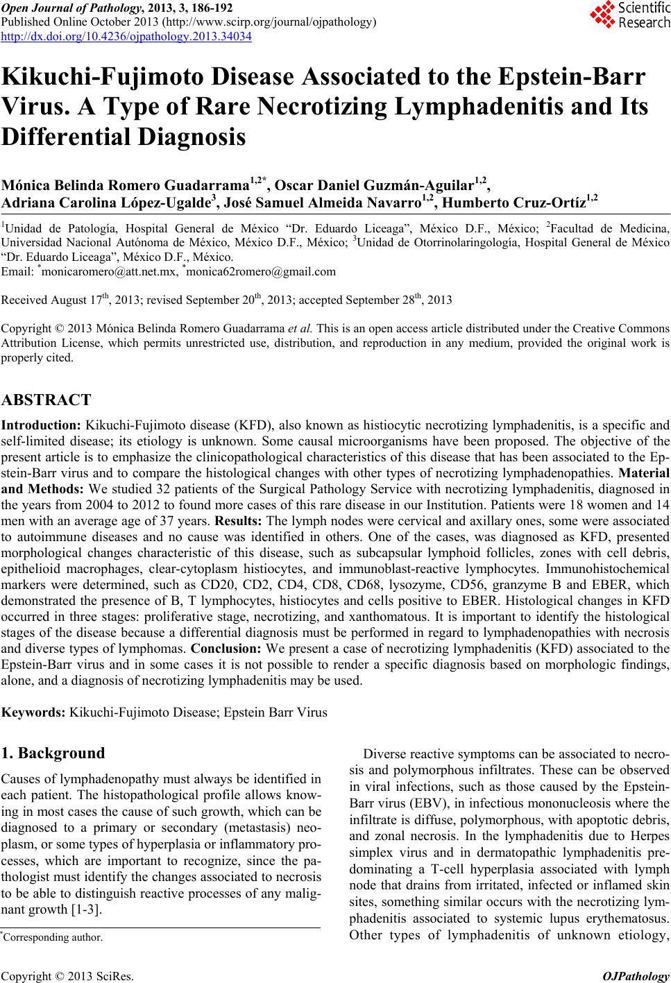
 Open Journal of Pathology, 2013, 3, 186-192 Published Online October 2013 (http://www.scirp.org/journal/ojpathology) http://dx.doi.org/10.4236/ojpathology.2013.34034 Copyright © 2013 SciRes. OJPathology Kikuchi-Fujimoto Disease Associated to th e Epstein-Barr Virus. A Type of Rare Necrotizing Lymphadenitis and Its Differential Diagnosis Mónica Belinda Romero Guadarrama1,2*, Oscar Daniel Guzmán-Aguilar1,2, Adriana Carolina López-Ugalde3, José Samuel Almeida Navarro1,2, Humberto Cruz-Ortíz1,2 1Unidad de Patología, Hospital General de México “Dr. Eduardo Liceaga”, México D.F., México; 2Facultad de Medicina, Universidad Nacional Autónoma de México, México D.F., México; 3Unidad de Otorrinolaringología, Hospital General de México “Dr. Eduardo Liceaga”, México D.F., México. Email: *monicaromero@att.net.mx, *monica62romero@gmail.com Received August 17th, 2013; revised September 20th, 2013; accepted September 28th, 2013 Copyright © 2013 Mónica Belinda Romero Guadarrama et al. This is an open access article distributed under the Creative Commons Attribution License, which permits unrestricted use, distribution, and reproduction in any medium, provided the original work is properly cited. ABSTRACT Introduction: Kikuchi-Fujimoto disease (KFD), also known as histiocytic necrotizing lymphadenitis, is a specific and self-limited disease; its etiology is unknown. Some causal microorganisms have been proposed. The objective of the present article is to emphasize th e clinicopathological characteristics of this disease that has been associated to the Ep- stein-Barr virus and to compare the histological changes with other types of necrotizing lymphadenopathies. Material and Methods: We studied 32 patients of the Surgical Pathology Service with necrotizing lymphadenitis, diagnosed in the years from 2004 to 2012 to found more cases of this rare disease in our Institutio n. Patients were 18 women and 14 men with an average age of 37 years. Results: The lymph nodes were cervical and axillary ones, so me were associated to autoimmune diseases and no cause was identified in others. One of the cases, was diagnosed as KFD, presented morphological changes characteristic of this disease, such as subcapsular lymphoid follicles, zones with cell debris, epithelioid macrophages, clear-cytoplasm histiocytes, and immunoblast-reactive lymphocytes. Immunohistochemical markers were determined, such as CD20, CD2, CD4, CD8, CD68, lysozyme, CD56, granzyme B and EBER, which demonstrated the presence of B, T lymphocytes, histiocytes and cells positive to EBER. Histological changes in KFD occurred in three stages: proliferative stage, necrotizing, and xanthomatous. It is important to identify the histological stages of the disease because a differential diagnosis must be performed in regard to lymphadenopathies with necrosis and diverse types of lymphomas. Conclusion: We present a case of necrotizing lymphadenitis (KFD) associated to the Epstein-Barr virus and in some cases it is not possible to render a specific diagnosis based on morphologic findings, alone, and a diagno sis of necrotizin g lymphadenitis may be used. Keywords: Kikuchi-Fujimoto Disease; Epstein Barr Virus 1. Background Causes of lymphadenopath y must always be identified in each patient. The histopathological profile allows know- ing in most cases the cause of such growth, which can be diagnosed to a primary or secondary (metastasis) neo- plasm, or some types of hyperplasia or inflammatory pro- cesses, which are important to recognize, since the pa- thologist must iden tify the changes associated to necrosis to be able to distinguish reactive processes of any malig- nant growth [1 -3] . Diverse reactive symptoms can be associated to necro- sis and polymorphous infiltrates. These can be observed in viral infections, such as those caused by the Epstein- Barr virus (EBV), in infectious mononucleosis where the infiltrate is diffuse, polymorph ous, with apoptotic debris, and zonal necrosis. In the lymphadenitis due to Herpes simplex virus and in dermatopathic lymphadenitis pre- dominating a T-cell hyperplasia associated with lymph node that drains from irritated, infected or inflamed skin sites, something similar occurs with the necrotizing lym- phadenitis associated to systemic lupus erythematosus. Other types of lymphadenitis of unknown etiology, *Corresponding author. 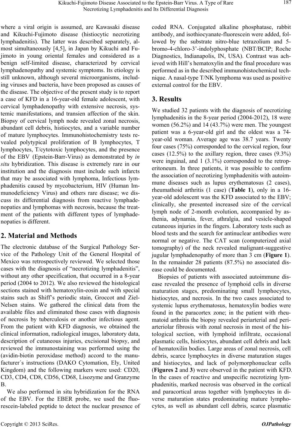 Kikuchi-Fujimoto Disease Associated to the Epstein-Barr Virus. A Type of Rare Necrotizing Lymphadenitis and Its Differential Diagnosis 187 where a viral origin is assumed, are Kawasaki disease and Kikuchi-Fujimoto disease (histiocytic necrotizing lymphadenitis). The latter was described separately, al- most simultaneously [4,5], in Japan by Kikuchi and Fu- jimoto in young oriental females and considered as a benign self-limited disease, characterized by cervical lymphadenopathy and systemic symptoms. Its etiology is still unknown, although several microorganisms, includ- ing viruses and bacteria, have been proposed as causes of the disease. The objectiv e of the presen t study is to repor t a case of KFD in a 16-year-old female adolescent, with cervical lymphadenopathy with extensive necrosis, sys- temic manifestations, and transien affection of the skin. Biopsy of cervical lymph node revealed zonal necrosis, abundant cell debris, histiocytes, and a variable number of mature lymphocytes. Immunohistochemistry tests re- vealed polytypical proliferation of B lymphocytes, T lymphocytes, T/cytotoxic lymphocytes, and the presence of the EBV (Epstein-Barr-Virus) as demonstrated by in situ hybridization. This disease is extremely rare in our institution and the diagnosis must include such infarcts that may be associated with lymphoma, Infectious lym- phadenitis caused by mycobacterium, HIV (Human Im- munodeficiency Virus) and others rare disease; we dis- cuss its differential diagnosis from reactive lymphade- nopaties and lymphomas with necrosis, because the treat- ment of the patients with different types of lymphade- nopaties is different. 2. Material and Methods The electronic database of the Surgical Pathology Ser- vice of the Pathology Unit of the General Hospital of Mexico was retrospectively reviewed. We selected those cases with the diagnosis of “necrotizing lymphadenitis”, without any other specification, that occurred in a 8-year period (2004 to 2012). We also reviewed the histological sections stained with hematoxylin-eosin and with special stains such as Shiff’s periodic stain, Groccot and Ziel- Nelsen stains. We gathered the clinical data from the available files and eliminated those cases with diagnosis of necrosis by tuberculosis or another infectious agent. From the patient with KFD diagnosis, we obtained the clinical information, radiological images, laboratory data, description of cutaneous injuries, excisional biopsy, and reviewed the immunostaining was performed using the (avidin-biotin peroxidase method) accord to the manu- facturer´s instructions (DAKO Cytomation, Ely, United Kingdom) and the following markers were used: CD20, CD3, CD4, CD8, CD56, CD68, Lisozyme and Granzyme B. We also performed in situ hybridization for the RNA of the EBV. For the EBER probe, we used the fluo- rescein-labeled peptide to detect the nuclear presence of coded RNA. Conjugated alkaline phosphatase, rabbit antibody, and isothiocyanate-fluorescein were added, fol- lowed by the substrate nitro-blue tetrazolium and 5- bromo-4-chloro-3’-indolyphosphate (NBT/BCIP; Roche Diagnostics, Indianapolis, IN, USA). Contrast was ach- ieved wi th Hil l’s he mat oxyli n and t he fi nal pro cedure was perform ed a s i n the desc ri bed im m unohi stoc hem i cal t ech- nique. A nasal-type T/NK lymphoma was used as positive external control for the EBV. 3. Results We studied 32 patients with the diagnosis of necrotizing lymphadenitis in th e 8-year period (2004-2012), 18 were women (56.2%) and 14 (43.7%) were men. The youngest patient was a 6-year-old girl and the oldest was a 74- year-old woman. Average age was 38.7 years. Twenty four cases (75%) corresponded to the cervical region, four cases (12.5%) to the axillary region, three cases (9.3%) were inguinal, and 1 (3.1%) corresponded to the retrop- eritoneum. In three patients, it was possible to confirm the association of necrotizing lympha denitis with autoim- mune diseases such as lupus erythematosus (2 cases), rheumathoid arthritis (1 case) (Table 1), only in a 16- year-old adolescent was the KFD associated to the EBV; clinically, she presented increased size of the cervical lymph node of 2-month evolution, accompanied by as- thenia, adynamia, fever, athralgia, and vesicle-shaped cutaneous injuries in the fingers. Laboratory tests such as blood tests and the search for antinuclear antibodies were normal or negative. The CAT scan (computerized axial tomography) of the neck revealed malignant-suggestive jugular lymphadenopathy of more than 3 cm (Figure 1). In the remainder 28 patients (87.5%) no associated dis- ease could be documented. Biopsies of patients with associated autoimmune dis- ease revealed the presence of lymphoid cells in diverse maturation stages, predominating small lymphocytes, histiocytes, and necrosis. In the two cases associated to systemic lupus erythematosus, hematoxylin bodies were found in the paracortex zone; in the patient with rheu- matoid arthritis the biopsy revealed periarterial and peri- arteriolar fibrosis with zonal necrosis in most of the his- tological section, with lymphoid infiltrate, occasional plasmatic cells, histiocytes, abundant cell debris and lack of hematoxilin bod ies. Large areas of zonal necro sis, cell debris, scarce lymphocytes in diverse maturation stages and histiocytes, and lack of polymorphonuclear cells (Figures 2 and 3) were observed in the patient with KFD. In the cases of reactive and unspecific necrotizing lym- phadenitis, marked necrosis was observed in the cortical and paracortical areas together with lymphocytes in di- verse maturation states predominating mature lympho- cytes, as well as abundant cell debris, scarce plasmatic Copyright © 2013 SciRes. OJPathology 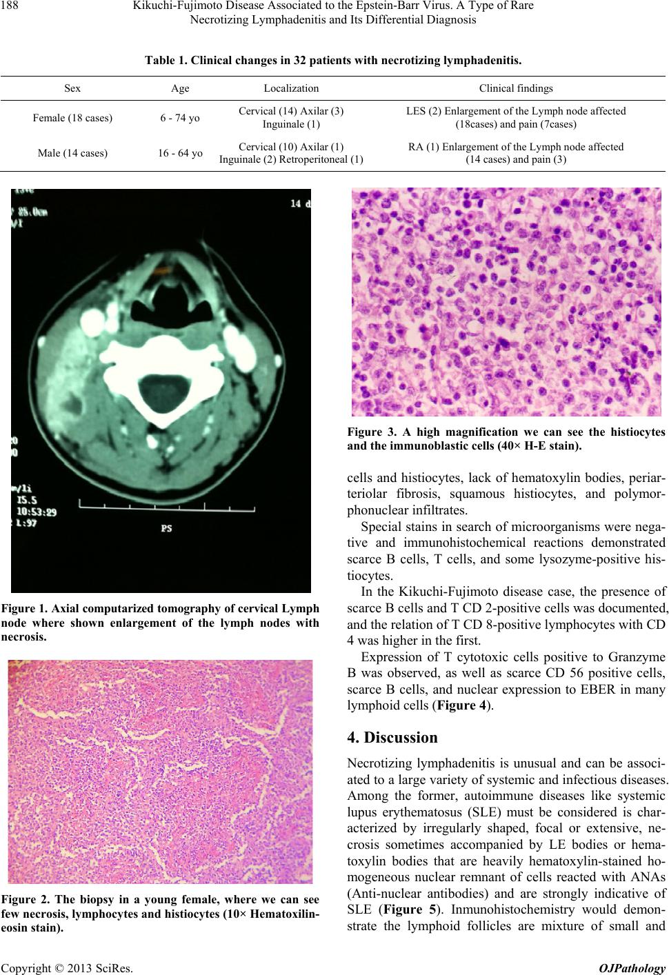 Kikuchi-Fujimoto Disease Associated to the Epstein-Barr Virus. A Type of Rare Necrotizing Lymphadenitis and Its Differential Diagnosis Copyright © 2013 SciRes. OJPathology 188 Table 1. Clinical changes in 32 patients with necrotizing lymphadenitis. Sex Age Localization Clinical findings Female (18 c as e s) 6 - 74 yo Cervical (14) Axilar (3) Inguinale (1) LES (2) Enlargement of the Lymph node affected (18cases) and pain (7cases) Male (14 cases) 16 - 64 yo Cervical (10) Axilar (1) Inguinale (2) Retroperitoneal (1)RA (1) Enlargement of the Lymph node affected (14 cases) and pain (3) Figure 3. A high magnification we can see the histiocytes and the immunoblastic cells (40× H-E stain). cells and histiocytes, lack of hematoxylin bodies, periar- teriolar fibrosis, squamous histiocytes, and polymor- phonuclear infiltrates. Special stains in search of microorganisms were nega- tive and immunohistochemical reactions demonstrated scarce B cells, T cells, and some lysozyme-positive his- tiocytes. In the Kikuchi-Fujimoto disease case, the presence of scarce B cells and T CD 2-positive cells was documented, and the relation of T CD 8-positive lymphocytes with CD 4 was higher in the first. Figure 1. Axial computari zed tomography of c ervical Lymph node where shown enlargement of the lymph nodes with necrosis. Expression of T cytotoxic cells positive to Granzyme B was observed, as well as scarce CD 56 positive cells, scarce B cells, and nuclear expression to EBER in many lymphoid cells (Figure 4). 4. Discussion Necrotizing lymphadenitis is unusual and can be associ- ated to a large variety of systemic and infectious diseases. Among the former, autoimmune diseases like systemic lupus erythematosus (SLE) must be considered is char- acterized by irregularly shaped, focal or extensive, ne- crosis sometimes accompanied by LE bodies or hema- toxylin bodies that are heavily hematoxylin-stained ho- mogeneous nuclear remnant of cells reacted with ANAs (Anti-nuclear antibodies) and are strongly indicative of SLE (Figure 5). Inmunohistochemistry would demon- strate the lymphoid follicles are mixture of small and Figure 2. The biopsy in a young female, where we can see few necrosis, lymphocy tes and histiocytes (10× Hematoxilin- eosin stain). 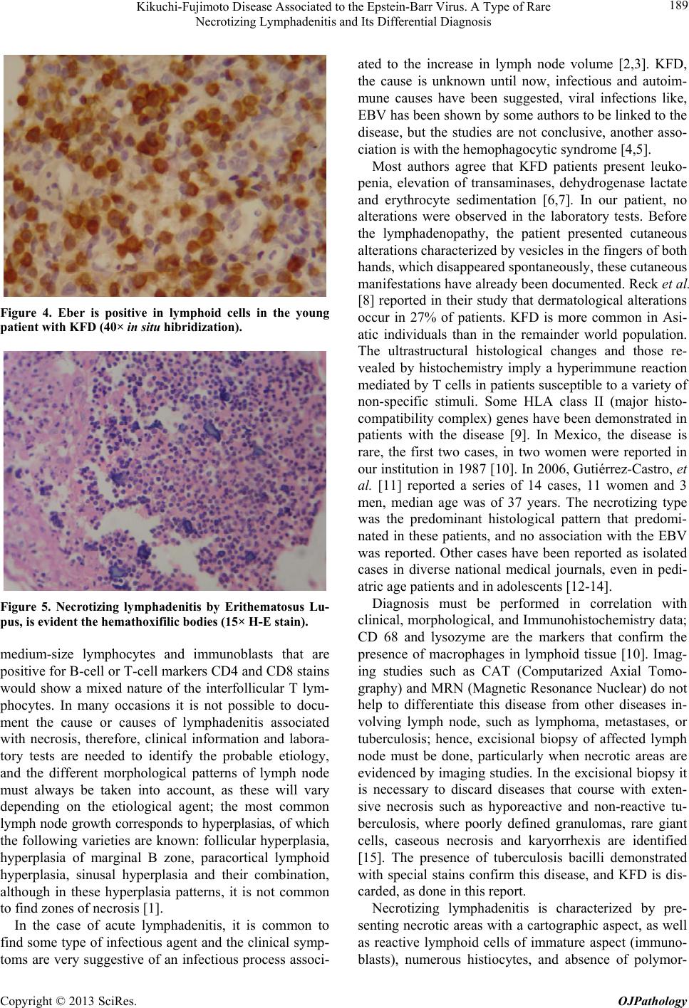 Kikuchi-Fujimoto Disease Associated to the Epstein-Barr Virus. A Type of Rare Necrotizing Lymphadenitis and Its Differential Diagnosis 189 Figure 4. Eber is positive in lymphoid cells in the young patient with KFD (40× in situ hibridization). Figure 5. Necrotizing lymphadenitis by Erithematosus Lu- pus, is evident the hemathoxifilic bodies (15× H-E stain). medium-size lymphocytes and immunoblasts that are positive for B-cell or T-cell markers CD4 and CD8 stains would show a mixed nature of the interfollicular T lym- phocytes. In many occasions it is not possible to docu- ment the cause or causes of lymphadenitis associated with necrosis, therefore, clinical information and labora- tory tests are needed to identify the probable etiology, and the different morphological patterns of lymph node must always be taken into account, as these will vary depending on the etiological agent; the most common lymph node growth corresponds to hyperplasias, of which the following varieties are known: follicular hyperplasia, hyperplasia of marginal B zone, paracortical lymphoid hyperplasia, sinusal hyperplasia and their combination, although in these hyperplasia patterns, it is not common to find zo n es of necrosi s [1 ]. In the case of acute lymphadenitis, it is common to find some type of infectious agent and the clinical symp- toms are very suggestive of an infectious process associ- ated to the increase in lymph node volume [2,3]. KFD, the cause is unknown until now, infectious and autoim- mune causes have been suggested, viral infections like, EBV has been shown by some authors to be linked to the disease, but the studies are not conclusive, another asso- ciation is with the hemophagocytic syn drome [4,5]. Most authors agree that KFD patients present leuko- penia, elevation of transaminases, dehydrogenase lactate and erythrocyte sedimentation [6,7]. In our patient, no alterations were observed in the laboratory tests. Before the lymphadenopathy, the patient presented cutaneous alterations characterized by vesicles in the fingers of both hands, which disappeared spontaneously, these cutaneous manifestations have already been documented. Reck et al. [8] reported in their study that dermatological alterations occur in 27% of patients. KFD is more common in Asi- atic individuals than in the remainder world population. The ultrastructural histological changes and those re- vealed by histochemistry imply a hyperimmune reaction mediated by T cells in patients su sceptible to a variety of non-specific stimuli. Some HLA class II (major histo- compatibility co mplex) genes h ave been demonstrated in patients with the disease [9]. In Mexico, the disease is rare, the first two cases, in two women were reported in our institution in 1987 [10]. In 2006 , Gutiérrez-Castro, et al. [11] reported a series of 14 cases, 11 women and 3 men, median age was of 37 years. The necrotizing type was the predominant histological pattern that predomi- nated in these patients, and no association with the EBV was reported. Other cases have been reported as isolated cases in diverse national medical journals, even in pedi- atric age patients and in adolescents [12-14]. Diagnosis must be performed in correlation with clinical, morphological, and Immunohistochemistry data; CD 68 and lysozyme are the markers that confirm the presence of macrophages in lymphoid tissue [10]. Imag- ing studies such as CAT (Computarized Axial Tomo- graphy) and MRN (Magnetic Resonance Nuclear) do not help to differentiate this disease from other diseases in- volving lymph node, such as lymphoma, metastases, or tuberculosis; hence, excisional biopsy of affected lymph node must be done, particularly when necrotic areas are evidenced by imaging studies. In the excisional biopsy it is necessary to discard diseases that course with exten- sive necrosis such as hyporeactive and non-reactive tu- berculosis, where poorly defined granulomas, rare giant cells, caseous necrosis and karyorrhexis are identified [15]. The presence of tuberculosis bacilli demonstrated with special stains confirm this disease, and KFD is dis- carded, as done in this report. Necrotizing lymphadenitis is characterized by pre- senting necrotic areas with a cartographic aspect, as well as reactive lymphoid cells of immature aspect (immuno- blasts), numerous histiocytes, and absence of polymor- Copyright © 2013 SciRes. OJPathology 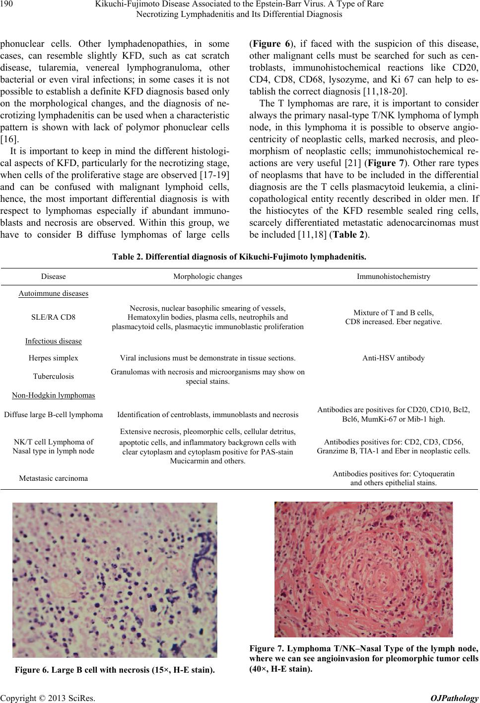 Kikuchi-Fujimoto Disease Associated to the Epstein-Barr Virus. A Type of Rare Necrotizing Lymphadenitis and Its Differential Diagnosis Copyright © 2013 SciRes. OJPathology 190 phonuclear cells. Other lymphadenopathies, in some cases, can resemble slightly KFD, such as cat scratch disease, tularemia, venereal lymphogranuloma, other bacterial or even viral infections; in some cases it is not possible to establish a definite KFD diagnosis based only on the morphological changes, and the diagnosis of ne- crotizing lymphadenitis can be used when a characteristic pattern is shown with lack of polymor phonuclear cells [16]. It is important to keep in mind the different histologi- cal aspects of KFD, particularly for the necrotizing stage, when cells of the proliferative stage are observed [17-19] and can be confused with malignant lymphoid cells, hence, the most important differential diagnosis is with respect to lymphomas especially if abundant immuno- blasts and necrosis are observed. Within this group, we have to consider B diffuse lymphomas of large cells (Figure 6), if faced with the suspicion of this disease, other malignant cells must be searched for such as cen- troblasts, immunohistochemical reactions like CD20, CD4, CD8, CD68, lysozyme, and Ki 67 can help to es- tablish the correct diagnosis [11,18-20]. The T lymphomas are rare, it is important to consider always the primary nasal-type T/NK lymphoma of lymph node, in this lymphoma it is possible to observe angio- centricity of neoplastic cells, marked necrosis, and pleo- morphism of neoplastic cells; immunohistochemical re- actions are very useful [21] (Figure 7). Other rare types of neoplasms that have to be included in the differential diagnosis are the T cells plasmacytoid leukemia, a clini- copathological entity recently described in older men. If the histiocytes of the KFD resemble sealed ring cells, scarcely differentiated metastatic adenocarcinomas must be include d [11,18] ( Table 2). Table 2. Differential diagnosis of Kikuchi-Fujimoto lymphadenitis. Disease Morphologic changes Immunohistochemistry Autoimmune diseases SLE/RA CD8 Necrosis, nuclear basophilic smearing of vessels, Hematoxylin bodies, plasma cells, neutrophils and plasmacytoid cells, plasmacytic immunoblastic proliferation Mixture of T and B cells, CD8 increased. Eber negative. Infectious disease Herpes simplex Viral inclusions must be demonstrate in tissue sections. Anti-HSV antibody Tuberculosis Granulomas with necrosis and microorganisms may show on special stains. Non-Hodgkin lym p h omas Diffuse large B-cell lymphoma Identification of centroblasts, immunoblasts and necrosi s Antibodies are positives for CD20, CD10, Bcl2, Bcl6, MumKi-67 o r Mib-1 high. NK/T cell Lymphoma of Nasal type in lymph node Extensive necrosis, pleomorphic cells, cellular detritus, apoptotic cells, and inflammatory backgrown cells with clear cytoplasm and cytoplasm positive for PAS-stain Mucicarmin and others. Antibodies positives for: CD2, CD3, CD56, Granzime B, TIA-1 and Eber in neoplastic cells. Metastasic carcinoma Antibodies positives for: Cytoqueratin and others epithelial stains. Figure 7. Ly mphoma T/NK–Nasal Type of the lymph node, where we can see angioinvasion for pleomorphic tumor cells (40×, H-E stain). Figure 6. Large B cell with necrosis (15×, H-E stain). 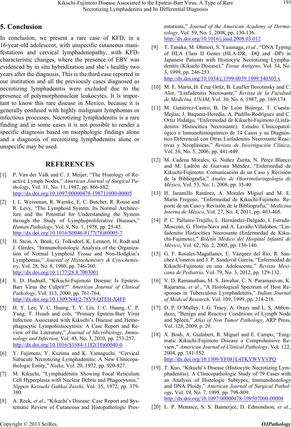 Kikuchi-Fujimoto Disease Associated to the Epstein-Barr Virus. A Type of Rare Necrotizing Lymphadenitis and Its Differential Diagnosis 191 5. Conclusion In conclusion, we present a rare case of KFD, in a 16-year-old adolescent, with unspecific cutaneous mani- festations and cervical lymphadenopathy, with KFD- characteristic changes, where the presence of EBV was evidenced by in situ hybridization and she’s healthy two years after the diagnosis. This is the third case reported in our institution and all the previously cases diagnosed as necrotizing lymphadenitis were excluded due to the presence of polymorphonuclear leukocytes. It is impor- tant to know this rare disease in Mexico, because it is generally confused with highly malignant lymphomas or infectious processes. Necrotizing lymphadenitis is a rare finding and in some cases it is not possible to render a specific diagnosis based on morphologic findings alone and a diagnosis of necrotizing lymphadenitis alone or unspecific may be used. REFERENCES [1] P. Van der Valk and C. J. Meijer, “The Histology of Re- active Lymph Nodes,” American Journal of Surgical Pa- thology, Vol. 11, No. 11, 1987, pp. 866-882. http://dx.doi.org/10.1097/00000478-198711000-00005 [2] I. L. Weissman, R. Warnke, E. C. Butcher, R. Rouse and R. Levy, “The Lymphoid System. Its Normal Architec- ture and the Potential for Understanding the System through the Study of Lymphoproliferative Diseases,” Human Pathology, Vol. 9, No. 1, 1978, pp. 25-45. http://dx.doi.org/10.1016/S0046-8177(78)80005-7 [3] H. Stein, A. Bonk, G. Tolksdorf, K. Lennert, H. Rodt and J. Gerdes, “Immunohistologic Analysis of the Organiza- tion of Normal Lymphoid Tissue and Non-Hodgkin’s Lymphomas,” Journal of Histochemistry & Cytochemis- try, Vol. 28, No. 8, 1980, pp. 746-760. http://dx.doi.org/10.1177/28.8.7003001 [4] S. D. Hudnall, “Kikuchi-Fujimoto Disease: Is Epstein- Barr Virus the Culprit?” American Journal of Clinical Pathology, Vol. 113, 2000, pp. 761-764. http://dx.doi.org/10.1309/N4E2-78V9-QTFH-X4FF [5] H. Y. Lee, Y. C. Huang, T. Y. Lin, J. C. Huang, C. P. Yang, T. Hsueh and cols, “Primary Epstein-Barr Virus Infection Associated with Kikuchi’s Disease and Hemo- phagocytic Lympohistiocytosis: A Case Report and Re- view of the Literature,” Journal of Microbiology, Immu- nology and Infection, Vol. 43, No. 3, 2010, pp. 253-257. http://dx.doi.org/10.1016/S1684-1182(10)60040-0 [6] Y. Fujimoto, Y. Kuzima and K. Yamaguchi, “Cervical Subacute Necrotizing Lymphadenitis: A New Clinicopa- thologic Entity,” Naika, Vol. 20, 1972, pp. 920-927. [7] M. Kikuchi, “Lymphadenitis Showing Focal Reticulum Cell Hyperplasia with Nuclear Debris and Phagocytosis,” Nippon Ketsueki Gakkai Zasshi, Vol. 35, 1972, pp. 379- 380. [8] A. Reck, et al., “Kikuchi’s Disease: Case Report and Sys- tematic Review of Cutaneous and Histopathologic Pres- entations,” Journal of the American Academy of Derma- tology, Vol. 59, No. 1, 2008, pp. 130-136. http://dx.doi.org/10.1016/j.jaad.2008.03.012 [9] T. Tanaka, M. Ohmori, S. Yasunaga, et al., “DNA Typing of HLA Class II Genes (HLA-DR, -DQ and -DP) in Japanese Patients with Histiocytc Necrotizing Lympha- denitis (Kikuchi Disease),” Tissue Antigens, Vol. 54, No. 3, 1999, pp. 246-253. http://dx.doi.org/10.1034/j.1399-0039.1999.540305.x [10] M. E. Marín, H. Cruz Ortíz, B. Lauffer Dorotinsky and C. Alor, “Linfadenitis Necrosante,” Revista de la Facultad de Medicina. UNAM, Vol. 30, No. 6, 1987, pp. 169-174. [11] M. Gutiérrez-Castro, B. De León Bojorge, T. Cuesta- Mejías, J. Baquera-Heredía, A. Padilla-Rodríguez and C. Ortíz Hidalgo, “Enfermedad de Kikuchi-Fujimoto (Linfa- denitis Histiocítica Necrosante). Estudio Clinicopatol- ógico e Inmunohistoquímico de 14 Casos y su Diagnós- tico Diferencial con Otras Linfadenitis Necrozante Reac- tivas y Neoplásicas,” Revista de Investigación Clínica, Vol. 58, No. 5, 2006, pp. 441-449. [12] M. Cadena Morales, G. Nuñez Zurita, N. Pérez Blanco and M. Ladrón de Guevara Méndez, “Enfermedad de Kikuchi-Fujimoto: Comunicación de un Caso y Revisión de la Bibliografía,” Anales de Otorrinolaringología de México, Vol. 53, No. 1, 2008, pp. 35-40. [13] H. Jaramillo Ramírez, A. Morales Miguel and M. E. Marín Fregoso, “Enfermedad de Kikuchi-Fujimoto. Re- porte de un Caso y Revisión de la Bibliografía,” Medicina Interna de México, Vol. 27, No. 4, 2011, pp. 403-405. [14] P. C. Pallares-Trujillo, L. Hernández-Delgado, I. Estrada- Moscoso, G. Flores-Nava and A. Lavalle-Villalobos, “Lin- fadenitis Histiocítica Necrosante (Enfermedad de Kiku- chi-Fujimoto),” Boletín Médico del Hospital Infantil de México, Vol. 62, No. 2, 2005, pp. 136-140. [15] G. F. Rosales-Magallanes, E. Vázquez del Río, R. Sán- chez-Cisneros and J. P. Sandoval García, “Enfermedad de Kikuchi-Fujimoto en una Adolescente,” Revista Mexi- cana de Pediatría, Vol. 79, No. 3, 2012, pp. 129-132. [16] V. D. Ramanathan, M. S. Jawahar, C. N. Paramasivan, K. Rajarama, et al., “A Histological Spectrum of Host Re- sponses in Tuberculous Lymphadenitis,” Indian Journal of Medical Research, Vol. 109, 1999, pp. 214-218. [17] D. P. O’Malley, I. G. Tracy, A. Orazy and L. S. Abbon- dazo, “Benign and Reactive Conditions of Lymph Node and Spleen,” Atlas of Non Tumor Pathology, ARP Press, Vol. 128, 2009, p. 29. [18] X. Bosh, A. Guilabert, R. Miguel and E. Campo, “Enig- matic Kikuchi-Fujimoto Disease a Comprehensive Re- view,” American Journal of Clinical Pathology, Vol. 122, 2004, pp. 141-152. http://dx.doi.org/10.1309/YF081L4TKYWVYVPQ [19] T. Kuo, “Kikuchi’s Disease (Histiocytic Necrotizing Lym- phadenitis). A Clinicopathologic Study of 79 Cases with an Analysis of Histologic Subtypes, Immunohistology and DNA Ploidy,” American Journal of Surgical Pathol- ogy, Vol. 19, No. 7, 1995, pp. 798-809. http://dx.doi.org/10.1097/00000478-199507000-00008 [20] L. P. Menasce, S. S. Bannerjee, D. Edmondson, et al., Copyright © 2013 SciRes. OJPathology  Kikuchi-Fujimoto Disease Associated to the Epstein-Barr Virus. A Type of Rare Necrotizing Lymphadenitis and Its Differential Diagnosis Copyright © 2013 SciRes. OJPathology 192 “Histiocytic Necrotizing Lymphadenitis (Kikuchi-Fuji- moto Disease): Continuing Diagnostic Difficulties,” His- topathol, Vol. 33, No. 3, 1998, pp. 248-254. [21] M. B. Romero-Guadarrama, M. A. Durán-Padilla, A. Alcántara Vásquez, M. Hernández González, K. Hopp García and S. Rivas Vera, “Linfomas no Hodgkin T Pri- marios de Ganglio Linfático. Análisis Morfológico, de Inmunofenotipo y su Asociación con Virus de Epstein- Barr en Pacientes del Hospital General de México,” Re- vista Médica del Hospital General de México, Vol. 70, No. 4, 2007, pp. 168-174.
|