Paper Menu >>
Journal Menu >>
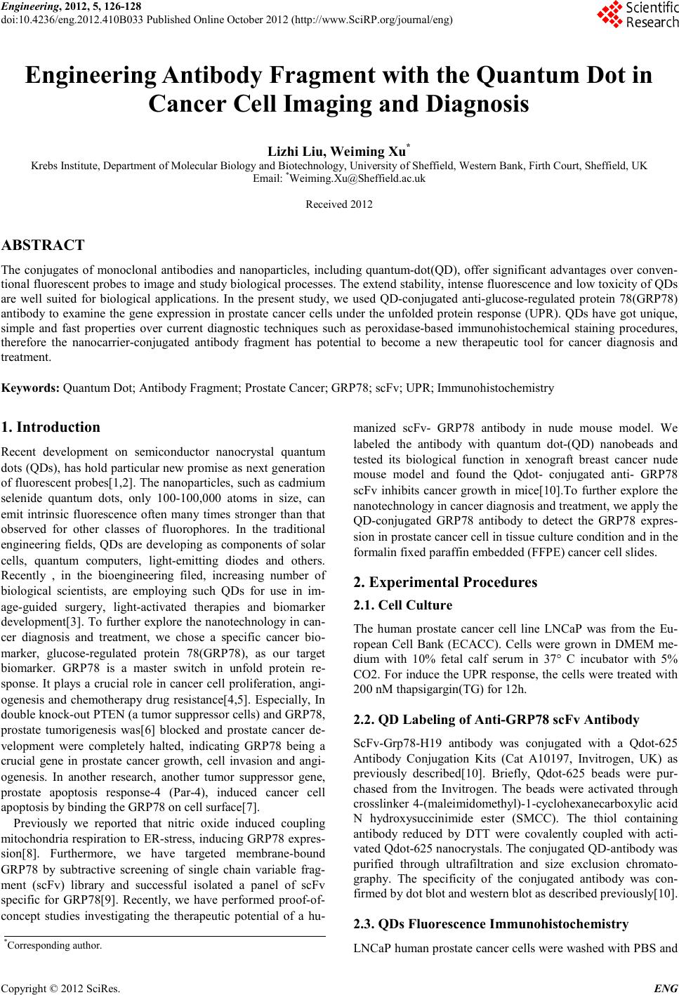 Engineering, 2012, 5, 126-128 doi:10.4236/eng.2012.410B033 Published Online October 2012 (http://www.SciRP.org/journal/eng) Copyright © 2012 SciRes. ENG Engineering Antibody Fragment with the Quantum Dot in Cancer Cell Imaging and Diagnosis Lizhi Liu, Weiming Xu* Krebs Institute, Department of Molecular Biology and Biotechnology, University of Sheffield, Western Bank, Firth Court, Sheffield, UK Email: *Weiming.Xu@Sheffield.ac.uk Received 2012 ABSTRACT The conjugates of monoclonal antibodies and nanoparticles, including quantum-dot(QD), offer significant advantages over conven- tional fluorescent probes to image and study biological processes. The extend stability, intense fluorescence and low toxicity of QDs are well suited for biological applications. In the present study, we used QD-conjugated anti-glucose-regulated protein 78(GRP78) antibody to examine the gene expression in prostate cancer cells under the unfolded protein response (UPR). QDs have got unique, simple and fast properties over current diagnostic techniques such as peroxidase-based immunohistochemical staining procedures, therefore the nanocarrier-conjugated antibody fragment has potential to become a new therapeutic tool for cancer diagnosis and treat ment . Keywords: Quantum Dot; Antibody Fragment; Pr ostate Cancer; GRP78; scFv; UPR; Immunohistochemistry 1. Introduction Recent development on semiconductor nanocrystal quantum dots (QDs), has hold parti cular new promise as n ext generat ion of fluorescent probes[1,2]. The nanoparticles, such as cadmium selenide quantum dots, only 100-100,000 atoms in size, can emit intrinsic fluorescence often many times stronger than that observed for other classes of fluorophores. In the traditional engineering fields, QDs are developing as components of solar cells, quantum computers, light-emitting diodes and others. Recently , in the bioengineering filed, increasing number of biological scientists, are employing such QDs for use in im- age-guided surgery, light-activated therapies and biomarker development[3]. To further explore the nanotechnology in can- cer diagnosis and treatment, we chose a specific cancer bio- marker, glucose-regulated protein 78(GRP78), as our target biomarker. GRP78 is a master switch in unfold protein re- sponse. It plays a crucial role in cancer cell proliferation, angi- ogenesis and chemotherapy drug resistance[4,5]. Especially, In double knock-out PTEN (a tumor suppressor cells) and GRP78, prostate tumorigenesis was[6] blocked and prostate cancer de- velopment were completely halted, indicating GRP78 being a crucial gene in prostate cancer growth, cell invasion and angi- ogenesis. In another research, another tumor suppressor gene, prostate apoptosis response-4 (Par-4), induced cancer cell apoptosis by binding the GRP78 on cell surface[7]. Previously we reported that nitric oxide induced coupling mitochondria respiration to ER-stress, inducing GRP78 expres- sion[8]. Furthermore, we have targeted membrane-bound GRP78 by subtractive screening of single chain variable frag- ment (scFv) library and successful isolated a panel of scFv specific for GRP78[9]. Recently, we have performed proof-of- concept studies investigating the therapeutic potential of a hu- manized scFv- GRP78 antibody in nude mouse model. We labeled the antibody with quantum dot-(QD) nanobeads and tested its biological function in xenograft breast cancer nude mouse model and found the Qdot- conjugated anti- GRP78 scFv inhibits cancer growth in mice[10].To further explore the nanotechnology in cancer diagnosis and treatment, we apply the QD-conjugated GRP78 antibody to detect the GRP78 expres- sion in prostate cancer cell in tissue culture condition and in the formalin fixed paraffin embedded (FFPE ) can cer cell slides. 2. Exp erimenta l Pro cedu r es 2.1. Cell Culture The human prostate cancer cell line LNCaP was from the Eu- ropean Cell Bank (ECACC). Cells were grown in DMEM me- dium with 10% fetal calf serum in 37° C incubator with 5% CO2. F or induce the UPR response, t he cells were treated with 200 nM thapsigargin(TG) for 12h. 2.2. QD Labeling of Anti-GRP78 scFv Antibody ScFv-Grp78-H19 antibody was conjugated with a Qdot-625 Antibody Conjugation Kits (Cat A10197, Invitrogen, UK) as previously described[10]. Briefly, Qdot-625 beads were pur- chased from the Invitrogen. The beads were activated through crosslinker 4-(maleimidomethyl)-1-cyclohexanecarboxylic acid N hydroxysuccinimide ester (SMCC). The thiol containing antibody reduced by DTT were covalently coupled with acti- vated Qdot-625 nanocrystals. The conjugated QD-antibody was purified through ultrafiltration and size exclusion chromato- graphy. The specificity of the conjugated antibody was con- firmed by dot blot and western blot as described p revio usly[10]. 2.3. QDs Fluorescence Immunohistochemistry LNCaP h uman p ro stat e cancer cel ls wer e wash ed with P BS and *Corresponding author. 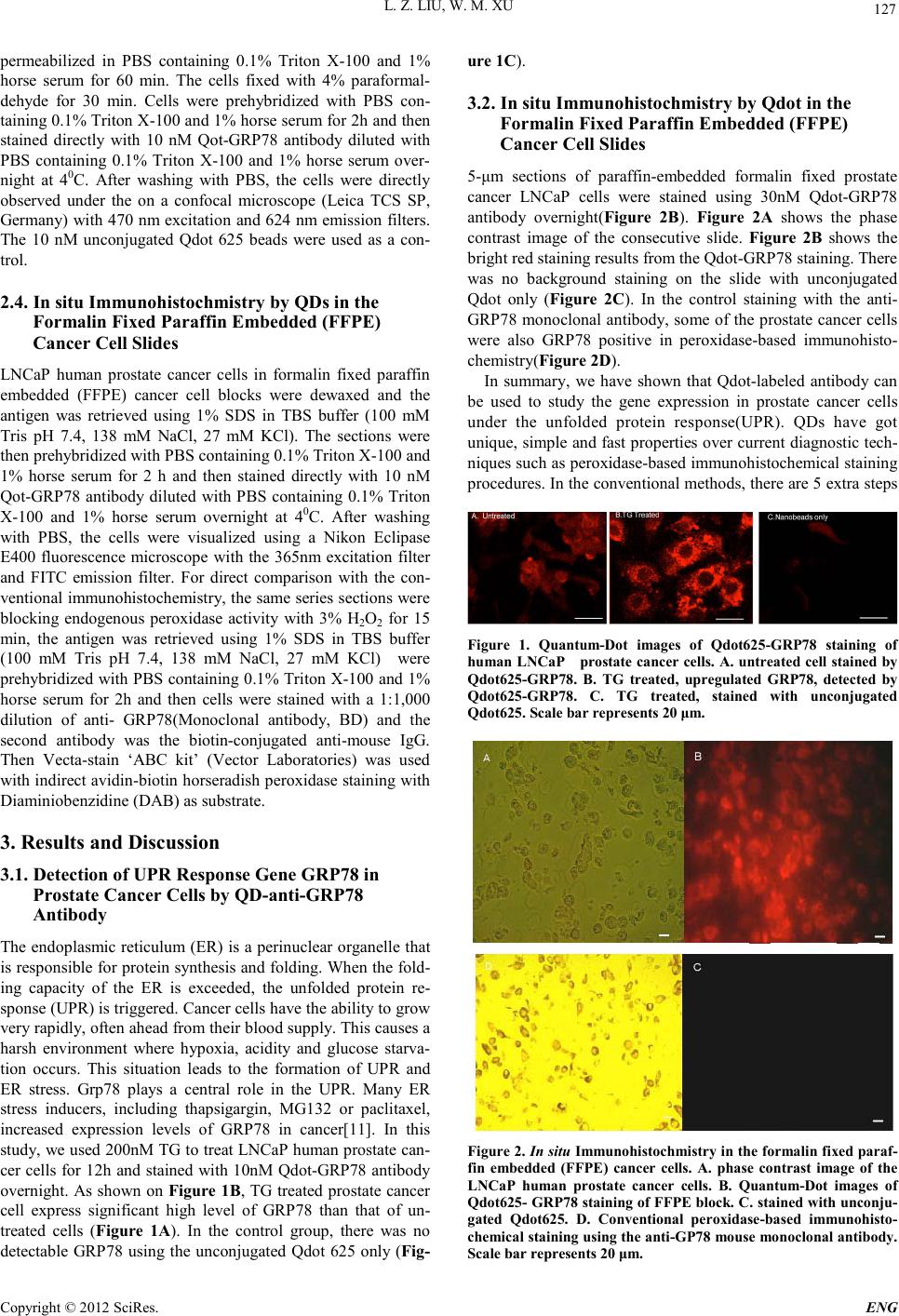 L. Z. LIU, W. M. XU Copyright © 2012 SciRes. E NG 127 permeabilized in PBS containing 0.1% Triton X-100 and 1% horse serum for 60 min. The cells fixed with 4% paraformal- dehyde for 30 min. Cells were prehybridized with PBS con- taining 0.1% Triton X-100 and 1% horse serum for 2h and then stained directly with 10 nM Qot-GRP78 antibody diluted with PBS containing 0.1% Triton X-100 and 1% horse serum over- night at 40C. After washing with PBS, the cells were directly observed under the on a confocal microscope (Leica TCS SP, Germany) with 470 nm excitation and 624 nm emission filters. The 10 nM unconjugated Qdot 625 beads were used as a con- trol. 2.4. In situ Immunohi stochmistry by QDs in the Formalin Fixed Paraffin Embedded (FFPE) Cancer Cell Slides LNCaP human prostate cancer cells in formalin fixed paraffin embedded (FFPE) cancer cell blocks were dewaxed and the antigen was retrieved using 1% SDS in TBS buffer (100 mM Tris pH 7.4, 138 mM NaCl, 27 mM KCl). The sections were then prehybridized with PBS containing 0.1% Triton X-100 and 1% horse serum for 2 h and then stained directly with 10 nM Qot-GRP78 antibody diluted with PBS containing 0.1% Triton X-100 and 1% horse serum overnight at 40C. After washing with PBS, the cells were visualized using a Nikon Eclipase E400 fluorescence microscope with the 365nm excitation filter and FITC emission filter. For direct comparison with the con- ventio nal immuno histochemistr y, the same series s ections wer e blocking endogenous peroxidase activity with 3% H2O2 for 15 min, the antigen was retrieved using 1% SDS in TBS buffer (100 mM Tris pH 7.4, 138 mM NaCl, 27 mM KCl) were prehybridized with PBS containing 0.1% Triton X-100 and 1% horse serum for 2h and then cells were stained with a 1:1,000 dilution of anti- GRP78(Monoclonal antibody, BD) and the second antibody was the biotin-conjugated anti-mouse IgG. Then Vecta-stain ‘ABC kit’ (Vector Laboratories) was used with indirect avidin-biotin horseradish peroxidase staining with Diaminiobenzidine (DAB) as substr ate. 3. Results and Discussion 3.1. Detection of UPR Response Gene GRP78 in Prostate Cancer Cells by QD-a nti-GRP78 Antibody The endo plasmic reticulu m (ER) i s a perinuclear o rganelle that is responsible for protein synthesis and folding. When the fold- ing capacity of the ER is exceeded, the unfolded protein re- spon se (UP R) is tr iggered. Can cer cell s have t he ab il ity to gro w very rapidly, often ahead from their blood supply. This causes a harsh environment where hypoxia, acidity and glucose starva- tion occurs. This situation leads to the formation of UPR and ER stress. Grp78 plays a central role in the UPR. Many ER stress inducers, including thapsigargin, MG132 or paclitaxel, increased expression levels of GRP78 in cancer[11]. In this study, we used 200nM TG to treat LNCaP human prostate can- cer cells for 12h and stained with 10nM Qdot-GRP78 antibody overnight. As shown on Figure 1B, TG treat ed prostate can cer cell express significant high level of GRP78 than that of un- treated cells (Figure 1A). In the control group, there was no detectable GRP78 using the unconjugated Qdot 625 only (Fig- ure 1C). 3.2. In situ Immunohi stochmistry by Qdot in the Formalin Fixed Paraffin Embedded (FFPE) Cancer Cell Slides 5-μm sections of paraffin-embedded formalin fixed prostate cancer LNCaP cells were stained using 30nM Qdot-GRP78 antibody overnight(Figure 2B). Figure 2A shows the phase contrast image of the consecutive slide. Figure 2B shows the bright red staining results from the Qdot-GRP78 staining. There was no background staining on the slide with unconjugated Qdot only (Figure 2C). In the control staining with the anti- GRP78 monoclonal antibody, some of the prostate cancer cells were also GRP78 positive in peroxidase-based immunohisto- chemistr y(Fig ure 2D). In summary, we have shown that Qdot-labeled antibody can be used to study the gene expression in prostate cancer cells under the unfolded protein response(UPR). QDs have got unique, simple and fast properties over current diagnostic tech- niques such as peroxidase-based immunohistochemical staining proced ures. In the con vention al method s, there are 5 extra s teps Figure 1. Quantum-Dot images of Qdot625-GRP78 staining of human LNCaP prostate cancer cells. A. untreated cell stained by Qdot625-GRP78. B. TG treated, upregulated GRP78, detected by Qdot625-GRP78. C. TG treated, stained with unconjugated Qdot625. Scale bar represents 20 μm. Figure 2. In situ Immunohistochmistry in the formalin f ixed paraf- fin embedded (FFPE) cancer cells. A. phase contrast image of the LNCaP human prostate cancer cells. B. Quantum-Dot images of Qdot625- GRP78 staining of FFPE block. C. sta ined with unconju- gated Qdot625. D. Conventional peroxidase-based immunohisto- chemical staining using the anti-GP78 mouse monoclonal antibody. Scale bar represents 20 μm. 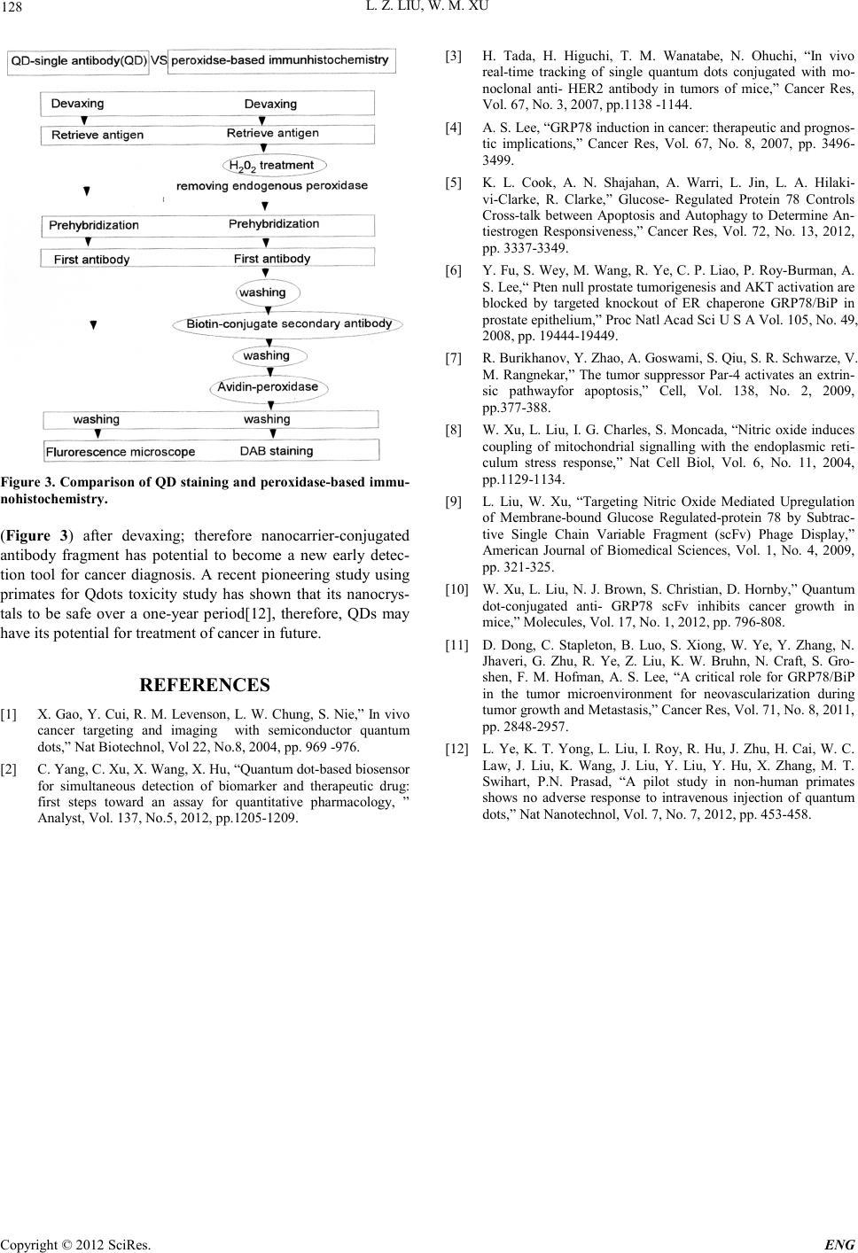 L. Z. LIU, W. M. XU Copyright © 2012 SciRes. ENG 128 Figure 3. Comparison of QD staining and peroxidase-based immu- nohistochemistry. (Figure 3) after devaxing; therefore nanocarrier-conjugated antibody fragment has potential to become a new early detec- tion tool for cancer diagnosis. A recent pioneering study using primates for Qdots toxicity study has shown that its nanocrys- tals to be safe over a one-year period[12], therefore, QDs may have its potenti al for treatment of cancer in future. REFERENCES [1] X. Gao, Y. Cui, R. M. Levenson, L. W. Chung, S. Nie,” In vivo cancer targeting and imaging with semiconductor quantum dots,” Nat Biotechnol, Vol 22, No.8, 2004, pp. 969 -976. [2] C . Yan g, C. Xu, X . Wan g, X. H u , “Qu an tu m dot -based biosensor for simultaneous detection of biomarker and therapeutic drug: first steps toward an assay for quantitative pharmacology, ” Analyst, Vol. 137, No.5, 201 2, pp.1205-1209. [3] H. Tada, H. Higuchi, T. M. Wanatabe, N. Ohuchi, “In vivo real-time tracking of single quantum dots conjugated with mo- noclonal anti- HER2 antibody in tumors of mice,” Cancer Res, Vol. 67, No. 3, 2007, pp.1138 -1144. [4] A. S. Lee, “GRP78 induction in cancer: therapeutic and prognos- tic implications,” Cancer Res, Vol. 67, No. 8, 2007, pp. 3496- 3499. [5] K. L. Cook, A. N. Shajahan, A. Warri, L. Jin, L. A. Hilaki- vi-Clarke, R. Clarke,” Glucose- Regulated Protein 78 Controls Cross-talk between Apoptosis and Autophagy to Determine An- tiestrogen Responsiveness,” Cancer Res, Vol. 72, No. 13, 2012, pp. 3337-3349. [6] Y. Fu, S. Wey, M. Wang, R. Ye, C. P. Liao, P. Roy-Burman, A. S. Lee,“ Pten null prosta te tumori genesis an d AKT activati on are blocked by targeted knockout of ER chaperone GRP78/BiP in pros t at e ep it h eli um ,” Pro c N at l Acad Sci U S A Vol. 105, No. 49, 2008, pp. 19444-19449. [7] R. Burikhanov, Y. Zhao, A. Goswami, S. Qiu, S. R. Schwarze, V. M. Rangnekar,” The tumor suppressor Par-4 activates an extrin- sic pathwayfor apoptosis,” Cell, Vol. 138, No. 2, 2009, pp.377-388. [8] W. Xu, L. Liu, I. G. Charles, S. Moncada, “Nitric oxide induces coupling of mitochondrial signalling with the endoplasmic reti- culum stress response,” Nat Cell Biol, Vol. 6, No. 11, 2004, pp.1129-1134. [9] L. Liu, W. Xu, “Targeting Nitric Oxide Mediated Upregulation of Membrane-bound Glucose Regulated-protein 78 by Subtrac- tive Single Chain Variable Fragment (scFv) Phage Display,” American Journal of Biomedical Sciences, Vol. 1, No. 4, 2009, pp. 321-325. [10] W. Xu , L. Liu , N. J. Brown, S. Christi an, D. Hornb y,” Quantum dot-conjugated anti- GRP78 scFv inhibits cancer growth in mice,” Molecules, Vol. 17, No. 1, 2012, pp. 796-808. [11] D. Dong, C. Stapleton, B. Luo, S. Xiong, W. Ye, Y. Zhang, N. Jhaveri, G. Zhu, R. Ye, Z. Liu, K. W. Bruhn, N. Craft, S. Gro- shen, F. M. Hofman, A. S. Lee, “A critical role for GRP78/BiP in the tumor microenvironment for neovascularization during tumor growth and Metastasis,” Cancer R es, Vol. 71, No. 8, 201 1, pp. 2848-2957. [12] L. Ye, K. T. Yong, L. Liu, I. Roy, R. Hu, J. Zhu, H. Cai, W. C. Law, J. Liu, K. Wang, J. Liu, Y. Liu, Y. Hu, X. Zhang, M. T. Swihart, P.N. Prasad, “A pilot study in non-human primates shows no adverse response to intravenous injection of quantum dots,” Nat Nanot echn o l , Vol. 7, No. 7, 2012, pp. 453-458. |